. 2020 Sep 1;23(9):101517.
doi: 10.1016/j.isci.2020.101517. eCollection 2020 Sep 25.

Identification of Small Molecules that Modulate Mutant p53 Condensation

Affiliations

Identification of Small Molecules that Modulate Mutant p53 Condensation

Clara Lemos et al. iScience. .

Abstract

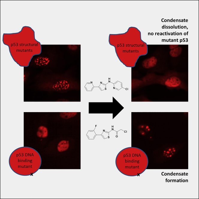
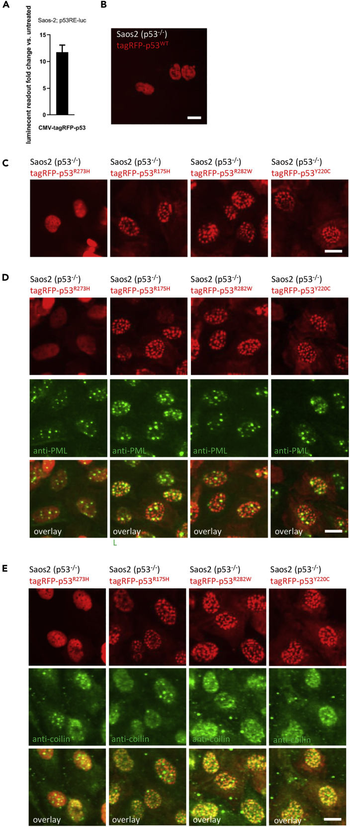
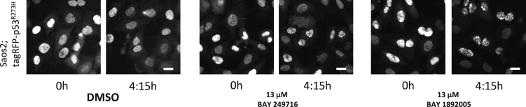
Structural mutants of p53 induce global p53 protein destabilization and misfolding, followed by p53 protein aggregation. First evidence indicates that p53 can be part of protein condensates and that p53 aggregation potentially transitions through a condensate-like state. We show condensate-like states of fluorescently labeled structural mutant p53 in the nucleus of living cancer cells. We furthermore identified small molecule compounds that interact with the p53 protein and lead to dissolution of p53 structural mutant condensates. The same compounds lead to condensation of a fluorescently tagged p53 DNA-binding mutant, indicating that the identified compounds differentially alter p53 condensation behavior depending on the type of p53 mutation. In contrast to p53 aggregation inhibitors, these compounds are active on p53 condensates and do not lead to mutant p53 reactivation. Taken together our study provides evidence for structural mutant p53 condensation in living cells and tools to modulate this process.

Keywords: Biochemistry Methods; Medical Biochemistry; Structural Biology.


Conflict of interest statement

All authors are or were employees of Bayer AG.

Figures

None
Graphical abstract
Figure 1
Figure 1
Condensate Formation of Fluorescent Structural Mutant p53 Variants (A) Saos2 containing a p53 activity reporter showed reporter activation after transduction of a tagRFP-p53 expression vector. Luminescent readout normalized to DMSO controls. Bars show mean with SD (n = 3). (B) Saos2 cells transiently transfected with a tagRFP-p53WT expression vector show homogeneous distribution of tagRFP-p53 in the nucleus. Scale bar, 10 μm. (C) Saos2 cells expressing fluorescently tagged structural mutants show p53 condensates in the nuclei. Scale bars, 10 μm. (D and E) Antibody staining of Saos2 cells expressing fluorescently tagged structural mutants with anti-PML or coilin antibodies. No co-localization of fluorescently tagged mutant p53 condensates with either PML (D) or Cajal bodies (E) in the nucleus is observable. Scale bars, 10 μm.
Figure 2
Figure 2
Identification of Compounds that Interact with p53 Proteins (A and B) (A) Hit compounds. (B) Deconvoluted spectra of intact mass analysis of p53 shows covalent binding of BAY 1892005 to p53R175H. DMSO control shows two peaks of p53R175H protein, one for the expected mass of 28,508 Da and one of 28,685 Da representing N-terminal gluconoylated (Gal) p53R175 (blue arrows). Incubation with BAY 1892005 showed mass shifts of 234 Da to both the apoprotein and the glyconoylated p53R175H (green arrows), indicative for covalent binding of BAY 1892005. An additional mass shift of 234 Da indicates a partial two-fold binding of BAY 1892005 to p53R175H. Exemplary data of multiple experiments are shown (n ≥ 2).
Figure 3
Figure 3
Condensate Formation of Fluorescent Structural Mutant p53 Variants and Their Dissolution by Aminothiazoles (A) Saos2 cells expressing fluorescently tagged structural mutants show p53 condensates in the nuclei, which are dissolved after aminothiazole treatment. Stills (0 and 4:15 h) from time-lapse Videos 2A, 2B, and 2C. Scale bars, 10 μm. (B) Kinetics of condensate dissolution in Saos2 cells expressing structural p53 mutants in response to treatment with aminothiazoles. Bars show mean with SD (n = 4). Exemplary data of multiple experiments are shown (n ≥ 2).
Figure 4
Figure 4
Aminothiazoles Induce Condensation of Fluorescent DNA-Binding Defective p53R273H Mutant Variant Saos2 cells expressing a fluorescently tagged DNA-binding domain mutant show homogeneous distribution of the fluorescent signal in the nuclei, and aminothiazole treatment leads to the formation of condensate-like structures in some cells of the population. Stills (0 and 4:15 h) from time-lapse Video 2D. Scale bars, 10 μm. Exemplary data of multiple experiments are shown (n ≥ 2).
Figure 5
Figure 5
Aminothiazoles Lead to Reduction of Nuclear Accumulation of Endogenous Structural p53 Mutants (A–E) p53R175H cell lines (A) SK-BR-3, (B) AU-565, (C) Cal-33, and (D) Detroit 562 and (E) p53Y220C cell line Huh7 were treated with aminothiazoles for 6 h and stained by IF for p53 (DO-7), and the intensity of the staining in the nucleus was quantified. ∗∗∗∗p value < 0.0001. Scale bar, 10 μm. Bars show mean with SD (n ≥ 5). Exemplary data of multiple experiments are shown (n ≥ 2).
Figure 6
Figure 6
Identified Hits Do Not Lead to Structural Mutant p53 Reactivation (A) U2OS (p53WT) cells containing the p53 activity reporter show nuclear accumulation of p53 upon 10 μM Nutlin-3 treatment and induction of the activity reporter. (B) Cal-33 (p53R175H) cells containing the p53 activity reporter show nuclear accumulation of p53R175H and no p53 activity reporter induction after Nutlin-3 treatment (data not shown) but induction after transduction of a CMV-driven p53 expression vector. (C) Saos2 (p53−/−) cells containing the p53 activity reporter showed no staining for p53 and no induction of the p53 activity reporter after Nutlin-3 treatment (data not shown) but induction of the p53 activity reporter after transduction of a CMV-driven p53 expression vector. Luminescent readout normalized to DMSO controls. Exemplary data of multiple experiments are shown (n ≥ 2) Scale bars, 10 μm. (D) p53 activity reporter activation upon aminothiazole treatment in cells with different p53 mutation status. Luminescent readout normalized to DMSO controls. Exemplary data of multiple experiments are shown (n ≥ 2), Bars show mean with SD (n = 8). (E) Expression of structural mutant p53 in p53−/− cells does not enhance p53 activity reporter activation. Luminescent readout normalized to DMSO controls. Exemplary data of multiple experiments are shown (n ≥ 2), Bars show mean with SD (n = 8). (F) p53 knockdown validated by IF and western blot; partial p73 knockdown shown in western blot. Exemplary data of multiple experiments are shown (n ≥ 2); full western blot data show in Figure S6. Scale bar, 10 μm. (G) p53 knockdown does not prevent p53 activity reporter activation in Cal-33 cells, whereas p73 knockdown shows suppression of p53 activity reporter activation. Luminescent readout normalized to DMSO controls. Exemplary data of multiple experiments are shown (n ≥ 2), Bars show mean with SD (n = 10). ∗∗∗p value < 0.001; ns, not significant.

References

    1. Alberti S., Gladfelter A., Mittag T. Considerations and challenges in studying liquid-liquid phase separation and biomolecular condensates. Cell. 2019;176:419–434. - PMC - PubMed
    1. Anbo H., Sato M., Okoshi A., Fukuchi S. Functional segments on intrinsically disordered regions in disease-related proteins. Biomolecules. 2019;9:88. - PMC - PubMed
    1. Ang H.C., Joerger A.C., Mayer S., Fersht A.R. Effects of common cancer mutations on stability and DNA binding of full-length p53 compared with isolated core domains. J. Biol. Chem. 2006;281:21934–21941. - PubMed
    1. Banani S.F., Lee H.O., Hyman A.A., Rosen M.K. Biomolecular condensates: organizers of cellular biochemistry. Nat. Rev. Mol. Cell Biol. 2017;18:285–298. - PMC - PubMed
    1. Boija A., Klein I.A., Sabari B.R., Dall'Agnese A., Coffey E.L., Zamudio A.V., Li C.H., Shrinivas K., Manteiga J.C., Hannett N.M. Transcription factors activate genes through the phase-separation capacity of their activation domains. Cell. 2018;175:1842–1855 e16. - PMC - PubMed