. 2020 Sep 10;12(9):1010.
doi: 10.3390/v12091010.

Family Level Phylogenies Reveal Relationships of Plant Viruses within the Order Bunyavirales

Affiliations

Family Level Phylogenies Reveal Relationships of Plant Viruses within the Order Bunyavirales

Venura Herath et al. Viruses. .

Abstract

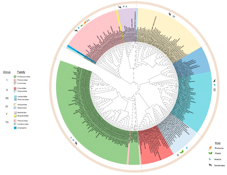
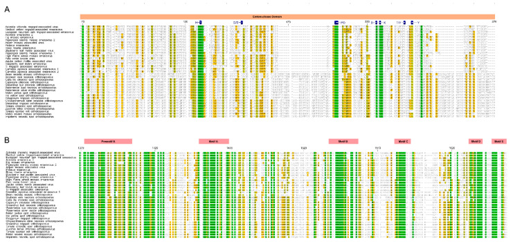
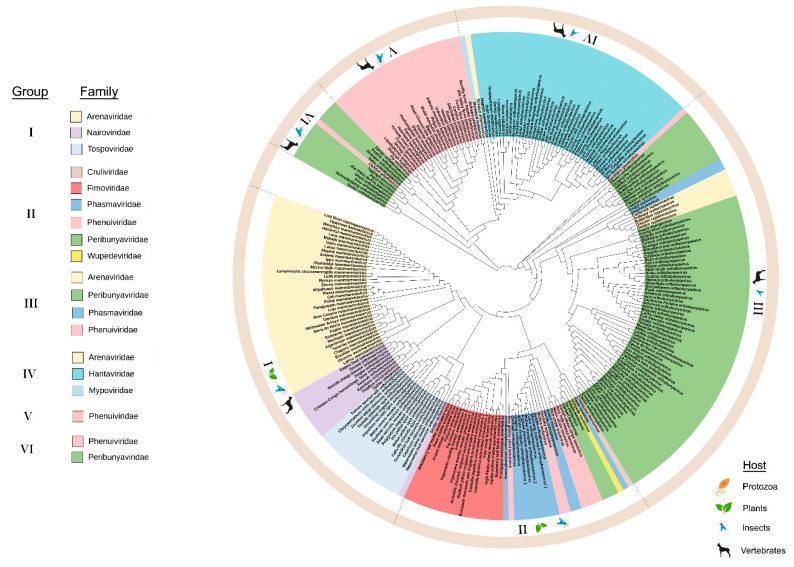
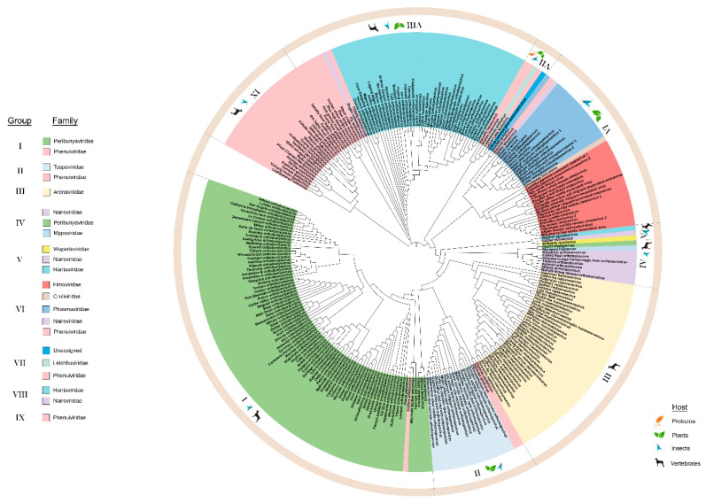
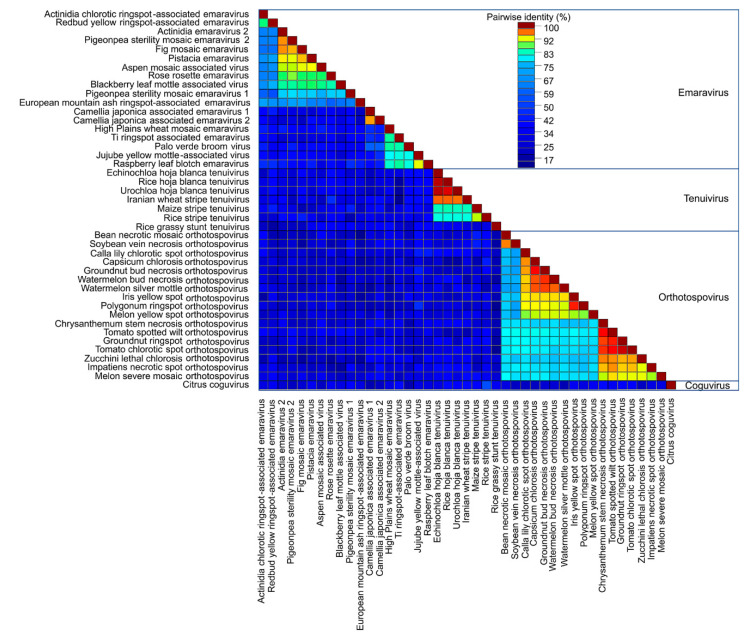
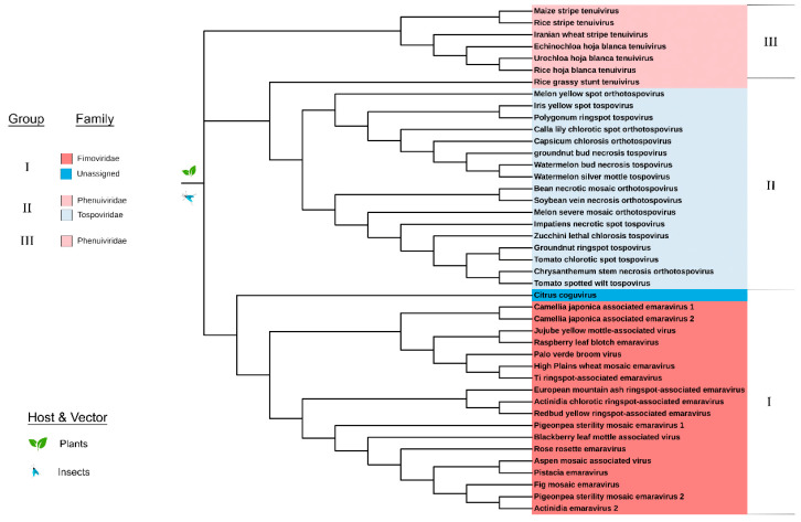
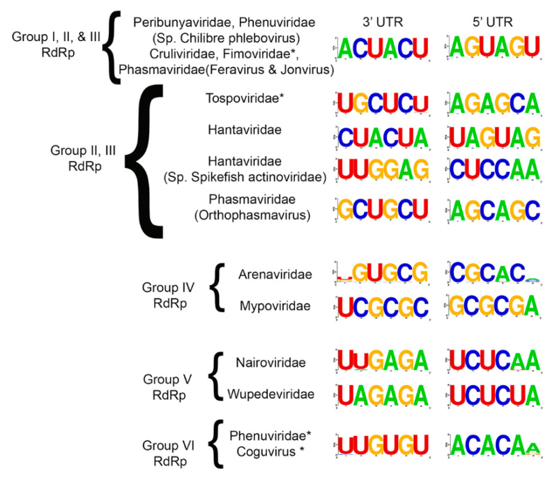
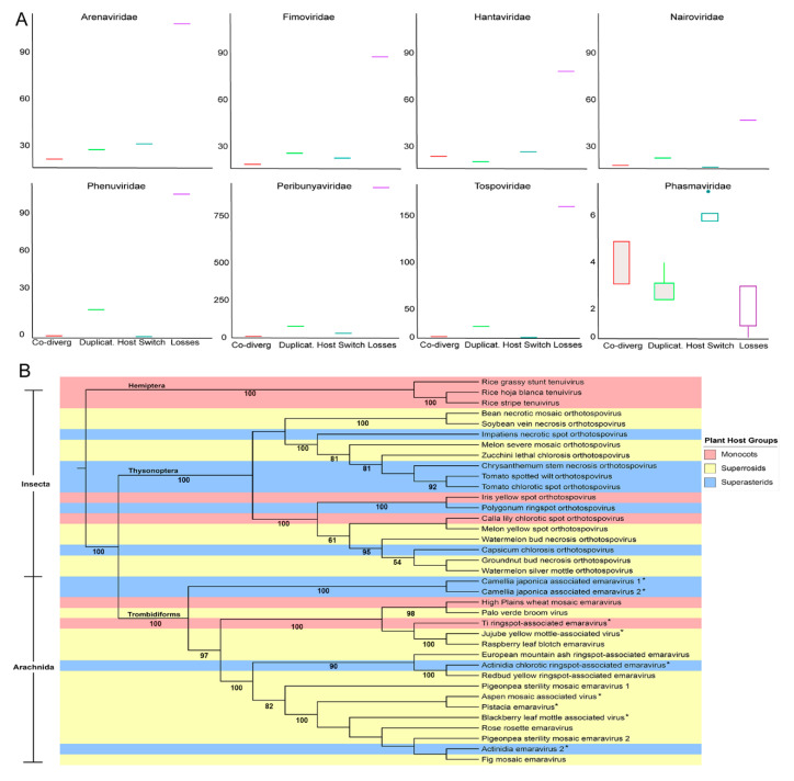
Bunyavirales are negative-sense segmented RNA viruses infecting arthropods, protozoans, plants, and animals. This study examines the phylogenetic relationships of plant viruses within this order, many of which are recently classified species. Comprehensive phylogenetic analyses of the viral RNA dependent RNA polymerase (RdRp), precursor glycoprotein (preGP), the nucleocapsid (N) proteins point toward common progenitor viruses. The RdRp of Fimoviridae and Tospoviridae show a close evolutional relationship while the preGP of Fimoviridae and Phenuiviridae show a closed relationship. The N proteins of Fimoviridae were closer to the Phasmaviridae, the Tospoviridae were close to some Phenuiviridae members and the Peribunyaviridae. The plant viral movement proteins of species within the Tospoviridae and Phenuiviridae were more closely related to each other than to members of the Fimoviridae. Interestingly, distal ends of 3' and 5' untranslated regions of species within the Fimoviridae shared similarity to arthropod and vertebrate infecting members of the Cruliviridae and Peribunyaviridae compared to other plant virus families. Co-phylogeny analysis of the plant infecting viruses indicates that duplication and host switching were more common than co-divergence with a host species.

Keywords: Bunyavirale; RNA virus; cophylogeny; emerging virus; hallmark genes; plant virus; virus evolution.


Conflict of interest statement

The authors declare no conflict of interest. The funders had no role in the design of the study; in the collection, analyses, or interpretation of data; in the writing of the manuscript, or in the decision to publish the results.

Figures

Figure 1
Figure 1
Maximum likelihood phylogenetic tree of the amino acid sequences of the RNA-dependent RNA polymerase (RdRp). The virus families are color-coded and the hosts for viruses within each group are indicated in the outermost circle. The six groups are identified in the legend and the boundaries of these groups are indicated in the outer ring of the phylogeny. Group I: Peribunyaviridae, Phenuiviridae, and Cruliviridae. Group II: Fimoviridae and Tospoviridae. Group III: Hantaviridae and Phasmaviridae. Group IV: Arenaviridae and Mypoviridae. Group V: Nairoviridae and Wupedeviridae. Group VI: Phenuiviridae, Leishbuviridae, and unassigned species. Clade validation is based on the approximate likelihood ratio test (aLRT)-Shimodaira–Hasegawa (SH)-like test values.
Figure 2
Figure 2
Amino acid alignment showing conserved motifs of the RdRp within Fimoviridae and Tospoviridae. (A). The endonuclease domain is indicated by pink bar and active site motifs are identified in blue. (B). The polymerase function motifs are named in the red bars as preA motif through E motif. The alignment colored based on the sequence similarity.
Figure 3
Figure 3
Maximum likelihood phylogenetic tree of the amino acid sequences of the glycoprotein precursor (preGP). Six groups were identified based on clustering from the most distant node. The legend identifies the lineage groups and colors used to identify taxonomic families as in Figure 1. Group I: Arenaviridae, Nairoviridae and Tospoviridae. Group II: Cruliviridae, Fimoviridae, Phasmaviridae, Phenuiviridae, Peribunyaviridae, and Wupedeviridae. Group III: Arenaviridae, Peribunyaviridae, Phasmaviridae, and Phenulviridae. Group IV: Arenaviridae, Hantaviridae, and Mypoviridae. Group V: Phenuiviridae. Group VI: Phenuiviridae and Peribunyaviridae. Families are color-coded and the hosts for viruses within each group are indicated in the outermost circle. Clade validation is based on the aLRT-SH-like test values.
Figure 4
Figure 4
Maximum likelihood phylogenetic tree of the amino acid sequences of the nucleocapsid (N). Ten lineage groups were identified based on clustering from the most distant node. Group I: Peribunyavirdae and Phenuiviridae. Group II: Tospoviridae and Phenuiviridae. Group III: Arenaviridae. Group IV: Nairoviridae, Peribunyaviridae, and Mypoviridae. Group V: Wupedeviridae, Nairoviridae, and Hantaviridae. Group VI: Fimoviridae, Cruliviridae, Phasmaviridae, Nairoviridae, and Phenuiviridae. Group VII: Leishbuviridae, Phenuiviridae, and unassigned species. Group VIII: Hantaviridae and Nairoviridae. Group IX: Phenuiviridae. Group X: Phenuiviridae. Families are color-coded and the hosts for viruses within each group are indicated in the outermost circle. Clade validation is based on the aLRT-SH-like test values.
Figure 5
Figure 5
Pairwise sequence alignment of movement proteins (MP) for plant viruses within Bunyavirales. Sequence analysis was conducted for all available plant virus within Bunyavirales. The plant virus families are Emaravirus, Tenuivirus, Orthotospovirus, and Coguvirus. The alignment is colored based on the sequence similarity.
Figure 6
Figure 6
Maximum likelihood phylogenetic tree of the amino acid sequences of the movement protein (MP) belonging to plant viruses. Three groups were identified based on clustering from the most distant node: Group I: Fimoviridae and an unassigned species. Group II: Phenuiviridae and Tospoviridae. Group III: Phenuiviridae. All viruses of Bunyavirales with an available MP sequence have plant hosts. Clade validation is based on the aLRT-SH-like test values.
Figure 7
Figure 7
Consensus nucleotide sequence of the 3′ and 5′ termini for each genomic segment of Bunyaviriales. The consensus sequences were generated using the 6 most distal nucleotides on each end of the viral genomic segments. Each of the analyzed regions was located within a UTR. Families that contain plant viruses are highlighted with an asterisk.
Figure 8
Figure 8
Estimation of co-phylogenetic events of the nucleic acid sequence of plant-infecting virus families within Bunyavirales. (A). The cophylogeny relationship is based on the RdRp sequences and analyzed using Jane ver. 4.0.1. (B). A neighbor joining tree generated using concatenated genomic segments containing RdRp, NC, GP, and MP. Color was used to identify host plant taxonomies and insect vector taxonomy is provided along the branches. Bootstrap values are provided.

References

    1. Wichgers Schreur P.J., Kormelink R., Kortekaas J. Genome packaging of the Bunyavirales. Curr. Opin. Virol. 2018;33:151–155. doi: 10.1016/j.coviro.2018.08.011. - DOI - PubMed
    1. Abudurexiti A., Adkins S., Alioto D., Alkhovsky S.V., Avšič-Županc T., Ballinger M.J., Bente D.A., Beer M., Bergeron É., Blair C.D., et al. Taxonomy of the order Bunyavirales: Update 2019. Arch. Virol. 2019;164:1949–1965. doi: 10.1007/s00705-019-04253-6. - DOI - PMC - PubMed
    1. Navarro B., Minutolo M., De Stradis A., Palmisano F., Alioto D., Di Serio F. The first phlebo-like virus infecting plants: A case study on the adaptation of negative-stranded RNA viruses to new hosts. Mol. Plant Pathol. 2018;19:1075–1089. doi: 10.1111/mpp.12587. - DOI - PMC - PubMed
    1. Navarro B., Zicca S., Minutolo M., Saponari M., Alioto D., Di Serio F. A negative-stranded RNA virus infecting citrus trees: The second member of a new genus within the order bunyavirales. Front. Microbiol. 2018;9:1–11. doi: 10.3389/fmicb.2018.02340. - DOI - PMC - PubMed
    1. Li C.X., Shi M., Tian J.H., Lin X.D., Kang Y.J., Chen L.J., Qin X.C., Xu J., Holmes E.C., Zhang Y.Z. Unprecedented genomic diversity of RNA viruses in arthropods reveals the ancestry of negative-sense RNA viruses. Elife. 2015;2015:1–26. doi: 10.7554/eLife.05378. - DOI - PMC - PubMed

Publication types