Review
. 2020 Sep 10;21(18):6615.
doi: 10.3390/ijms21186615.

State of the Art Review on Genetics and Precision Medicine in Arrhythmogenic Cardiomyopathy

Affiliations
Review

State of the Art Review on Genetics and Precision Medicine in Arrhythmogenic Cardiomyopathy

Viraj Patel et al. Int J Mol Sci. .

Abstract

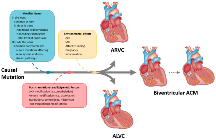
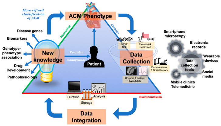
Arrhythmogenic cardiomyopathy (ACM) is an inherited cardiomyopathy characterised by ventricular arrhythmia and an increased risk of sudden cardiac death (SCD). Numerous genetic determinants and phenotypic manifestations have been discovered in ACM, posing a significant clinical challenge. Further to this, wider evaluation of family members has revealed incomplete penetrance and variable expressivity in ACM, suggesting a complex genotype-phenotype relationship. This review details the genetic basis of ACM with specific genotype-phenotype associations, providing the reader with a nuanced perspective of this condition; whilst also proposing a future roadmap to delivering precision medicine-based management in ACM.

Keywords: arrhythmogenic cardiomyopathy; arrhythmogenic right ventricular cardiomyopathy; cardiac arrhythmia; desmosome; genetics; genotype phenotype correlation; sudden cardiac death.


Conflict of interest statement

The authors declare no conflict of interest.

Figures

Figure 1
Figure 1
Explanted heart images showing pathological features of different phenotypes in ACM, adapted from Calkins (2013) [10], Thiene et al. (2016) [11], Corrado et al. (2020) [12]. (A) Gross specimen shows dilated thin RV, fatty replacement of entire RV free wall epicardium and thin fibrotic endocardium (white arrow). (B) Gross specimen shows little evidence of RV involvement, however subepicardial grey band of fibrotic tissue (black arrow) is seen in the posterolateral section of the LV. (C) Gross specimen shows fibro-fatty biventricular involvement (black arrows). (D) Histology image shows fat replacement extending from epicardium to endocardium. (E) Histology image with trichrome staining identifies fibrous scars within fat tissue. LV = left ventricle; RV = right ventricle.
Figure 2
Figure 2
Spectrum of clinical presentations of right ventricular disease, left ventricular disease, and dilated cardiomyopathy. (A) ACM and ALVC show overlapping phenotypes with ARVC, but the pathology is seen primarily in the left ventricle (although right ventricle is also usually affected). (B) Timeline showing the clinical features of ACM. Hypokinetic non-dilated cardiomyopathy and isolated-ischaemic scar pertain to the pathology where there is marked ventricular scarring as identified by cardiac MRI. Modified from Elliott et al. (2019) [22] and Pinto et al. (2016) [24]. ACM = arrhythmogenic cardiomyopathy; aDCM = arrhythmogenic dilated cardiomyopathy; ALVC = arrhythmogenic left ventricular cardiomyopathy; ARVC = arrhythmogenic right ventricular cardiomyopathy; BiV = biventricular; BivACM = biventricular arrhythmogenic cardiomyopathy; CMP = cardiomyopathy; DCM = dilated cardiomyopathy; LV = left ventricle; PVC = premature ventricular contraction; RV = right ventricle; RWMA = regional wall motion abnormality; SCD = sudden cardiac death.
Figure 3
Figure 3
Genetic mutations associated with arrhythmogenic cardiomyopathy. Overlapping genetic determinant implicated in ACM identified from our clinical experience. ACM = arrhythmogenic cardiomyopathy; ALVC = arrhythmogenic left ventricular cardiomyopathy; ARVC = arrhythmogenic right ventricular cardiomyopathy; DCM = dilated cardiomyopathy.
Figure 4
Figure 4
Exemplar MRI and ECG finding found in ACM patients with PKP2 mutations. (A) CMR of a patient with PKP2 c1664del T mutation reveals moderate enlargement of the right ventricle; (B) yellow arrow shows apical fibrosis in LGE mode;.(C) 12-Lead ECG of same patient showing ventricular tachycardia with left bundle branch block morphology and inferior axis; (D) resting 12-Lead ECG upon admission to our hospital showing sinus rhythm, and T-wave inversion is present in V1-V4. Reproduced with permission from Trenkwalder et al. (2015) [34].
Figure 5
Figure 5
Exemplar MRI and ECG finding found in ACM patients with DSG2 mutations. (A) CMR of a patient with DSG2 p.Leu237Ter mutation showing dilation of both ventricles; (B) a representative ECG of the same patient showing T wave inversion in V1-V3. Reproduced with permission from Chen et al. (2020) [35].
Figure 6
Figure 6
Exemplar MRI and ECG finding found in ACM patients with DSP mutations. (A) CMR of a patient with DSP mutation showing left ventricular lateral wall fatty infiltration (red arrow); (B) LGE MRI of the same patient showing fibrosis/scaring of both left ventricular lateral wall and septum; (C) 12-lead ECG showing T-wave inversion in V5-V6 (anterolateral leads). Reproduced with permission from Mattesi et al. (2020) [36].
Figure 7
Figure 7
Exemplar MRI and ECG finding found in ACM patients with DSC2 mutations. (A,B) CMR of a patient with DSC2 p.Arg49His mutation showing biventricular involvement in the LGE mode (red arrows); (C) 12-lead ECG showing a reduction of R waves amplitude in left precordial leads (left involvement) and inverted T waves in V1-V6 (RV strain pattern); together they indicate biventricular involvement. Reproduced with permission from Gaido et al. (2017) [37].
Figure 8
Figure 8
Exemplar MRI and ECG finding found in ACM patients with JUP mutations. (A,B) CMR of a patient with JUP mutation showing LGE of the RV free wall, secondary to fibrosis (white arrow); (C) ECG findings in a different patient with JUP mutation at 3 different times (separated by two vertical lines into 1. right panel = sinus rhythm with T waves inversion in V1–V6, and 2. middle and 3.left panels = ventricular tachycardia). (A,B) were reproduced with permission from Mavrogeni et al. (2012) [38]; (C) was reproduced with permission from Protonotarios et al. (1986) [27].
Figure 9
Figure 9
Exemplar MRI and ECG finding found in ACM patients with TTN mutations. (A) CMR of a patient with TTN mutation, the white arrows show mid-wall scars in the inferior septum and inferior wall; (B) ECG of a different patient with TTN Thr2896Ile mutation showing epsilon waves. (A) was reproduced from Augusto et al. (2019) [39]; (B) from Taylor et al. (2011) [40] with permissions.
Figure 10
Figure 10
Exemplar MRI and ECG finding found in ACM patients with LMNA mutations. (A) CMR of a patient with LMNA mutation showing a mid-wall (LGE) scar in the septum and inferior RV; (B) 12-lead ECG of a different patient with a p.Gly382Val LMNA mutation, which shows sinus bradycardia, poor R-wave progression, T-wave inversion in V1–V3 leads. (A) reproduced with permission from Augusto et al. (2019) [39]; and (B) reproduced with permission from Quarta et al. (2012) [41].
Figure 11
Figure 11
Exemplar MRI and ECG finding found in ACM patients with DES mutations. (A) CMR of a patient with DES mutation (deletion of guanine at position 735) showing extensive mid-wall fibrosis (LGE short-axis view); (B) 12-lead ECG of a different patient with p.Gly382Val mutation of the LMNA gene showing sinus tachycardia, inverted T waves in V1–V3 ad poor R wave progression. Reproduced from Koitka et al. (2017) with permission [42].
Figure 12
Figure 12
Exemplar MRI and ECG finding found in ACM patients with FLNC mutations. (A,B) CMR of a patient with p.Arg991X (nonsense) mutation of the FLNC gene; the arrows show basal lateral subepicardial fibrosis in the LGE mode (white arrows); (C) 12-lead ECG of the same patient showing T-wave inversion in III and aVF (inferior leads). Reproduced with permission from Hall et al. (2020) [43].
Figure 13
Figure 13
Exemplar MRI and ECG finding found in ACM patients with TJP1 mutations. (A,B) CMR of a patient with TJP1 p.Tyr669Cys mutation showing right ventricular dilatation (white arrows); (C,D) LGE shows mid-mural fibrosis in the inferior-lateral wall of the left ventricle (empty arrows); (E) 12-lead ECG of the same patient showing shows sinus rhythm, intra-ventricular conduction delay, and T wave inversion in V1 to V5. Reproduced with permission from De Bortoli et al. (2018) [44].
Figure 14
Figure 14
Exemplar MRI and ECG finding found in ACM patients with CTNNA3 mutations. (A) CMR of a patient with CTNNA3 p.val94asp mutation showing marked right ventricular dilatation; (B) 12-lead ECG of the same patient showing a first degree heart block and T-wave inversion in V1–V4; (C) sustained ventricular tachycardia, with left bundle-branch block and left axis deviation. Reproduced with permission from van Hengel et al. (2013) [45].
Figure 15
Figure 15
Exemplar MRI and ECG finding found in ACM patients with TMEM43 mutations. (AD): CMR images of a male subject with TMEM43 p.S358L mutation. (A) Biventricular dilatation, wall motion abnormalities; (B) asynchronous contraction (red arrows); (C,D) LGE showing severe and almost concentric intra-myocardial lesions (red arrows); (E) a representative 12-lead ECG of a different patient with the same TMEM43 mutation, showing poor R-wave progression with 1-mV R-wave voltage in lead V3 and widened QRS complex. Reproduced with permission from Dominguez et al. (2020) [46].
Figure 16
Figure 16
Exemplar MRI and ECG finding found in ACM patients with PLN mutations. (A) CMR image from a patient with PLN p.Arg14del mutation showing inferolateral wall thinning and late-gadolinium enhancement of the LV inferolateral wall; (B) 12-lead ECG of the same patient showing normal sinus rhythm with low voltages in all leads (<0.5 mV) and flattened or inverted T-waves in V1-V6 and inferior leads (II, III, aVF). Reproduced with permission from te Rijdt et al. (2019) [47].
Figure 17
Figure 17
Exemplar MRI and ECG finding found in ACM patients with RYR2 mutations. (A) CMR of a patient with RYR2 p.Trp98Ter mutation showing dilated cardiomyopathy; (B,C) 12-lead ECG showing inverted T waves in leads II, III, aVF, V3–V6, and two premature ventricular complexes originating from the anterobasal left ventricle. Reproduced with permission from Costa et al. (2020) [48].
Figure 18
Figure 18
Exemplar MRI and ECG finding found in ACM patients with SCN5A mutations. (A,B) CMR of a female patient with SCN5A mutation (p.Arg1898His) showing end-diastolic and end-systolic state, respectively. The white arrow shows an enlarged right ventricle and dyskinesia in the RV outflow tract; (C) a 12-lead ECG of the same patient showed T-wave inversion in V1–2. Reproduced with permission from te Riele et al. (2017) [49].
Figure 19
Figure 19
Exemplar MRI and ECG finding found in ACM patients with NKX2-5 mutations. (A) A cardiac MRI of a patient with NKX2-5 c.471_472delCA variant showing non-dilated hypokinetic ventricle, no valvular heart disease, no LGE; (B) ECG finding of a different patient with the same NKX2-5 mutation as in (A). CMR = cardiovascular magnetic resonance; ECG = electrocardiogram; LGE = late gadolinium enhancement; LV = left Ventricle; RV = right Ventricle. Gene location images taken from https://www.ncbi.nlm.nih.gov/gene/.
Figure 20
Figure 20
Select proteins implicated in the pathogenesis of ACM at the cardiomyocyte junction. BAG3 = BAG family molecular chaperone regulator 3; Nav1.5 = voltage-gated Na+ channel 1.5; NKX2-5 = NK2 homeobox 5; RBM20 = RNA-binding motif protein 20; SERCA = sarcoplasmic reticulum Ca2+ ATPase; SR = sarcoplasmic reticulum; TGF-𝛽3 = transforming growth factor 𝛽3; TJP1 = tight junction protein. * = location is still unclear.
Figure 21
Figure 21
Schematic view of the possible modifier genes, post-translational, epigenetic, and environmental factors contributing to the pathogenesis of ACM including ARVC, ALVC, and biventricular ACM. Modified from Watkins et al. (2011) [131]. ACM = arrhythmogenic cardiomyopathy; ALVC = arrhythmogenic left ventricular cardiomyopathy ARVC = arrhythmogenic right ventricular cardiomyopathy.
Figure 22
Figure 22
An integrated precision medicine systems model in ACM. The goal of precision medicine is to redefine the classification of diseases such as ACM into more refined classes. Multidimensional longitudinal data is collected by clinicians, clinical researchers and patients; technological advances in the last decade have provided opportunities to deeply phenotype and rapidly genotype patients—we describe some innovative data collection tools in our diagram. Data is curated, analysed and stored known as data processing by bioinformaticians using computational methods. Processed data can be used to discover new knowledge about a disease phenotype such as biomarkers, new disease genes etc. (the field of research). These insights can be translated to provide more personalised management to patients. As disease phenotypes such as ACM become more refined, e.g., from morphofunctional classes to molecular classes, more targeted therapies will be discovered. ACM = arrhythmogenic cardiomyopathy.

Similar articles

Cited by

References

    1. Towbin J.A., McKenna W.J., Abrams D.J., Ackerman M.J., Calkins H., Darrieux F.C.C., Daubert J.P., de Chillou C., DePasquale E.C., Desai M.Y., et al. 2019 HRS expert consensus statement on evaluation, risk stratification, and management of arrhythmogenic cardiomyopathy. Heart Rhythm. 2019;16:e301–e372. doi: 10.1016/j.hrthm.2019.05.007. - DOI - PubMed
    1. Asatryan B., Medeiros-Domingo A. Translating emerging molecular genetic insights into clinical practice in inherited cardiomyopathies. J. Mol. Med. 2018;96:993–1024. doi: 10.1007/s00109-018-1685-y. - DOI - PubMed
    1. Marcus F.I., Fontaine G.H., Guiraudon G., Frank R., Laurenceau J.L., Malergue C., Grosgogeat Y. Right ventricular dysplasia: A report of 24 adult cases. Circulation. 1982;65:384–398. doi: 10.1161/01.CIR.65.2.384. - DOI - PubMed
    1. Basso C., Corrado D., Marcus F.I., Nava A., Thiene G. Arrhythmogenic right ventricular cardiomyopathy. Lancet. 2009;373:1289–1300. doi: 10.1016/S0140-6736(09)60256-7. - DOI - PubMed
    1. Marcus F.I., McKenna W.J., Sherrill D., Basso C., Bauce B., Bluemke D.A., Calkins H., Corrado D., Cox M.G., Daubert J.P., et al. Diagnosis of arrhythmogenic right ventricular cardiomyopathy/dysplasia: Proposed modification of the task force criteria. Circulation. 2010;121:1533–1541. doi: 10.1161/CIRCULATIONAHA.108.840827. - DOI - PMC - PubMed

MeSH terms