. 2020 Sep 10;12(9):863.
doi: 10.3390/pharmaceutics12090863.

Encapsulation of Nedaplatin in Novel PEGylated Liposomes Increases Its Cytotoxicity and Genotoxicity against A549 and U2OS Human Cancer Cells

Affiliations

Encapsulation of Nedaplatin in Novel PEGylated Liposomes Increases Its Cytotoxicity and Genotoxicity against A549 and U2OS Human Cancer Cells

Salma El-Shafie et al. Pharmaceutics. .

Abstract

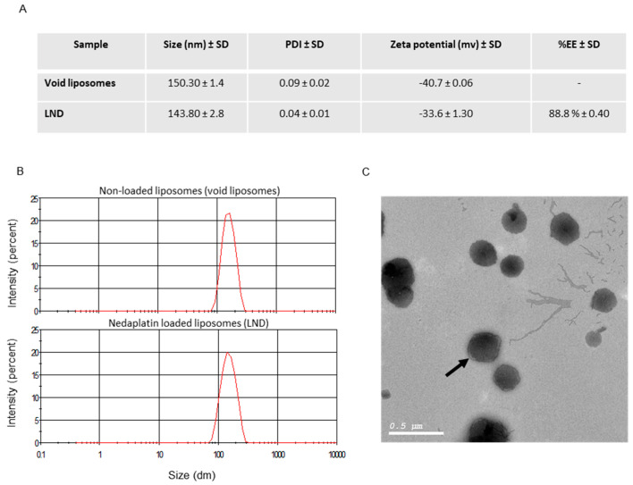
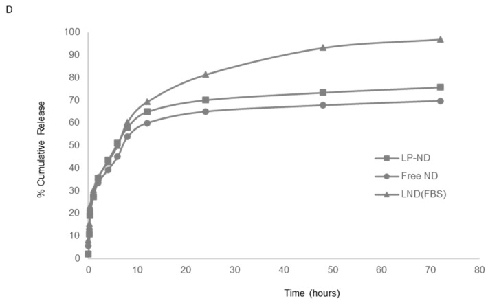
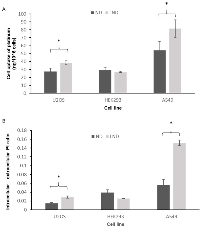
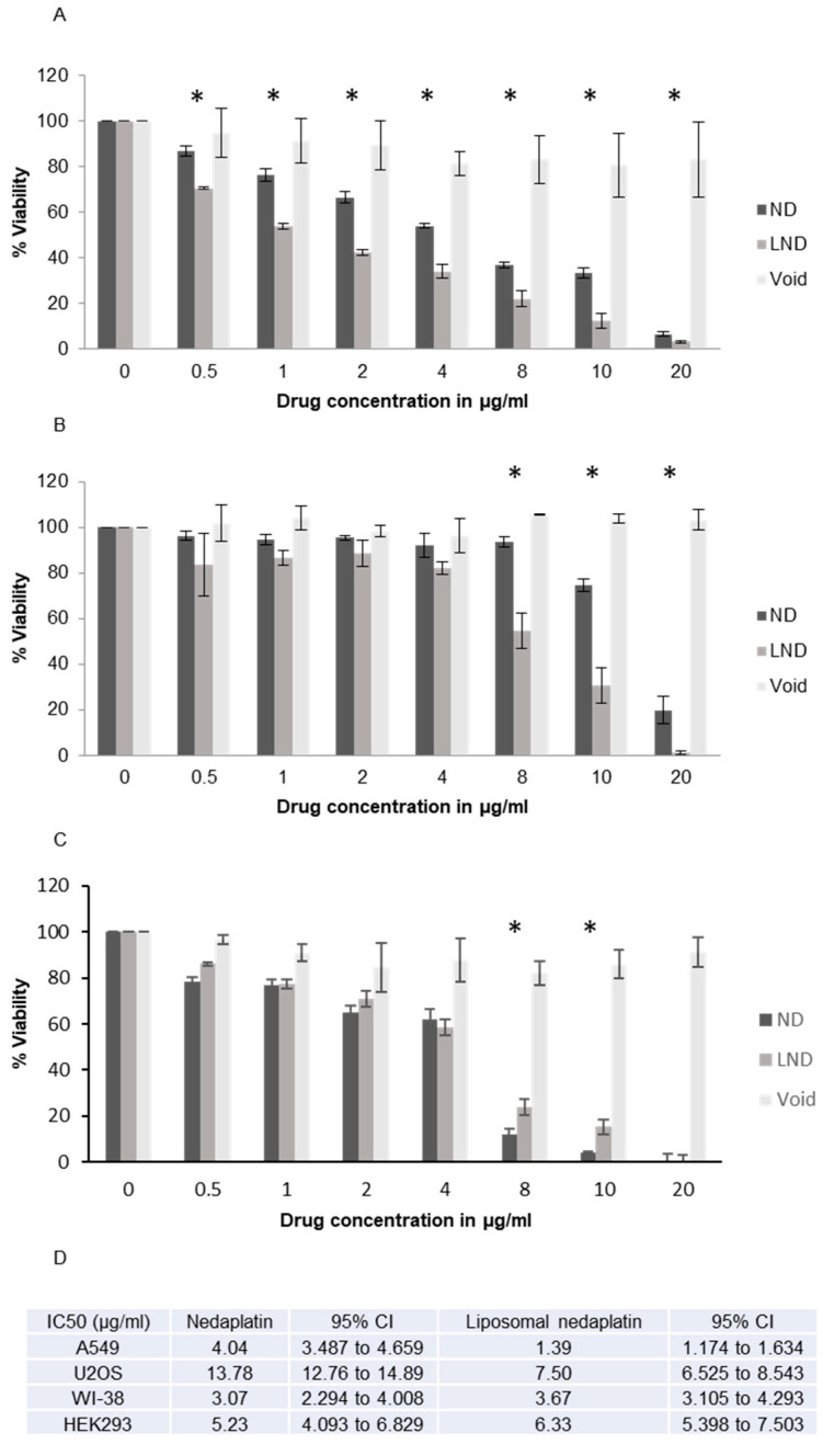
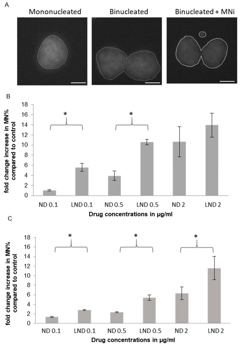
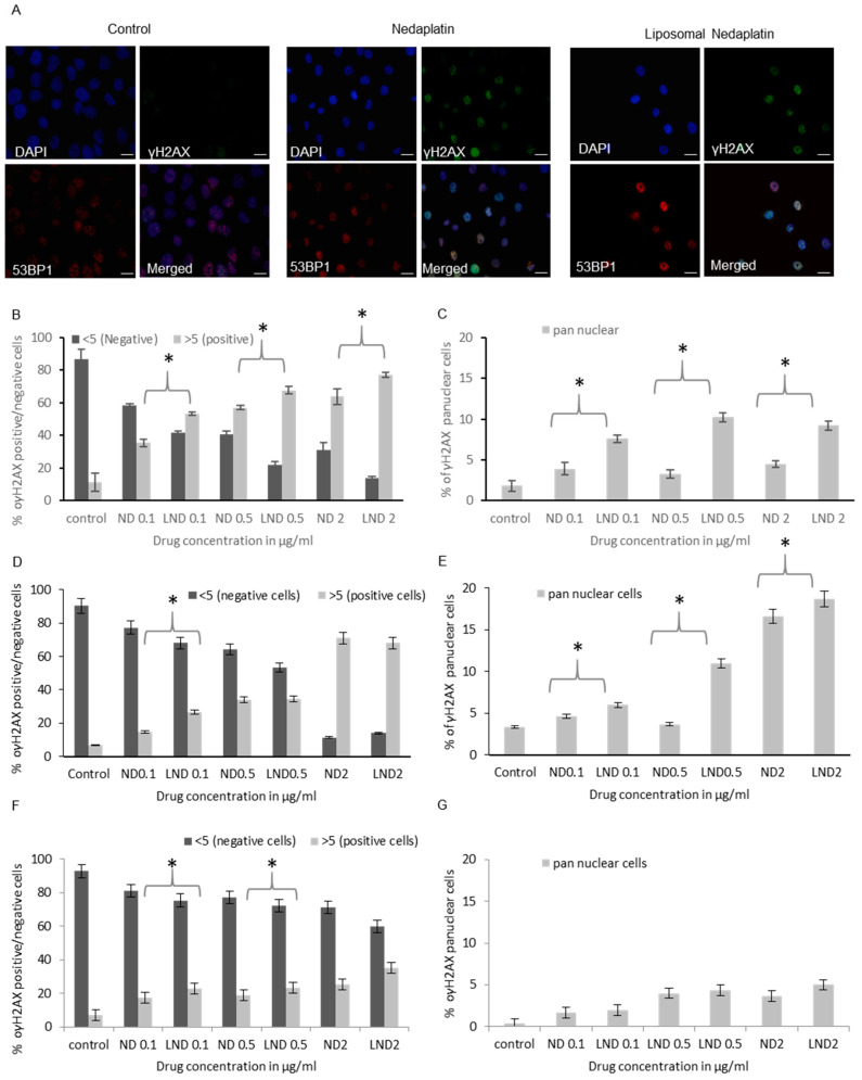
Following the discovery of cisplatin over 50 years ago, platinum-based drugs have been a widely used and effective form of cancer therapy, primarily causing cell death by inducing DNA damage and triggering apoptosis. However, the dose-limiting toxicity of these drugs has led to the development of second and third generation platinum-based drugs that maintain the cytotoxicity of cisplatin but have a more acceptable side-effect profile. In addition to the creation of new analogs, tumor delivery systems such as liposome encapsulated platinum drugs have been developed and are currently in clinical trials. In this study, we have created the first PEGylated liposomal form of nedaplatin using thin film hydration. Nedaplatin, the main focus of this study, has been exclusively used in Japan for the treatment of non-small cell lung cancer, head and neck, esophageal, bladder, ovarian and cervical cancer. Here, we investigate the cytotoxic and genotoxic effects of free and liposomal nedaplatin on the human non-small cell lung cancer cell line A549 and human osteosarcoma cell line U2OS. We use a variety of assays including ICP MS and the highly sensitive histone H2AX assay to assess drug internalization and to quantify DNA damage induction. Strikingly, we show that by encapsulating nedaplatin in PEGylated liposomes, the platinum uptake cytotoxicity and genotoxicity of nedaplatin was significantly enhanced in both cancer cell lines. Moreover, the enhanced platinum uptake as well as the cytotoxic/antiproliferative effect of liposomal nedaplatin appears to be selective to cancer cells as it was not observed on two noncancer cell lines. This is the first study to develop PEGylated liposomal nedaplatin and to demonstrate the superior cell delivery potential of this product.

Keywords: DNA repair; cancer treatment; chemotherapeutics; liposomes; nedaplatin; platinum drugs.


Conflict of interest statement

The authors have no competing interests.

Figures

Figure 1
Figure 1
Liposome preparation and characterization. (A) Liposome characterization. (B) Size distribution plots as obtained at a scan rate of 140 counts per second at 25 °C. Top panel is for nonloaded liposomes while the bottom panel is for nedaplatin-loaded liposomes. (C) TEM image for LND formulation showing uniform spherical structures. The coating PEG layers are also shown as lighter outer circles (see arrow). (D) Nedaplatin and formulated nedaplatin release profiles in PBS and FBS media. Circles represent free nedaplatin in PBS, squares are liposomal nedaplatin in PBS and triangles represent liposomal nedaplatin in FBS.
Figure 1
Figure 1
Liposome preparation and characterization. (A) Liposome characterization. (B) Size distribution plots as obtained at a scan rate of 140 counts per second at 25 °C. Top panel is for nonloaded liposomes while the bottom panel is for nedaplatin-loaded liposomes. (C) TEM image for LND formulation showing uniform spherical structures. The coating PEG layers are also shown as lighter outer circles (see arrow). (D) Nedaplatin and formulated nedaplatin release profiles in PBS and FBS media. Circles represent free nedaplatin in PBS, squares are liposomal nedaplatin in PBS and triangles represent liposomal nedaplatin in FBS.
Figure 2
Figure 2
Uptake of platinum by cancer and noncancer cells. (A) Total platinum uptake (ng) in samples of one million treated cells accurately quantified by ICP MS. (B) Platinum uptake ratios (intracellular: extracellular platinum) in cancer and noncancer cells. In both cases, a statistically significant increase in cellular uptake of platinum with PEGylated liposomal nedaplatin (LND) compared to ND was observed specifically in the cancer cells lines (p-value < 0.05). Multiple pair-wise t-tests showed the cell lines where a significant difference in platinum uptake between LND and ND was observed. These points are indicated by asterisks * (p < 0.05) in the charts above.
Figure 3
Figure 3
Evaluating cytotoxicity of free and liposomal nedaplatin after 72 h of drug exposure using MTT assay in (A) A549, (B) U2OS, and (C) WI-38. (D) IC50 table. An overall statistically significant difference in cell viability was observed between LND and ND in A549 and U2OS (p-value < 0.05) with LND being more cytotoxic. This was not observed in WI-38. Multiple pair-wise t-tests show the concentrations at which a significant difference between both drugs was observed. These points are indicated by asterisks * (p < 0.05).
Figure 4
Figure 4
Genotoxicity of free and liposomal ND in different cell lines assessed through micronucleus formation induction. (A) Representative image of MNi. (B) A549; (C) U2OS and (D) WI-38 shows a significant increase in MNi induction between free and liposomal ND in cancer cell lines A and B, but not in normal cell lines, D. Multiple pair-wise t-tests show the concentrations at which a significant difference between both drugs was observed. These points are indicated by asterisks * (p < 0.05). Scale bars are 5 µm.
Figure 4
Figure 4
Genotoxicity of free and liposomal ND in different cell lines assessed through micronucleus formation induction. (A) Representative image of MNi. (B) A549; (C) U2OS and (D) WI-38 shows a significant increase in MNi induction between free and liposomal ND in cancer cell lines A and B, but not in normal cell lines, D. Multiple pair-wise t-tests show the concentrations at which a significant difference between both drugs was observed. These points are indicated by asterisks * (p < 0.05). Scale bars are 5 µm.
Figure 5
Figure 5
Genotoxicity of free and liposomal ND in cancer and noncancer cell lines assessed through γH2AX induction. (A) Representative immunofluorescence images of control, ND and LND treated A549 cells. Quantification of γH2AX positive and γH2AX pan nuclear cells, respectively, in (B,C) A549, (D,E) U2OS, (F,G) WI-38 cells. While an overall statistically significant increase in the percentage of cells positive for DNA damage observed with LND compared to ND was shown in all cell lines, a statistically significant increase in the γH2AX pan-nuclear signal was only observed with LND in the cancer cell lines (p-value < 0.05). Multiple pair-wise t-tests showed the concentrations of treatment where a significant difference between LND and ND DNA damage is observed. These points are indicated by asterisks * (p < 0.05) in the charts above. Scale bars are 10 µm.

References

    1. Hang Z., Cooper M.A., Ziora Z.M. Platinum-based anticancer drugs encapsulated liposome and polymeric micelle formulation in clinical trials. Biochem. Compd. 2016;4:1. doi: 10.7243/2052-9341-4-2. - DOI
    1. Latosińska J.N., Latosińska M. Anticancer Drug Discovery—From Serendipity to Rational Design. IntechOpen Limited; London, UK: 2013. - DOI
    1. Kelland L. The resurgence of platinum-based cancer chemotherapy. Nat. Rev. Cancer. 2007;7:573–584. doi: 10.1038/nrc2167. - DOI - PubMed
    1. Chaney S.G., Campbell S.L., Temple B., Bassett E., Wu Y., Faldu M. Protein interactions with platinum–DNA adducts: From structure to function. J. Inorg. Biochem. 2004;98:1551–1559. doi: 10.1016/j.jinorgbio.2004.04.024. - DOI - PubMed
    1. Barry M.A., Behnke C.A., Eastman A. Activation of programmed cell death (apoptosis) by cisplatin, other anticancer drugs, toxins and hyperthermia. Biochem. Pharmacol. 1990;40:2353–2362. doi: 10.1016/0006-2952(90)90733-2. - DOI - PubMed

LinkOut - more resources