Review
. 2020 Dec;40(1_suppl):S6-S24.
doi: 10.1177/0271678X20951995. Epub 2020 Sep 14.

Regulation of blood-brain barrier integrity by microglia in health and disease: A therapeutic opportunity

Affiliations
Review

Regulation of blood-brain barrier integrity by microglia in health and disease: A therapeutic opportunity

Patrick T Ronaldson et al. J Cereb Blood Flow Metab. 2020 Dec.

Abstract

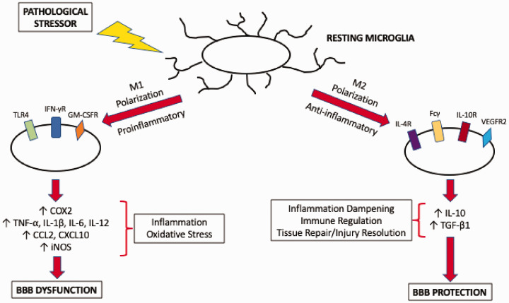
The blood-brain barrier (BBB) is a critical regulator of CNS homeostasis. It possesses physical and biochemical characteristics (i.e. tight junction protein complexes, transporters) that are necessary for the BBB to perform this physiological role. Microvascular endothelial cells require support from astrocytes, pericytes, microglia, neurons, and constituents of the extracellular matrix. This intricate relationship implies the existence of a neurovascular unit (NVU). NVU cellular components can be activated in disease and contribute to dynamic remodeling of the BBB. This is especially true of microglia, the resident immune cells of the brain, which polarize into distinct proinflammatory (M1) or anti-inflammatory (M2) phenotypes. Current data indicate that M1 pro-inflammatory microglia contribute to BBB dysfunction and vascular "leak", while M2 anti-inflammatory microglia play a protective role at the BBB. Understanding biological mechanisms involved in microglia activation provides a unique opportunity to develop novel treatment approaches for neurological diseases. In this review, we highlight characteristics of M1 proinflammatory and M2 anti-inflammatory microglia and describe how these distinct phenotypes modulate BBB physiology. Additionally, we outline the role of other NVU cell types in regulating microglial activation and highlight how microglia can be targeted for treatment of disease with a focus on ischemic stroke and Alzheimer's disease.

Keywords: Alzheimer’s disease; blood–brain barrier; inflammation; ischemic stroke; microglia; neurovascular unit; oxidative stress; paracellular permeability; tight junctions.


Conflict of interest statement

Declaration of conflicting interests: The author(s) declared no potential conflicts of interest with respect to the research, authorship, and/or publication of this article.

Figures

Figure 1.
Figure 1.
Perivascular microglial cells. This image illustrates the proximity of microglia to cerebral capillaries in the adult rat hindbrain. A 30-µm rat brain section was stained for the microglial marker IBA1 (red) and major histocompatibility complex (MHC) II (green), which is upregulated in activated microglia but also stains endothelial cells. Nuclei were stained with 4ʹ,6-diamindino-2-phenylindole (DAPI; blue). The white arrow highlights a surveillance microglia and the arrowhead highlights an activated microglia that has increased expression of MHCII. Note the capillary between the two microglia. This image is a maximum intensity projection of a 10-µm segment of the brain slice and was processed for brightness, contrast, and RGB levels. Adapted from Herndon JM, Tome ME and Davis TP. Development and maintenance of the blood-brain barrier. In: LR Caplan, J Biller, MC Leary, et al. (eds) Primer on cerebrovascular diseases, 2nd ed. San Diego: Elsevier Academic Press, 2017.
Figure 2.
Figure 2.
Polarization of microglia in response to pathological stressors. In the absence of pathological mediators, microglia maintain a resting phenotype where they perform surveillance of the brain extracellular milieu. In the presence of a stressor, microglia are activated and can assume one of two activation states. Polarization to an M1 state is mediated by TLR4, IFN-γ receptors, or GM-CSF receptors and leads to increased production of proinflammatory cytokines and chemokines as well as increased expression of COX2 and iNOS. This results in increased inflammation and oxidative stress, processes that cause dysfunction of the BBB. In contrast, M2 microglia perform inflammation dampening, immune regulation, and tissue repair/injury resolution functions. Polarization of microglia to a M2 activation state involves IL-4 receptors, Fcγ, IL-10 receptors, or VEGFR2. M2 microglia secrete anti-inflammatory mediators such as IL-10 and TGF-β1 and help to protect the BBB in the setting of neurological disease.
Figure 3.
Figure 3.
Involvement of triggering receptor expressed on myeloid cells (TREM) signaling on microglial phagocytosis in Alzheimer’s disease. Amyloid-beta (Aβ) oligomers or apolipoprotein E (APOE) isoforms can activate TREM1 or TREM2 at the microglia cell surface. Activation of TREM receptors results in formation of heterologous protein complex with DAP12, thereby enabling recruitment of Syk protein tyrosine kinase and subsequent signal transduction. Downstream effectors of the TREM/DAP12 complex include ERK, phospholipase C-gamma (PLC-γ), the Vav signaling pathway, and phosphoinositide 3-kinase (PI3K). These pathways activate phagocytotic mechanisms in microglia, thereby enabling these cells to participate in clearance of Aβ plaques from the brain.

References

    1. Haseloff RF, Dithmer S, Winkler L, et al. Transmembrane proteins of the tight junctions at the blood-brain barrier: structural and functional aspects. Semin Cell Dev Biol 2015; 38: 16–25. - PubMed
    1. Liebner S, Dijkhuizen RM, Reiss Y, et al. Functional morphology of the blood-brain barrier in health and disease. Acta Neuropathol 2018; 135: 311–336. - PMC - PubMed
    1. Abdullahi W, Tripathi D, Ronaldson PT. Blood-brain barrier dysfunction in ischemic stroke: targeting tight junctions and transporters for vascular protection. Am J Physiol Cell Physiol 2018; 315: C343–C356. - PMC - PubMed
    1. Nitta T, Hata M, Gotoh S, et al. Size-selective loosening of the blood-brain barrier in claudin-5-deficient mice. J Cell Biol 2003; 161: 653–660. - PMC - PubMed
    1. Saitou M, Furuse M, Sasaki H, et al. Complex phenotype of mice lacking occludin, a component of tight junction strands. Mol Biol Cell 2000; 11: 4131–4142. - PMC - PubMed

Publication types

LinkOut - more resources