. 2020;18(12):1266-1278.
doi: 10.2174/1570159X18666200914161231.

The Updated Role of the Blood Brain Barrier in Subarachnoid Hemorrhage: From Basic and Clinical Studies

Affiliations

The Updated Role of the Blood Brain Barrier in Subarachnoid Hemorrhage: From Basic and Clinical Studies

Sheng Chen et al. Curr Neuropharmacol. 2020.

Abstract

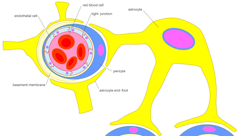
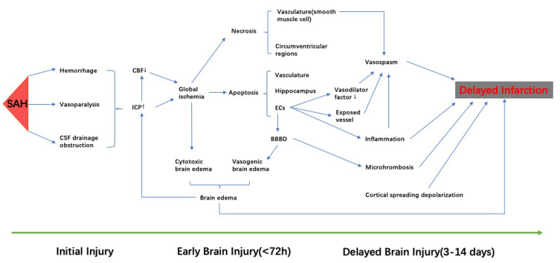
Subarachnoid hemorrhage (SAH) is a type of hemorrhagic stroke associated with high mortality and morbidity. The blood-brain-barrier (BBB) is a structure consisting primarily of cerebral microvascular endothelial cells, end feet of astrocytes, extracellular matrix, and pericytes. Post-SAH pathophysiology included early brain injury and delayed cerebral ischemia. BBB disruption was a critical mechanism of early brain injury and was associated with other pathophysiological events. These pathophysiological events may propel the development of secondary brain injury, known as delayed cerebral ischemia. Imaging advancements to measure BBB after SAH primarily focused on exploring innovative methods to predict clinical outcome, delayed cerebral ischemia, and delayed infarction related to delayed cerebral ischemia in acute periods. These predictions are based on detecting abnormal changes in BBB permeability. The parameters of BBB permeability are described by changes in computed tomography (CT) perfusion and magnetic resonance imaging (MRI). Kep seems to be a stable and sensitive indicator in CT perfusion, whereas Ktrans is a reliable parameter for dynamic contrast-enhanced MRI. Future prediction models that utilize both the volume of BBB disruption and stable parameters of BBB may be a promising direction to develop practical clinical tools. These tools could provide greater accuracy in predicting clinical outcome and risk of deterioration. Therapeutic interventional exploration targeting BBB disruption is also promising, considering the extended duration of post-SAH BBB disruption.

Keywords: Subarachnoid hemorrhage; blood brain barrier; clinical trial; delayed cerebral ischemia; early brain injury; imaging.


Figures

Fig. (1)
Fig. (1)
Components of blood brain barrier: Cerebral microvascular endothelial cells connected by tight junctions form the internal layer of the microvessel wall, which is embedded in the extracellular matrix, known as the basement membrane. Pericytes insert into the basement membrane and encircle the internal layer of microvessel wall and part of the basement membrane. The end-feet from different astrocytes cover the surface of pericytes, forming the outer layer of the microvessel wall. The functions of endothelial cells and astrocytes can be integrated by the pericyte though polarization of the astrocytic end-feet. (A higher resolution / colour version of this figure is available in the electronic copy of the article).
Fig. (2)
Fig. (2)
The pathophysiological events occurred after SAH. Initial injury damages brain. Increased ICP and decreased CBF leads to global ischemia, which contributes to the cytotoxic brain edema. Cell apoptosis, resulting from ischemic damage, can be observed in the vasculature, hippocampus, and blood brain barrier, and was the major cell death after SAH. Apoptosis of endothelial cells results in disruption of blood-brain barrier, which contributes directly to vasogenic brain edema. Brain edema, including cytotoxic and vasogenic, aggressively increases ICP. Apoptosis of endothelial cells reduces the secretion of vasodilation factors and exposes the vessel to vasoactive and toxic metabolites, which aggravates vasospasm. Necrosis is not the primary route of cell death in SAH, but the necrosis of smooth muscle cells partially contributes to vasospasm. Cortical spreading depolarization, microthrombosis, inflammation, brain edema, and vasospasm are thought to be a part of delayed brain injury, and may develop to delayed infarction. SAH: subarachnoid hemorrhage; CBF: cerebral blood flow; ICP: intracranial pressure. (A higher resolution / colour version of this figure is available in the electronic copy of the article).

Similar articles

Cited by

References

    1. Lovelock C.E., Rinkel G.J., Rothwell P.M. Time trends in outcome of subarachnoid hemorrhage: Population-based study and systematic review. Neurology. 2010;74(19):1494–1501. doi: 10.1212/WNL.0b013e3181dd42b3. - DOI - PMC - PubMed
    1. Nieuwkamp D.J., Setz L.E., Algra A., Linn F.H., de Rooij N.K., Rinkel G.J. Changes in case fatality of aneurysmal subarachnoid haemorrhage over time, according to age, sex, and region: a meta-analysis. Lancet Neurol. 2009;8(7):635–642. doi: 10.1016/S1474-4422(09)70126-7. - DOI - PubMed
    1. Al-Khindi T., Macdonald R.L., Schweizer T.A. Cognitive and functional outcome after aneurysmal subarachnoid hemorrhage. Stroke. 2010;41(8):e519–e536. doi: 10.1161/STROKEAHA.110.581975. - DOI - PubMed
    1. Suzuki H. What is early brain injury? Transl. Stroke Res. 2015;6(1):1–3. doi: 10.1007/s12975-014-0380-8. - DOI - PubMed
    1. Eagles M.E., Jaja B.N.R., Macdonald R.L. Incorporating a modified graeb score to the modified fisher scale for improved risk prediction of delayed cerebral ischemia following aneurysmal subarachnoid hemorrhage. Neurosurgery. 2018;82(3):299–305. doi: 10.1093/neuros/nyx165. - DOI - PubMed

MeSH terms