. 2020 Sep 14;20(1):298.
doi: 10.1186/s12876-020-01438-1.

Clinical characteristics of adult T-cell leukemia/lymphoma infiltration in the gastrointestinal tract

Affiliations

Clinical characteristics of adult T-cell leukemia/lymphoma infiltration in the gastrointestinal tract

Tadashi Miike et al. BMC Gastroenterol. .

Abstract

Background: Adult T-cell leukemia/lymphoma (ATLL) is a peripheral T-cell malignancy caused by human T-cell leukemia virus type 1. The clinical course of ATLL is very heterogeneous, and many organs, including the gastrointestinal (GI) tract, can be involved. However, there are few detailed reports on ATLL infiltration in the GI tract. We investigated the clinical characteristics of ATLL infiltration in the GI tract.

Methods: This retrospective observational single-center study included 40 consecutive ATLL patients who underwent GI endoscopy. The patients' demographic and clinical characteristics and endoscopic findings were analyzed retrospectively. Patients with ATLL who were diagnosed by histological examination were divided into two groups based on GI tract infiltration.

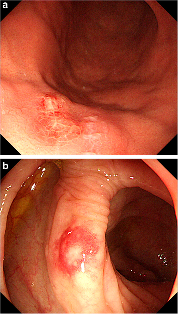
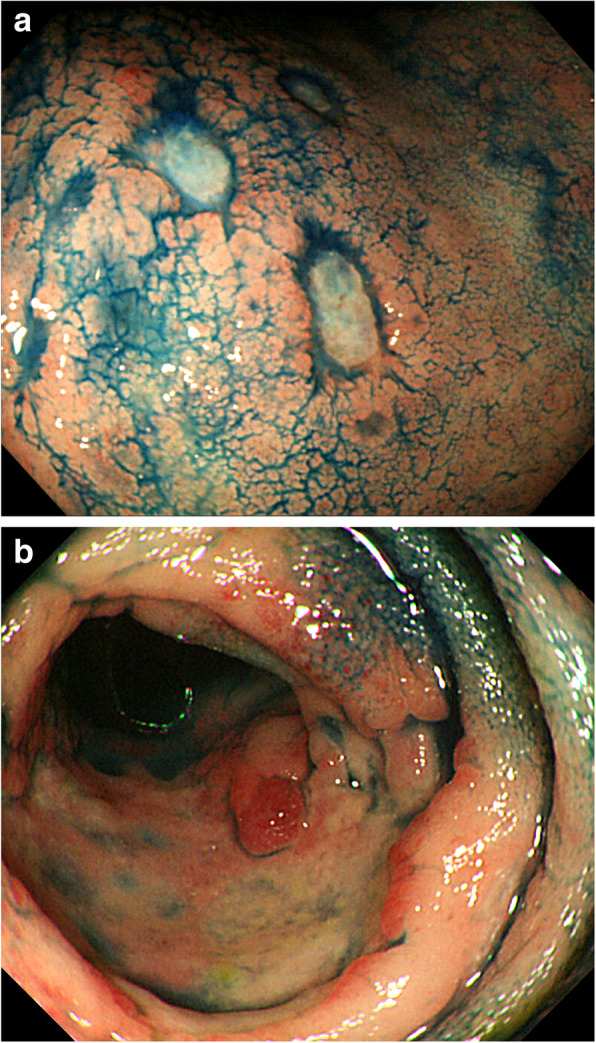
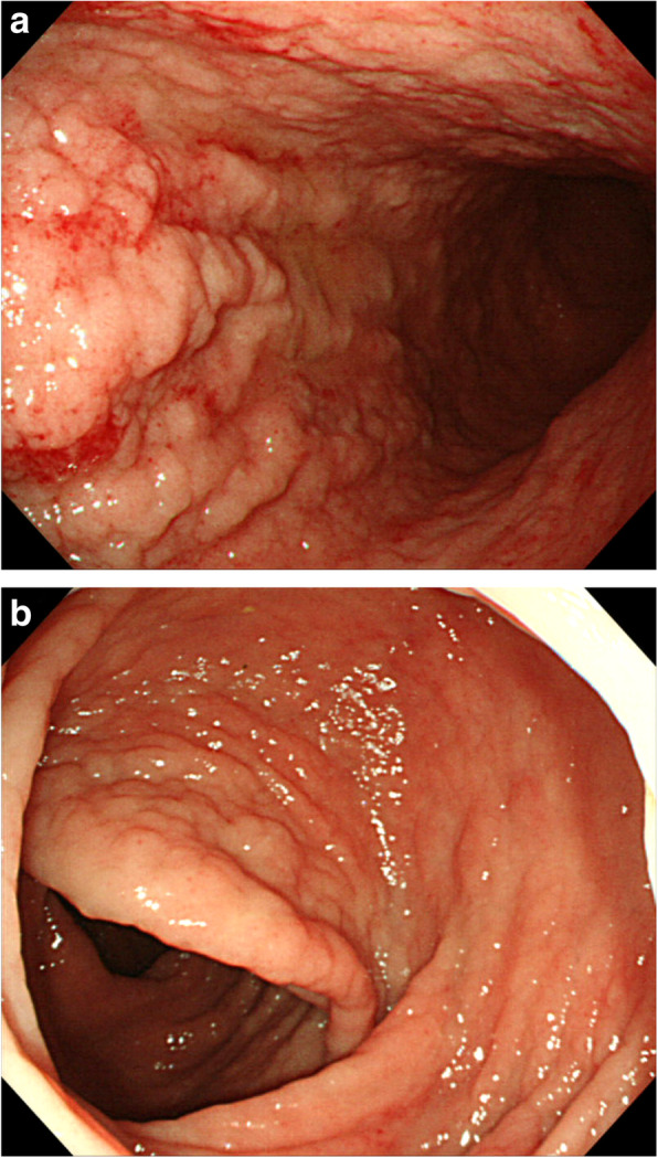
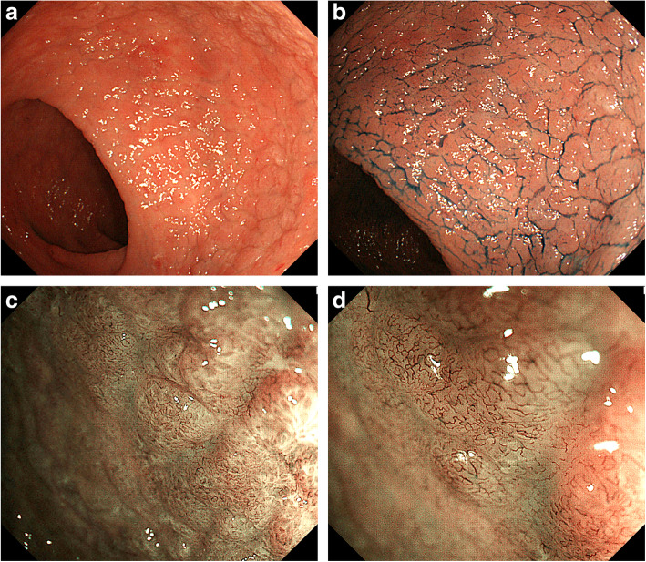
Results: Multivariate analysis revealed that the absence of skin lesions was significantly associated with GI infiltration (P < 0.05). Furthermore, the infiltration group tended to have similar macroscopic lesions in the upper and lower GI tracts, such as diffuse type, tumor-forming type, and giant-fold type.

Conclusions: GI endoscopy may be considered for ATLL patients without skin lesions.

Keywords: Adult T-cell leukemia/lymphoma; GI tract infiltration; Irregular blood vessels; Obscure glandular structures; Skin lesion.


Conflict of interest statement

The authors declare no conflicts of interest for this article.

Figures

Fig. 1
Fig. 1
Macroscopic gastrointestinal findings. a A 60s-year-old person underwent endoscopy for screening purposes. Upper gastrointestinal (GI) endoscopy shows scattered reddish mucosal protrusions on the greater curvature of the gastric body. b Colonoscopy image shows sporadic protrusions
Fig. 2
Fig. 2
Macroscopic gastrointestinal findings. a A 70s-year-old person underwent endoscopy due to a tarry stool. Upper GI endoscopy shows diffusely ulcerative mucosa on the anterior wall of the gastric body. b Colonoscopy image shows sporadic ulceration
Fig. 3
Fig. 3
Macroscopic gastrointestinal findings. a A 70s-year-old person underwent endoscopy due to diarrhea. Esophagogastroduodenoscopy shows diffuse areas of coarse mucosal granularity on the greater curvature of the gastric body. b Colonoscopy image shows sporadic diffuse granularity
Fig. 4
Fig. 4
Magnified endoscopic images on Image-enhanced endoscopy. a A 70s-year-old person underwent endoscopy due to epigastralgia and diarrhea. Colonoscopy shows diffuse areas of coarse mucosal granularity. b Chromoendoscopy with indigo carmine clearly shows coarse mucosal granularity. c Magnified endoscopic examination with narrow-band imaging (NBI) reveals obscure glandular structures. d Magnified endoscopic examination with NBI reveals irregular bifurcated meandering blood vessels

Similar articles

References

    1. Uchiyama T, Yodoi J, Sagawa K, Takatsuki K, Uchino H. Adult T-cell leukemia: clinical and hematologic features of 16 cases. Blood. 1977;50:481–492. doi: 10.1182/blood.V50.3.481.481. - DOI - PubMed
    1. Poiesz BJ, Ruscetti FW, Gazdar AF, Bunn PA, Minna JD, Gallo RC. Detection and isolation of type C retrovirus particles from fresh and cultured lymphocytes of a patient with cutaneous T-cell lymphoma. Proc Natl Acad Sci U S A. 1980;77:7415–7419. doi: 10.1073/pnas.77.12.7415. - DOI - PMC - PubMed
    1. Ishitsuka K, Tamura K. Human T-cell leukemia virus type I and adult T-cell leukaemia-lymphoma. Lancet Oncol. 2014;15:e517–e526. doi: 10.1016/S1470-2045(14)70202-5. - DOI - PubMed
    1. Nonaka K, Ohata K, Matsuhashi N, Shimizu M, Arai S, Hiejima Y, et al. Is narrow-band imaging useful for histological evaluation of gastric mucosa-associated lymphoid tissue lymphoma after treatment? Dig Endosc. 2014;26:358–364. doi: 10.1111/den.12169. - DOI - PubMed
    1. Norimura D, Fukuda E, Yamao T, Niino D, Haraguchi M, Ozawa E, et al. Education and Imaging. Gastrointestinal: gastric mucosa-associated lymphoid tissue (MALT) lymphoma observed by magnifying endoscopy with narrow band imaging. J Gastroenterol Hepatol. 2012;27:987. doi: 10.1111/j.1440-1746.2012.07105.x. - DOI - PubMed