Case Reports
. 2020 Sep 14;20(1):200.
doi: 10.1186/s12905-020-01055-w.

A case report of Multiple Symmetric Lipomatosis (MSL) in an East Asian Female

Affiliations
Case Reports

A case report of Multiple Symmetric Lipomatosis (MSL) in an East Asian Female

Kyunghun Jung et al. BMC Womens Health. .

Abstract

Background: Multiple Symmetric Lipomatosis (MSL) is a rare disorder related to fat metabolism and lipid storage. The condition results in characteristic depositions of fat, especially around the cephalic, cervical, and upper thoracic subcutaneous. It is much more common in adult males who live in the Mediterranean region and has only rarely been reported in Asian females. In this report, we present a case of an Asian female with MSL and also review the clinical features of the condition, including radiological and histological findings required for proper diagnosis and management.

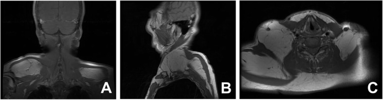
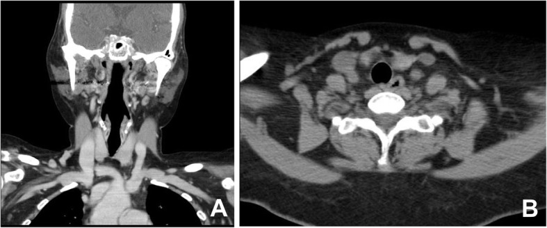
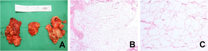
Case presentation: A 59-year-old Korean female came in with a chief complaint of palpable mass present in shoulder and upper back regions. Images showed diffuse non-encapsulated adipose tissue in the subcutaneous layer of the suboccipital, posterior neck area. The patient wanted to remove the mass for cosmetic reasons and discomfort. Excisional biopsy was planned. Preoperative blood analyses showed deteriorated liver function, and the computed tomography findings were consistent with liver cirrhosis. Detailed history taking revealed that she consumed highly levels of alcohol. Lipectomy was performed and the histological findings demonstrated large dystrophic adipocyte morphology. The patient was recovered uneventfully.

Conclusion: When patients have multiple symmetric lipomatous lesions, clinicians should suspect MSL and survey possible associated conditions, such as alcoholism, liver cirrhosis, dyspnea, and neuropathy in detail.

Keywords: Alcohol; Female; Lipoma; Multiple; Symmetric.


Conflict of interest statement

The authors declare that they have no competing interests.

Figures

Fig. 1
Fig. 1
Preoperative distribution of masses. There was symmetrical swelling caused by masses of adipose tissue in both shoulders, the back, and the posterior neck, but the anterior neck was relatively spared. Anterior view (a), posterior view (b), neck lateral view (c)
Fig. 2
Fig. 2
Ultrasonography findings. Ultrasonography indicated excessive adipose tissue (white lines) located in the subcutaneous layer of the shoulders (a, 5.73 × 2.12 × 4.32 cm), posterior neck (b, 4.7 × 7.7 × 2.0 cm), and back (c, 8.2 × 8.6 × 2.2 cm). All masses were compressible and had minimal vascularity
Fig. 3
Fig. 3
Magnetic resonance images of the patient’s neck. Coronal and sagittal T1 weighted sequences from the neck MRI revealed subcutaneous adipose tissue in both the shoulders (a) and posterior neck (b). Axial T1 weighted sequence findings also revealed diffuse subcutaneous fatty infiltration in both shoulders, but there was no mediastinal lipomatosis (c). MRI: Magnetic resonance image
Fig. 4
Fig. 4
Computed tomography image of the patient’s neck. CT images were taken to evaluate the trachea and esophagus. There was no apparent airway or esophageal obstruction on the coronal view (a) or the axial view (b). CT: Computed tomography
Fig. 5
Fig. 5
Gross photo and histologic finding of the excised mass. Specimens consisted of well-defined, pale yellow, fatty tissue. They had homogeneous, soft, solid, greasy cut surfaces without hemorrhage or necrosis. They were 8 × 4.5 × 2 cm (left), 8 × 4 × 2 cm (middle), and 6 × 4.5 × 1.5 cm (right) (a). Hematoxylin and eosin staining of the biopsy sample showed fatty tissue without any signs of malignant transformation. The adipocytes had significantly larger cross-sectional surface areas of the lipid vacuoles, compared with those from normal regions (40× (b), 200× (c)). The estimated weights were 64.8 g (Left), 57.6 g (Middle), 40.5 g (Right)

References

    1. Busetto L, Strater D, Enzi G, Coin A, Sergi G, Inelmen EM, Pigozzo S. Differential clinical expression of multiple symmetric lipomatosis in men and women. Int J Obes Relat Metab Disord. 2003;27(11):1419–1422. doi: 10.1038/sj.ijo.0802427. - DOI - PubMed
    1. Frikh R, Sedrati O. Images in clinical medicine. Keratoacanthoma. N Engl J Med. 2008;359(16):1717. doi: 10.1056/NEJMicm073453. - DOI - PubMed
    1. Wollina U, Heinig B. Madelung's disease - case series and treatment by tumescent liposuction or Lipectomy. Open Access Maced J Med Sci. 2017;5(4):427–431. doi: 10.3889/oamjms.2017.114. - DOI - PMC - PubMed
    1. Fernández-Vozmediano J, Armario-Hita J. Benign symmetric lipomatosis (Launois–Bensaude syndrome) Int J Dermatol. 2005;44(3):236–237. doi: 10.1111/j.1365-4632.2004.02489.x. - DOI - PubMed
    1. Donhauser G, Vieluf D, Ruzicka T, Braun-Falco O. Benign symmetric Launois-Bensaude type III lipomatosis and bureau-Barriere syndrome. Hautarzt. 1991;42(5):311–314. - PubMed

Publication types