

Mesenchymal stem cell-based Smad7 gene therapy for experimental liver cirrhosis
- PMID: 32928296
- PMCID: PMC7489041
- DOI: 10.1186/s13287-020-01911-4
Mesenchymal stem cell-based Smad7 gene therapy for experimental liver cirrhosis
Abstract
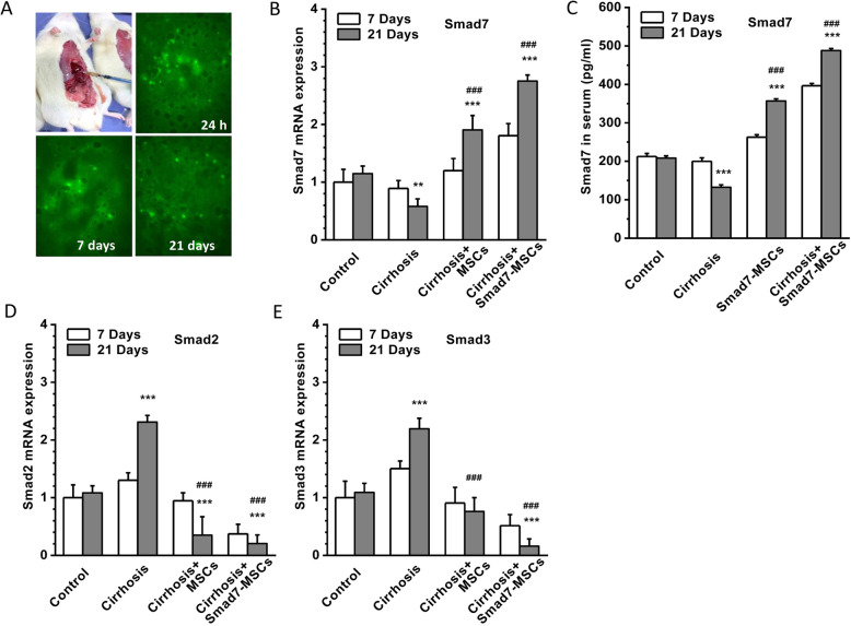
Background: Bone mesenchymal stem cells (MSCs) can promote liver regeneration and inhibit inflammation and hepatic fibrosis. MSCs also can serve as a vehicle for gene therapy. Smad7 is an essential negative regulatory gene in the TGF-β1/Smad signalling pathway. Activation of TGF-β1/Smad signalling accelerates liver inflammation and fibrosis; we therefore hypothesized that MSCs overexpressing the Smad7 gene might be a new cell therapy approach for treating liver fibrosis via the inhibition of TGF-β1/Smad signalling.
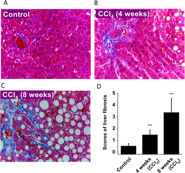
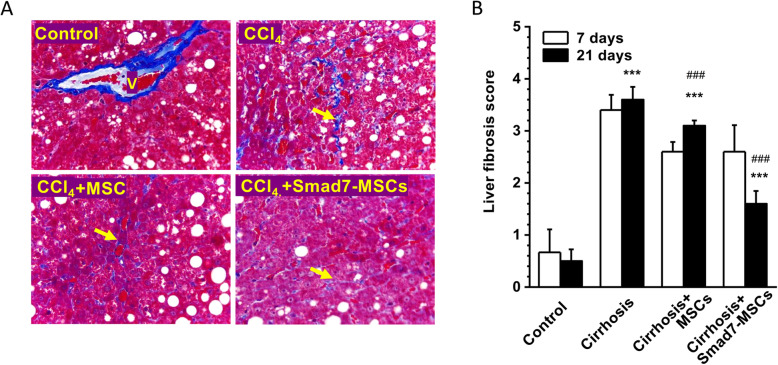
Methods: MSCs were isolated from 6-week-old Wistar rats and transduced with the Smad7 gene using a lentivirus vector. Liver cirrhosis was induced by subcutaneous injection of carbon tetrachloride (CCl4) for 8 weeks. The rats with established liver cirrhosis were treated with Smad7-MSCs by direct injection of cells into the main lobes of the liver. The expression of Smad7, Smad2/3 and fibrosis biomarkers or extracellular matrix proteins and histopathological change were assessed by quantitative PCR, ELISA and Western blotting and staining.
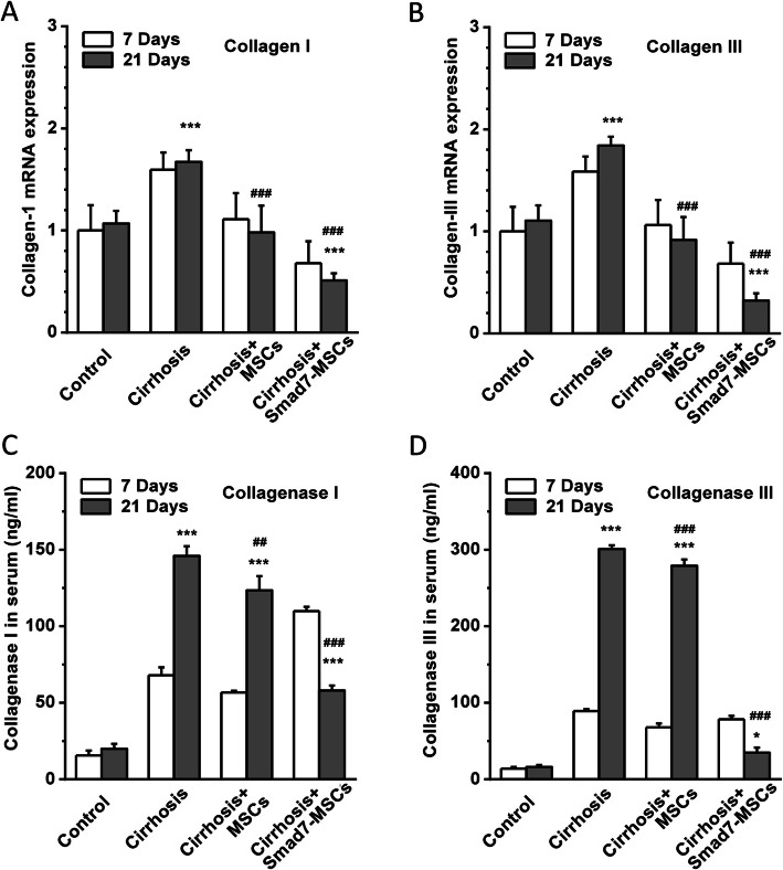
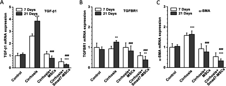
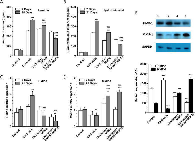
Results: The mRNA and protein level of Smad7 in the recipient liver and serum were increased after treating with Smad-MSCs for 7 and 21 days (P < 0.001). The serum levels of collagen I and III and collagenase I and III were significantly (P < 0.001) reduced after the treatment with Smad7-MSCs. The mRNA levels of TGF-β1, TGFBR1, α-SMA, TIMP-1, laminin and hyaluronic acid were decreased (P < 0.001), while MMP-1 increased (P < 0.001). The liver fibrosis score and liver function were significantly alleviated after the cell therapy.
Conclusions: The findings suggest that the MSC therapy with Smad7-MSCs is effective in the treatment of liver fibrosis in the CCl4-induced liver cirrhosis model. Inhibition of TGF-β1 signalling pathway by enhancement of Smad-7 expression could be a feasible cell therapy approach to mitigate liver cirrhosis.
Keywords: Fibrosis; Gene therapy; Liver cirrhosis; Mesenchymal stem cells; Smad7; TGF-β.
Conflict of interest statement
The authors declare no conflict of interest.
Figures
Similar articles
-
[Dynamic expression of TGF-beta1/Smad protein in CCl4-induced liver fibrosis and its significance in rats].Zhonghua Shi Yan He Lin Chuang Bing Du Xue Za Zhi. 2011 Oct;25(5):334-7. Zhonghua Shi Yan He Lin Chuang Bing Du Xue Za Zhi. 2011. PMID: 22338217 Chinese.
-
Shugan Huoxue Huayu Fang attenuates carbon tetrachloride-induced hepatic fibrosis in rats by inhibiting transforming growth factor-β1/Smad signaling.J Tradit Chin Med. 2022 Feb;42(1):65-72. doi: 10.19852/j.cnki.jtcm.20210624.001. J Tradit Chin Med. 2022. PMID: 35294124 Free PMC article.
-
Effect of Human Umbilical Cord Mesenchymal Stem Cells Transfected with HGF on TGF-β1/Smad Signaling Pathway in Carbon Tetrachloride-Induced Liver Fibrosis Rats.Stem Cells Dev. 2020 Nov 1;29(21):1395-1406. doi: 10.1089/scd.2020.0060. Epub 2020 Sep 24. Stem Cells Dev. 2020. PMID: 32867602
-
Role of TGF-Beta and Smad7 in Gut Inflammation, Fibrosis and Cancer.Biomolecules. 2020 Dec 27;11(1):17. doi: 10.3390/biom11010017. Biomolecules. 2020. PMID: 33375423 Free PMC article. Review.
-
Mesenchymal stromal cells in hepatic fibrosis/cirrhosis: from pathogenesis to treatment.Cell Mol Immunol. 2023 Jun;20(6):583-599. doi: 10.1038/s41423-023-00983-5. Epub 2023 Feb 24. Cell Mol Immunol. 2023. PMID: 36823236 Free PMC article. Review.
Cited by
-
IFN-β Overexpressing Adipose-Derived Mesenchymal Stem Cells Mitigate Alcohol-Induced Liver Damage and Gut Permeability.Int J Mol Sci. 2024 Aug 4;25(15):8509. doi: 10.3390/ijms25158509. Int J Mol Sci. 2024. PMID: 39126076 Free PMC article.
-
Stem cells for treatment of liver fibrosis/cirrhosis: clinical progress and therapeutic potential.Stem Cell Res Ther. 2022 Jul 26;13(1):356. doi: 10.1186/s13287-022-03041-5. Stem Cell Res Ther. 2022. PMID: 35883127 Free PMC article. Review.
-
Cell mediated ECM-degradation as an emerging tool for anti-fibrotic strategy.Cell Regen. 2023 Sep 1;12(1):29. doi: 10.1186/s13619-023-00172-9. Cell Regen. 2023. PMID: 37653282 Free PMC article. Review.
-
Advance of Mesenchymal Stem Cells in Chronic End-Stage Liver Disease Control.Stem Cells Int. 2022 Oct 7;2022:1526217. doi: 10.1155/2022/1526217. eCollection 2022. Stem Cells Int. 2022. PMID: 36248254 Free PMC article. Review.
-
Immunomodulatory role of mesenchymal stem cell therapy in liver fibrosis.Front Immunol. 2023 Jan 4;13:1096402. doi: 10.3389/fimmu.2022.1096402. eCollection 2022. Front Immunol. 2023. PMID: 36685534 Free PMC article. Review.
References
-
- Seto WK, Lo YR, Pawlotsky JM, Yuen MF. Chronic hepatitis B virus infection. Lancet. 2018;392(10161):2313–2324. - PubMed
-
- Liu J, Zhang S, Wang Q, Shen H, Zhang M, Zhang Y, Yan D, Liu M. Seroepidemiology of hepatitis B virus infection in 2 million men aged 21-49 years in rural China: a population-based, cross-sectional study. Lancet Infect Dis. 2016;16(1):80–86. - PubMed
-
- Tang LSY, Covert E, Wilson E, Kottilil S. Chronic hepatitis B infection: a review. JAMA. 2018;319(17):1802–1813. - PubMed
-
- Newsome PN, Fox R, King AL, Barton D, Than NN, Moore J, Corbett C, Townsend S, Thomas J, Guo K, et al. Granulocyte colony-stimulating factor and autologous CD133-positive stem-cell therapy in liver cirrhosis (REALISTIC): an open-label, randomised, controlled phase 2 trial. Lancet Gastroenterol Hepatol. 2018;3(1):25–36. - PMC - PubMed
Publication types
MeSH terms
Substances
LinkOut - more resources
Full Text Sources
Research Materials
Miscellaneous
