Review
. 2022 Feb 1;12(2):a038406.
doi: 10.1101/cshperspect.a038406.

Emerging HxNy Influenza A Viruses

Affiliations
Review

Emerging HxNy Influenza A Viruses

William J Liu et al. Cold Spring Harb Perspect Med. .

Abstract

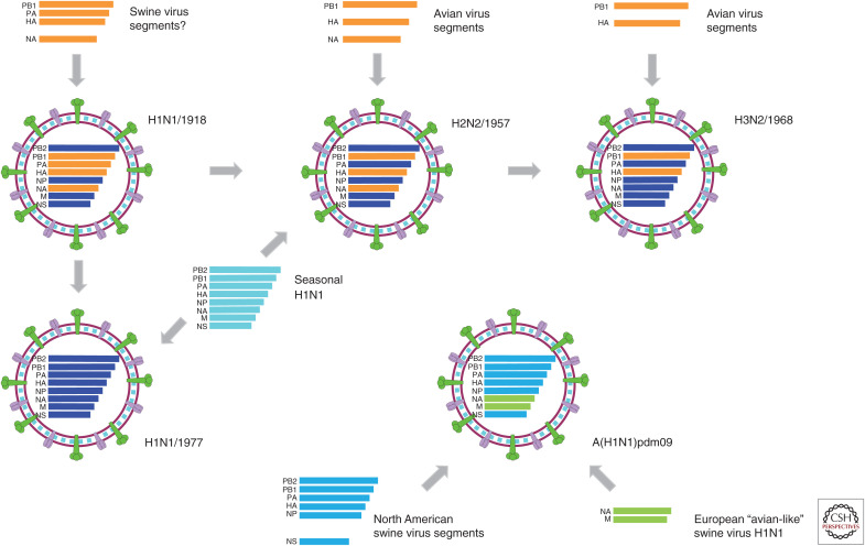
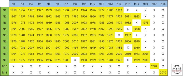
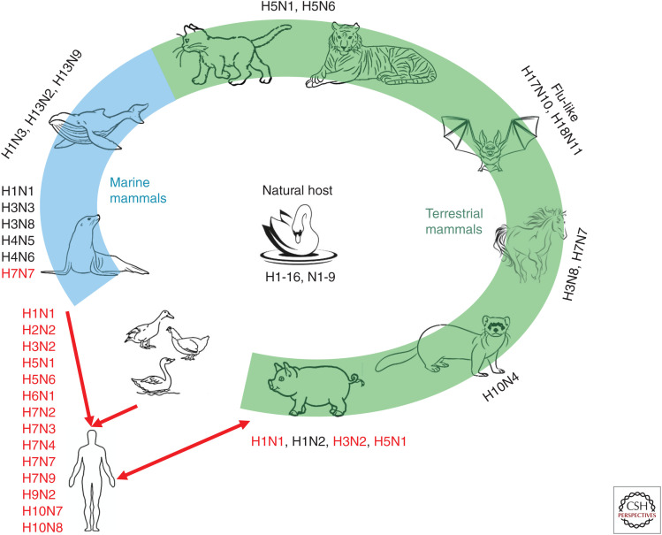
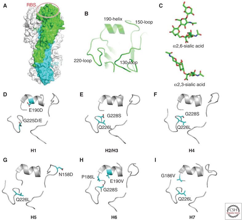
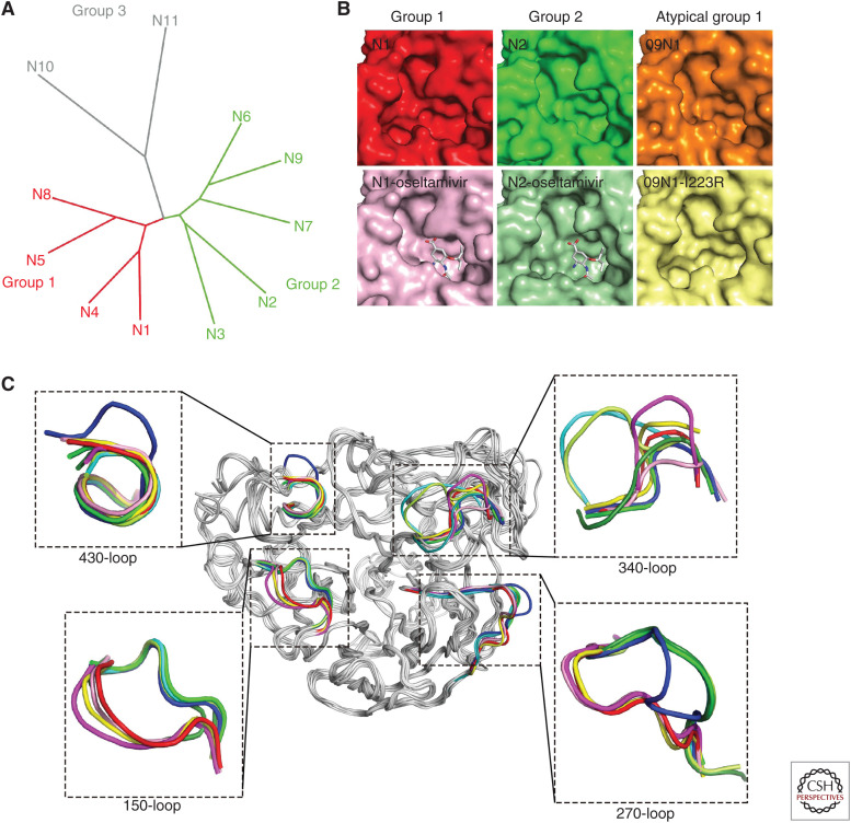
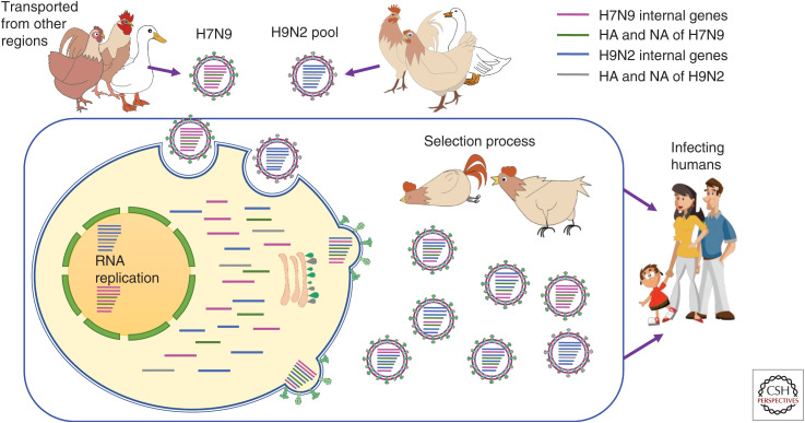
The continuous emergence and reemergence of diverse subtypes of influenza A viruses, which are known as "HxNy" and are mediated through the reassortment of viral genomes, account for seasonal epidemics, occasional pandemics, and zoonotic outbreaks. We summarize and discuss the characteristics of historic human pandemic HxNy viruses and diverse subtypes of HxNy among wild birds, mammals, and live poultry markets. In addition, we summarize the key molecular features of emerging infectious HxNy influenza viruses from the perspectives of the receptor binding of Hx, the inhibitor-binding specificities and drug-resistance features of Ny, and the matching of the gene segments. Our work enhances our understanding of the potential threats of novel reassortant influenza viruses to public health and provides recommendations for effective prevention, control, and research of this pathogen.


Figures

Figure 1.
Figure 1.
Graphical representation of emerging pandemic influenza viruses. The possible reassortant pathway of pandemic influenza viruses. Gene segments, such as PB2, PB1, PA, HA, NP, NA, M, and NS, are ordered according to the gene lengths from top to bottom within the virus particle diagrams. For the gene segments of H1N1/1918, H2N2/1957, H3N2/1968, and H1N1/1977, the human influenza–derived segments are shown in blue, whereas the animal-, including swine- and avian-, derived segments are shown in orange. For H2N2/1957 and H1N1/1977, distinct seasonal H1N1 viruses (represented in cyan) at that time were also involved for the possible reassortment.
Figure 2.
Figure 2.
Currently known subtypes of influenza A viruses and their earliest year of identification. To show the influenza A viruses that have naturally occurred with different pairing of HA and NA, H1-H18 and N1-N11 were paired in a matrix. The earliest year of identification for each natural influenza A virus is shown in yellow cells. The influenza A viruses that have not been identified are indicated by an “X.”
Figure 3.
Figure 3.
Natural reservoir and mammalian hosts of influenza A viruses. Animals with green and blue backgrounds are terrestrial and marine mammals, respectively. Subtypes of influenza marked next to the animal are currently known to infect that animal. The red font indicates that the virus can infect humans.
Figure 4.
Figure 4.
Molecular determinants for receptor binding properties of different hemagglutinin (HA) subtypes. (A) Structure of HA trimer. One of the protomers is colored by subunits with HA1 in green and HA2 in cyan. The receptor binding site (RBS) is indicated by a dashed oval. (B) Close-up view of the structural motifs within RBS. (C) Structures of different sialic acid receptors. (DI) The key determinant residues (sticks) for the receptor binding properties of different HA subtypes.
Figure 5.
Figure 5.
Comprehensive analysis of 11 known neuraminidases of influenza A virus. (A) The phylogenetic tree of neuraminidases (NAs) from 11 known influenza A virus subtypes. (B) Comparison of the active sites of typical group 1, group 2, and atypical group 1 NAs. All the NA molecules are presented in surface representation. Free VN04N1 (PDB code 2HTY, red), free N2 (PDB code 4K1H, green), 09N1 (PDB code 3NSS, orange), oseltamivir carboxylate-bound VN04N1 (PDB code 2HU4, pink), oseltamivir carboxylate-bound N2 with half-open form of 150-loop (PDB code 4K1K, light green), and 09N1-I223R (PDB code 4B7M, light yellow). (C) Superimposition of NA monomers with emphasis on the 150-loop, 270-loop, 340-loop, and 430-loop. The colors of different NAs are VN04N1 (PDB code 2HTY, red), N2 (PDB code 4K1H, green), N3 (PDB code 4HZV, blue), N4 (PDB code 2HTV, yellow), N5 (PDB code 3SAL, magenta), N6 (PDB code 4QN4, cyan), N7 (PDB code 4QN3, lemon), N8 (PDB code 2HT5, pink), and N9 (PDB code 4MWJ, deep green).
Figure 6.
Figure 6.
The matching of internal gene segments within H7N9 and H9N2. Poultry act as a “mixing vessel” (in the blue box) for the emergence of different strains of H7N9. When H7N9 and H9N2 avian influenza viruses coinfect the same cell via HA glycoproteins their genomes replicate synchronously. If the packaging signals from viral ribonucleoprotein complexes (vRNPs) of the same segments are the same, the genomic segments from different viruses may assemble together. The progeny viruses with appropriate adaptability, transmissibility, and compatibilities will successfully survive and jump to the new host. Different colors represent different gene segments of H7N9 and H9N2 avian influenza viruses. (Magenta) Internal genes of H7N9, (green) HA and NA of H7N9, (blue) internal genes of H9N2, (gray) HA and NA of H9N2.

References

    1. Air GM, Laver WG. 1989. The neuraminidase of influenza virus. Proteins 6: 341–356. 10.1002/prot.340060402 - DOI - PubMed
    1. Anthony SJ, St Leger JA, Pugliares K, Ip HS, Chan JM, Carpenter ZW, Navarrete-Macias I, Sanchez-Leon M, Saliki JT, Pedersen J, et al. 2012. Emergence of fatal avian influenza in New England harbor seals. MBio 3: e00166-12. 10.1128/mBio.00166-12 - DOI - PMC - PubMed
    1. Bao CJ, Cui LB, Zhou MH, Hong L, Gao GF, Wang H. 2013. Live-animal markets and influenza A (H7N9) virus infection. N Engl J Med 368: 2337–2339. 10.1056/NEJMc1306100 - DOI - PubMed
    1. Bi Y, Chen Q, Wang Q, Chen J, Jin T, Wong G, Quan C, Liu J, Wu J, Yin R, et al. 2016. Genesis, evolution and prevalence of H5N6 avian influenza viruses in China. Cell Host Microbe 20: 810–821. 10.1016/j.chom.2016.10.022 - DOI - PubMed
    1. Bi YH, Tan SG, Yang Y, Wong G, Zhao M, Zhang QC, Wang Q, Zhao XN, Li LQ, Yuan J, et al. 2019. Clinical and immunological characteristics of human infections with H5N6 avian influenza virus. Clin Infect Dis 68: 1100–1109. 10.1093/cid/ciy681 - DOI - PubMed