. 2020 Nov 13;295(46):15454-15463.
doi: 10.1074/jbc.RA120.014814. Epub 2020 Sep 14.

Ferric uptake regulator (Fur) reversibly binds a [2Fe-2S] cluster to sense intracellular iron homeostasis in Escherichia coli

Affiliations

Ferric uptake regulator (Fur) reversibly binds a [2Fe-2S] cluster to sense intracellular iron homeostasis in Escherichia coli

Chelsey R Fontenot et al. J Biol Chem. .

Abstract

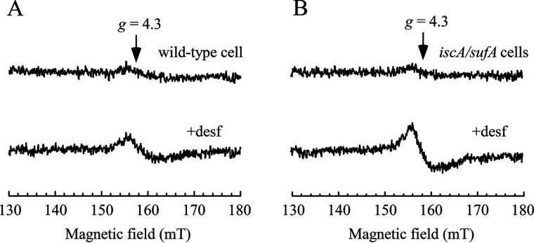
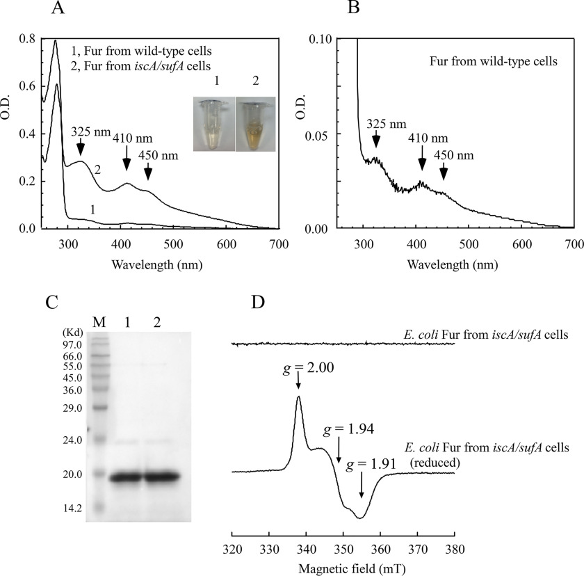
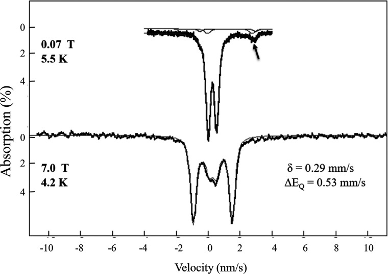
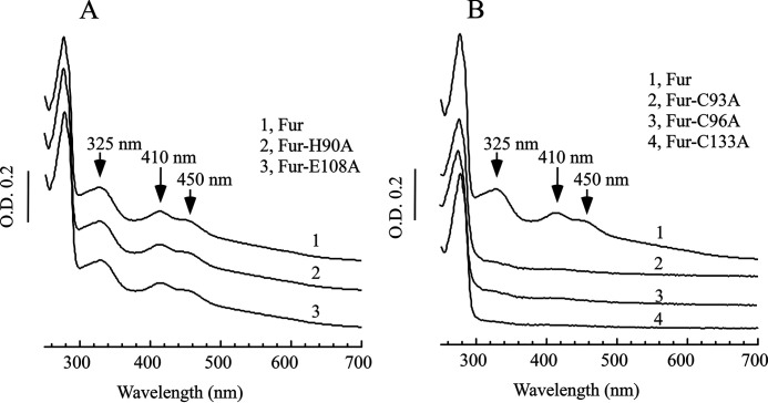
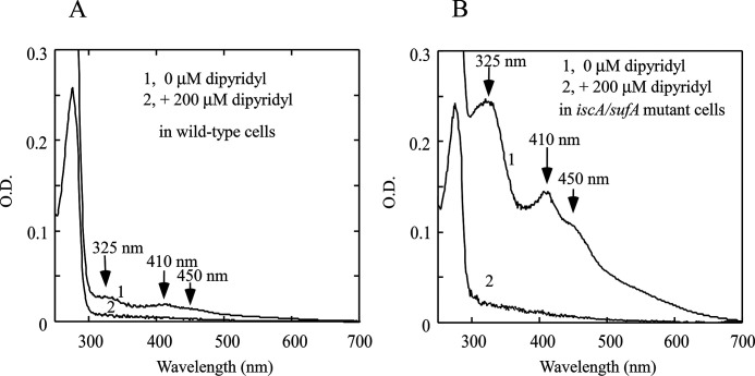
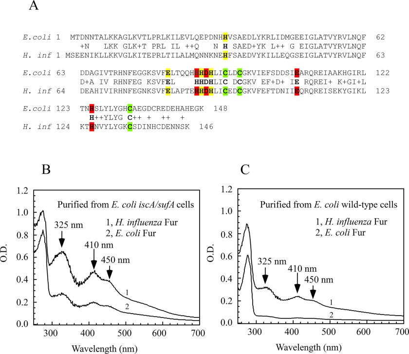
The ferric uptake regulator (Fur) is a global transcription factor that regulates intracellular iron homeostasis in bacteria. The current hypothesis states that when the intracellular "free" iron concentration is elevated, Fur binds ferrous iron, and the iron-bound Fur represses the genes encoding for iron uptake systems and stimulates the genes encoding for iron storage proteins. However, the "iron-bound" Fur has never been isolated from any bacteria. Here we report that the Escherichia coli Fur has a bright red color when expressed in E. coli mutant cells containing an elevated intracellular free iron content because of deletion of the iron-sulfur cluster assembly proteins IscA and SufA. The acid-labile iron and sulfide content analyses in conjunction with the EPR and Mössbauer spectroscopy measurements and the site-directed mutagenesis studies show that the red Fur protein binds a [2Fe-2S] cluster via conserved cysteine residues. The occupancy of the [2Fe-2S] cluster in Fur protein is ∼31% in the E. coli iscA/sufA mutant cells and is decreased to ∼4% in WT E. coli cells. Depletion of the intracellular free iron content using the membrane-permeable iron chelator 2,2´-dipyridyl effectively removes the [2Fe-2S] cluster from Fur in E. coli cells, suggesting that Fur senses the intracellular free iron content via reversible binding of a [2Fe-2S] cluster. The binding of the [2Fe-2S] cluster in Fur appears to be highly conserved, because the Fur homolog from Hemophilus influenzae expressed in E. coli cells also reversibly binds a [2Fe-2S] cluster to sense intracellular iron homeostasis.

Keywords: Escherichia coli (E. coli); IscA; Mossbauer spectroscopy; electron paramagnetic resonance (EPR); ferric uptake regulator (Fur); intracellular iron homeostasis; iron homeostasis; iron metabolism; iron–sulfur cluster; iron–sulfur protein; repressor protein.


Conflict of interest statement

Conflict of interest—The authors declare that they have no conflicts of interest with the contents of this article.

Figures

Figure 1.
Figure 1.
Deletion of IscA and SufA results in accumulation of the intracellular free iron content in E. coli cells. A, EPR spectra of E. coli WT cells treated with or without desferrioxamine (desf). B, EPR spectra of the E. coli iscA/sufA mutant cells treated with or without desferrioxamine. The experimental conditions are described under “Experimental procedures.” The data are representative of three independent experiments.
Figure 2.
Figure 2.
The E. coli Fur has a bright red color when expressed in the E. coli iscA/sufA mutant cells. Recombinant E. coli Fur was expressed in the E. coli WT and the E. coli iscA/sufA mutant cells grown in LB medium under aerobic conditions. A, UV-visible absorption spectra of purified Fur proteins. Spectrum 1, Fur protein purified from WT E. coli cells. Spectrum 2, Fur protein purified from the iscA/sufA mutant cells. The protein (100 μm) was dissolved in buffer containing 20 mm Tris (pH 8.0) and 500 mm NaCl. Inset, photographs of purified Fur proteins from WT (panel 1) and the iscA/sufA mutant (panel 2) cells. B, UV-visible absorption spectrum of the Fur protein purified from WT E. coli cells. Same as in A, except the y axis is expanded by 8-fold. C, SDS-PAGE gel of purified Fur proteins from WT E. coli cells (lane 1) and from the iscA/sufA mutant cells (lane 2). Lane M, molecular mass ladder. D, EPR spectra of the E. coli Fur protein purified from the iscA/sufA mutant cells. The Fur protein (500 μm) was reduced with freshly prepared sodium dithionite (10 mm) and immediately frozen in liquid nitrogen until the EPR measurement.
Figure 3.
Figure 3.
Variable-field Mössbauer spectra of the 57Fe-enriched Fur protein purified from the E. coli iscA/sufA mutant cells. The 57Fe-labeled E. coli Fur protein was purified from the E. coli iscA/sufA mutant cells growing in M9 minimum medium supplemented with 57Fe (10 μm). The protein concentration of 57Fe-labeled Fur was ∼1.0 mm. Mössbauer spectra were collected at cryogenic temperatures at 5.5 K and 70 mT (top spectrum) and 4.2 K and 7.0 T (bottom spectrum). The magnetic field was applied parallel to the observed γ-radiation. Hash marks are raw data, and lines are spectral simulations with the parameters shown and discussed in the text. The line widths (full width at half-maximum) are 0.33 mm/s. The arrow marks the high-energy line of the ferrous components discussed in the text.
Figure 4.
Figure 4.
The conserved cysteine residues in the E. coli Fur are the ligands for the [2Fe-2S] cluster. The Fur mutant proteins were constructed by the site-directed mutagenesis and purified from the E. coli iscA/sufA mutant cells. A, UV-visible absorption spectra of the WT Fur (spectrum 1), the mutant H190A (spectrum 2), and the mutant E108A (spectrum 3). B, UV-visible absorption spectra of the WT Fur (spectrum 1), the mutant C93A (spectrum 2), the mutant C96A (spectrum 3), and the mutant C133A (spectrum 4). Each spectrum was offset for clarity. The protein concentrations were ∼100 μm. The results are representative of three independent experiments.
Figure 5.
Figure 5.
Depletion of intracellular free iron content removes the [2Fe-2S] cluster from Fur protein in E. coli cells. A, UV-visible absorption spectra of the E. coli Fur protein. Fur proteins were purified from the WT E. coli cells treated with 0 μm (spectrum 1) or 200 μm (spectrum 2) of 2,2´-dipyridyl, respectively. B, UV-visible absorption spectra of the E. coli Fur proteins. Fur proteins were purified from the E. coli iscA/sufA mutant cells treated with 0 μm (spectrum 1) or 200 μm (spectrum 2) of 2,2´-dipyridyl, respectively. The concentrations of purified proteins were ∼60 μm. The results are representative of three independent experiments.
Figure 6.
Figure 6.
The H. influenzae Fur also binds a [2Fe-2S] cluster when expressed in E. coli cells. A, the sequence alignment of the H. influenzae Fur and the E. coli Fur. The three metal-binding sites in Fur proteins are highlighted in red (site 1), yellow (site 2), and green (site 3), respectively. B, UV-visible absorption spectra of the H. influenzae Fur (spectrum 1) and the E. coli Fur (spectrum 2) purified from the E. coli iscA/sufA mutant cells. C, UV-visible absorption spectra of the H. influenzae Fur (spectrum 1) and the E. coli Fur (spectrum 2) purified from the E. coli WT cells. The protein concentrations were 100 μm. The results are representative of three independent experiments.
Figure 7.
Figure 7.
A structure model of the full-length E. coli Fur. Full-length sequence of the E. coli Fur was modeled as described in Ref. . Site 1 is coordinated by His-87, Asp-89, Glu-108, and His-125. Site 2 is coordinated by His-33, Glu-81, His-88, and His-90. Site 3 is formed by Cys-93, Cys-96, and Cys-133. The structure model of the E. coli Fur was visualized using RasMol (57). The zinc-binding sites and the [2Fe-2S] cluster binding site are indicated.

Comment in

Similar articles

Cited by

References

    1. Andrews S., Norton I., Salunkhe A. S., Goodluck H., Aly W. S., Mourad-Agha H., and Cornelis P. (2013) Control of iron metabolism in bacteria. Met. Ions Life Sci. 12, 203–239 10.1007/978-94-007-5561-1_7 - DOI - PubMed
    1. Seo S. W., Kim D., Latif H., O'Brien E. J., Szubin R., and Palsson B. O. (2014) Deciphering Fur transcriptional regulatory network highlights its complex role beyond iron metabolism in Escherichia coli. Nat. Commun. 5, 4910 10.1038/ncomms5910 - DOI - PMC - PubMed
    1. Roncarati D., Pelliciari S., Doniselli N., Maggi S., Vannini A., Valzania L., Mazzei L., Zambelli B., Rivetti C., and Danielli A. (2016) Metal-responsive promoter DNA compaction by the ferric uptake regulator. Nat. Commun. 7, 12593 10.1038/ncomms12593 - DOI - PMC - PubMed
    1. Wofford J. D., Bolaji N., Dziuba N., Outten F. W., and Lindahl P. A. (2019) Evidence that a respiratory shield in Escherichia coli protects a low-molecular-mass Fe(II) pool from O2-dependent oxidation. J. Biol. Chem. 294, 50–62 10.1074/jbc.RA118.005233 - DOI - PMC - PubMed
    1. Fillat M. F. (2014) The FUR (ferric uptake regulator) superfamily: diversity and versatility of key transcriptional regulators. Arch. Biochem. Biophys. 546, 41–52 10.1016/j.abb.2014.01.029 - DOI - PubMed

Publication types

MeSH terms