. 2020 Nov 16;88(12):e00426-20.
doi: 10.1128/IAI.00426-20. Print 2020 Nov 16.

An Ixodes scapularis Protein Disulfide Isomerase Contributes to Borrelia burgdorferi Colonization of the Vector

Affiliations

An Ixodes scapularis Protein Disulfide Isomerase Contributes to Borrelia burgdorferi Colonization of the Vector

Yongguo Cao et al. Infect Immun. .

Abstract

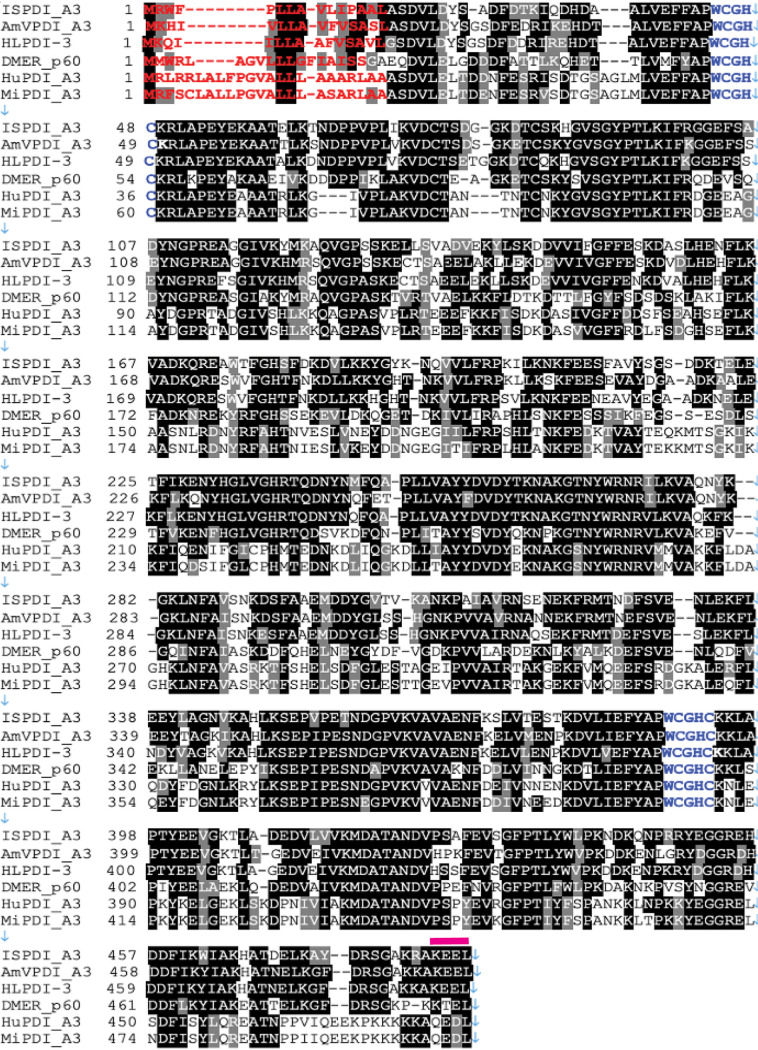
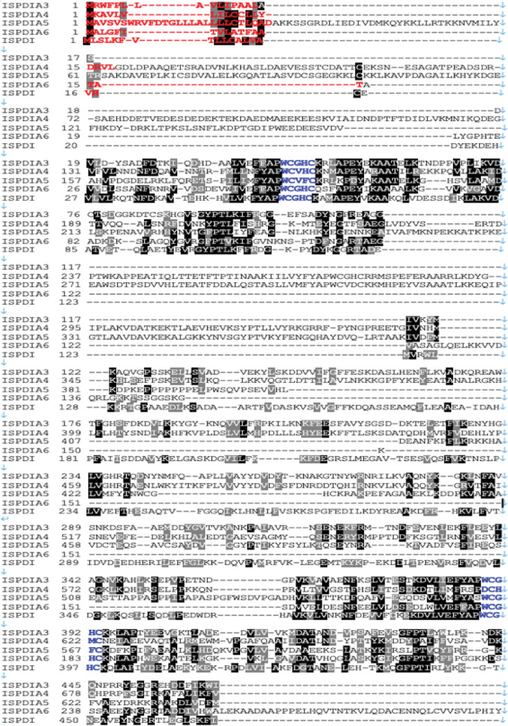
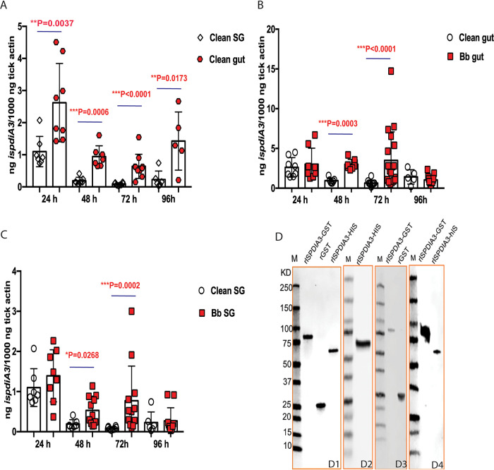
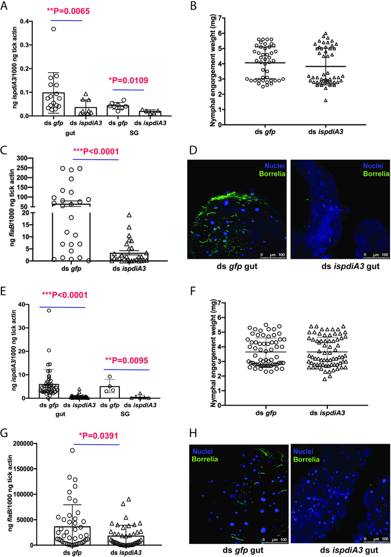
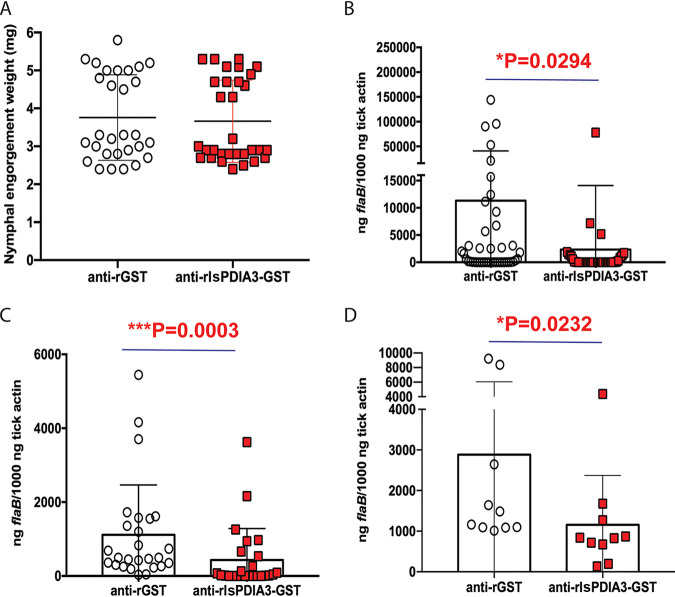
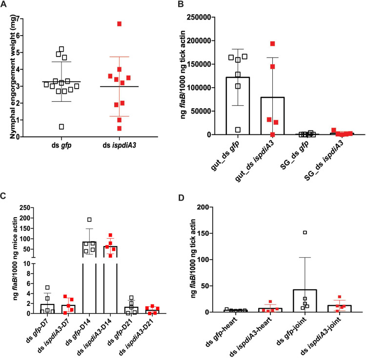
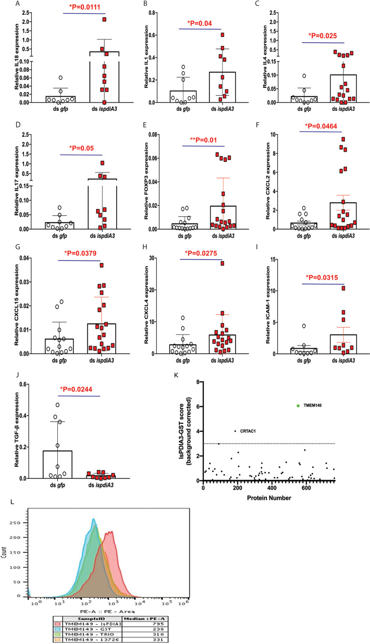
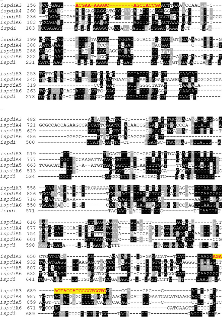
Borrelia burgdorferi causes Lyme disease, the most common tick-transmitted illness in North America. When Ixodes scapularis feed on an infected vertebrate host, spirochetes enter the tick gut along with the bloodmeal and colonize the vector. Here, we show that a secreted tick protein, I. scapularisprotein disulfide isomerase A3 (IsPDIA3), enhances B. burgdorferi colonization of the tick gut. I. scapularis ticks in which ispdiA3 has been knocked down using RNA interference have decreased spirochete colonization of the tick gut after engorging on B. burgdorferi-infected mice. Moreover, administration of IsPDIA3 antiserum to B. burgdorferi-infected mice reduced the ability of spirochetes to colonize the tick when feeding on these animals. We show that IsPDIA3 modulates inflammatory responses at the tick bite site, potentially facilitating spirochete survival at the vector-host interface as it exits the vertebrate host to enter the tick gut. These data provide functional insights into the complex interactions between B. burgdorferi and its arthropod vector and suggest additional targets to interfere with the spirochete life cycle.

Keywords: Borrelia burgdorferi; Ixodes scapularis; colonization; protein disulfide isomerase.


Figures

FIG 1
FIG 1
Protein sequence comparison of IsPDIA3. Multiple-sequence alignment of Ixodes scapularis IsPDI aligned with nine homologs of protein disulfide isomerase (PDI) in Amblyomma variegatum (AmVPDI_A3), Haemaphysalis longicornis (HLPDI_A3), Drosophila melanogaster (DMPDI_p60), Homo sapiens (HuPDI_A3), and Mus musculus (MiPDI_A3). Amino acids shown in black are identical. Amino acids shown in red denote the predicted signal peptide sequence, blue letters label the thioredoxin motif, and pink bars above the sequence indicate the endoplasmic reticulum retention signal.
FIG 2
FIG 2
Protein sequence comparison of IsPDIA3. Multiple-sequence alignment of I. scapularis IsPDI aligned with 4 paralogs of protein disulfide isomerase (PDI) in I. scapularis. Amino acids shown in black are identical. Amino acids shown in red denote the predicted signal peptide sequence, blue letters label the thioredoxin motif, and pink bars above the sequence indicate the endoplasmic reticulum retention signal.
FIG 3
FIG 3
Expression profile of IsPDIA3. (A) IsPDIA3 transcript expression profile in guts (gut) and salivary glands (SG) in pathogen-free (clean) nymphs after 24, 48, 72, and 96 h of feeding. Each data point represents a pool of three nymphal salivary glands or guts. (B) Gut and (C) SG of clean nymphs and Borrelia-infected (Bb) nymphs after 24, 48, 72, and 96 h of feeding. Each data point represents a pool of three nymphal salivary glands or guts. (D) Coomassie stain of rIsPDIA3-GST, rGST, rIsPDIA3-HIS (D1); Western blot assessment of: rIsPDIA3-His using anti-His antibody (D2), rIsPDIA3-GST using anti-GST antibody (D3); and rIsPDIA3-GST and rIsPDIA3-HIS using anti-rIsPDIA3-GST rabbit antiserum (D4). Results represent mean ± standard deviation (SD) of values.
FIG 4
FIG 4
Knockdown of ispdiA3 expression decreases B. burgdorferi colonization by I. scapularis ticks. mRNA transcription was knocked down in I. scapularis salivary glands or gut by body microinjection (A to D) or by anal pore injection (E to H) of ds ispdia3. Knockdown efficiency was measured in I. scapularis nymph salivary glands (SG) and gut (gut). Each data point represents a pool of three nymphal salivary glands or guts (A and E). The impact of ispdia3 knockdown on tick feeding was measured by engorgement weights. Each data point represents a replete nymph (B and F). B. burgdorferi burden in replete tick guts (C and G) was determined by qRT-PCR of flaB transcripts, and data were normalized to tick actin and visualized by immunofluorescence microscopy using rabbit polyclonal B. burgdorferi antibodies. Each data point represents a pool of three nymphal guts (D and H). Nuclei stained with 4′,6-diamidino-2-phenylindole (DAPI). Magnification is ×40. Data in panels A to H represent averages of 3 biological replicates. Results represent mean ± SD of values. Statistical significance was assessed using a nonparametric Mann-Whitney test; *, P < 0.05; **, P < 0.01; ***, P < 0.001.
FIG 5
FIG 5
Passive immunization against IsPDIA3 expression decreases B. burgdorferi colonization by I. scapularis ticks. I. scapularis nymphs were fed on mice passively immunized with polyclonal rabbit rIsPDIA3-GST antiserum or polyclonal rabbit anti-rGST rabbit serum and the impact on (A) feeding as measured by engorgement weights, with each data point representing a replete nymph; (B) B. burgdorferi burden in replete tick guts as measured by qRT-PCR, with each data point representing a pool of two nymphal guts. (C) Borrelia burden in pooled larvae fed on mice passively immunized with rIsPDIA3-GST rabbit antiserum or rGST rabbit antiserum, with each data point representing a pool of five larvae; and (D) qRT-PCR assessment of B. burgdorferi burden in nymphs molted from larvae fed on mice passively immunized with rIsPDIA3-GST rabbit antiserum or rGST rabbit antiserum, with each data point representing a pool of three nymphs. Data (A to D) are averages of 3 biological replicates. Results represent mean ± SD of values. Statistical significance was tested using a nonparametric Mann-Whitney test; *, P < 0.05; **, P < 0.01; ***, P < 0.001.
FIG 6
FIG 6
Knockdown of ispdia3 expression does not impact B. burgdorferi transmission by I. scapularis ticks. Borrelia-infected (Bb) nymphs microinjected by body with ds ispdia3 or ds gfp were fed on clean mice to assess transmission of the spirochete. (A) Impact of knockdown of ispdia3 on engorgement weights; each data point represents a replete nymph. (B) B. burgdorferi burden in salivary glands and guts of replete nymphs, with each data point representing a pool of three nymphal salivary glands and guts, and in (C) mouse skin at 7,14 and 21 days, and (D) and in heart and joint tissues at 21 days was determined by qPCR of flaB and normalized to tick or mouse β-actin. Horizontal bars represent the mean ± SD. Data in panels A to D are averages of 3 biological replicates. Statistical significance was assessed using a nonparametric Mann-Whitney test (*, P < 0.05; **, P < 0.01; ***, P < 0.001).
FIG 7
FIG 7
RNAi-mediated knockdown of IsPDIA3 results in an increased inflammatory response at the tick bite site. Clean nymphs microinjected with ds ispdia3 or ds gfp were fed on clean mice to assess expression profiles of cytokines and chemokines by qRT-PCR at 72 h post tick feeding. Expression profiles of (A) interleukin 18 (IL-18), (B) IL-1β, (C) IL-4, (D) IL-17, (E) FOXP3, (F) CXCL2, (G) CXCL15, (H) CXCL4, (I) ICAM-1, and (J) TGF-β. All of these cytokine and chemokine levels in skin tissues were normalized to mouse β-actin RNA levels according to threshold cycle (2−ΔCT) calculations. Data are averages of 3 biological replicates. Results represent mean ± SD of values. Statistical significance was assessed using a nonparametric Mann-Whitney test (*, P < 0.05; **, P < 0.01; ***, P < 0.001). (K) Screening for rIsPDIA3 interactants by yeast display and fluorescence-activated cell sorting (FACS) data show 2 proteins. TMEM149 and CRTAC1, that enriched with rIsPDIA3-GST. (L) FACS assessment of TMEM149 binding to rIsPDIA3-GST, rGST, and GST-tagged Anopheles gambiae salivary proteins TRIO (AGAP001374) or 13726 (AGAP013726).
FIG 8
FIG 8
DNA sequence of I. scapularis PDIs. T-Coffee alignment of ispdiA3 nucleotide sequence (ISCW016161) with other protein disulfide isomerases (PDIs) in I. scapularis. Nucleotide sequences shown in black are identical. The nucleic acid sequences in yellow show the sequences of dsRNA primers.

References

    1. Hair JA, Bowman J, Sauer J. 1986. Morphology, physiology and behavioural biology of ticks. Ellis Horwood, Chichester, UK.
    1. Anderson JF, Magnarelli LA. 2008. Biology of ticks. Infect Dis Clin North Am 22:195–215, v. doi:10.1016/j.idc.2007.12.006. - DOI - PubMed
    1. Ribeiro JM, Alarcon-Chaidez F, Francischetti IM, Mans BJ, Mather TN, Valenzuela JG, Wikel SK. 2006. An annotated catalog of salivary gland transcripts from Ixodes scapularis ticks. Insect Biochem Mol Biol 36:111–129. doi:10.1016/j.ibmb.2005.11.005. - DOI - PubMed
    1. Ribeiro JM, Francischetti IM. 2003. Role of arthropod saliva in blood feeding: sialome and post-sialome perspectives. Annu Rev Entomol 48:73–88. doi:10.1146/annurev.ento.48.060402.102812. - DOI - PubMed
    1. Wikel SK. 1999. Tick modulation of host immunity: an important factor in pathogen transmission. Int J Parasitol 29:851–859. doi:10.1016/s0020-7519(99)00042-9. - DOI - PubMed

Publication types

MeSH terms