. 2020 Sep 29;117(39):24464-24474.
doi: 10.1073/pnas.2007814117. Epub 2020 Sep 14.

Microglia depletion exacerbates demyelination and impairs remyelination in a neurotropic coronavirus infection

Affiliations

Microglia depletion exacerbates demyelination and impairs remyelination in a neurotropic coronavirus infection

Alan Sariol et al. Proc Natl Acad Sci U S A. .

Abstract

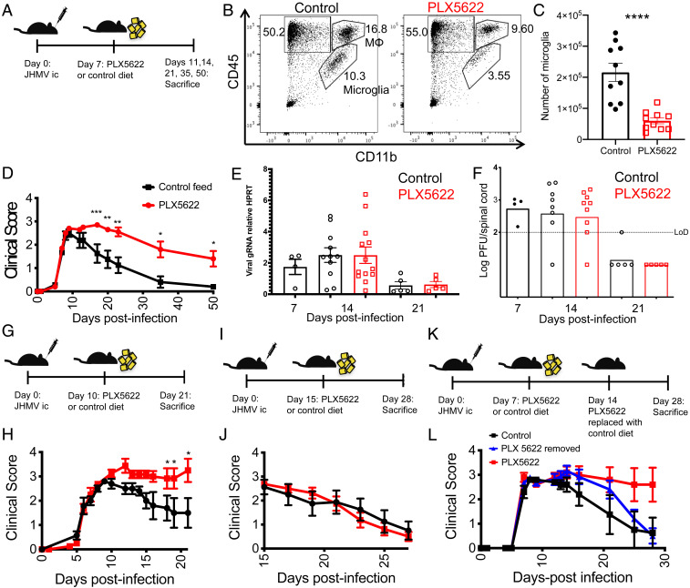
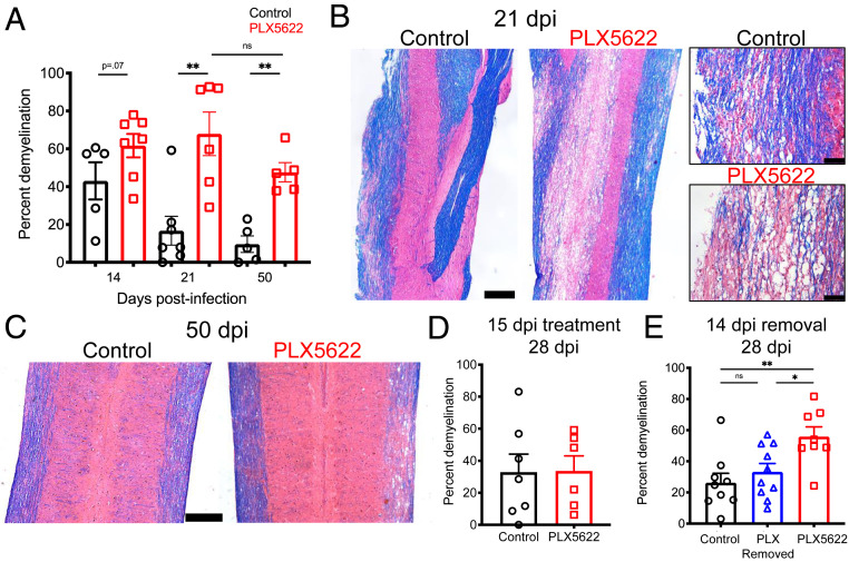
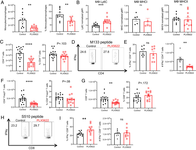
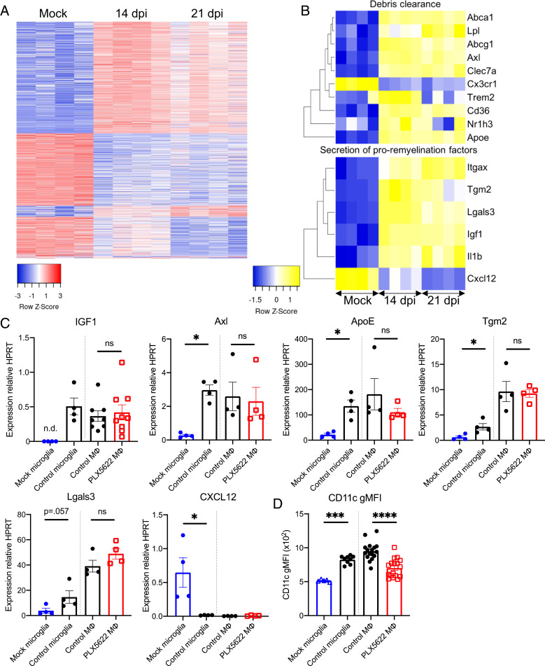
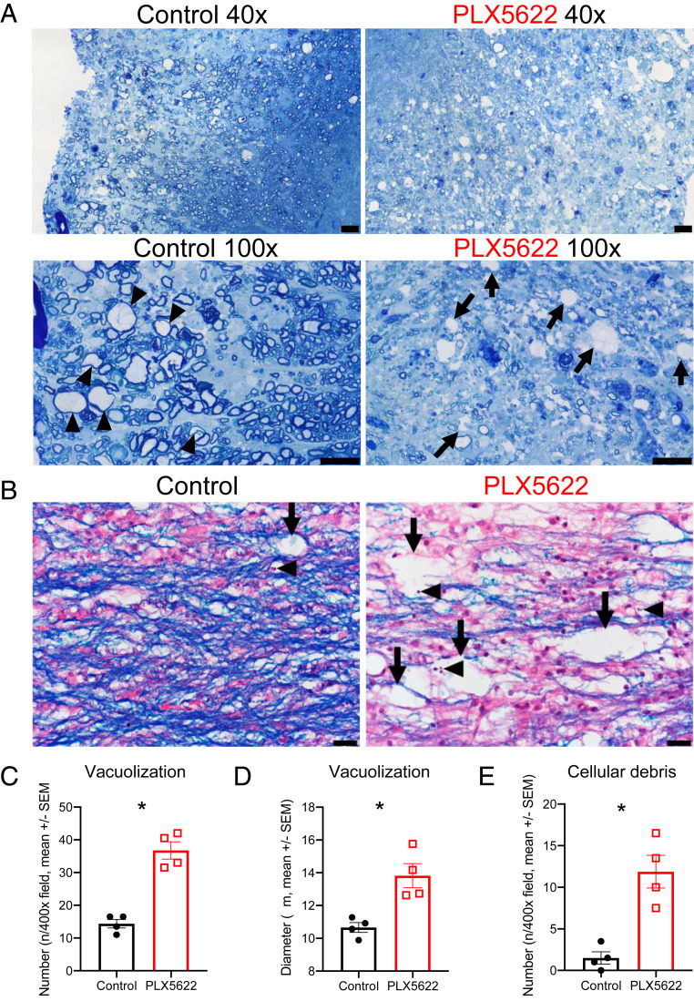
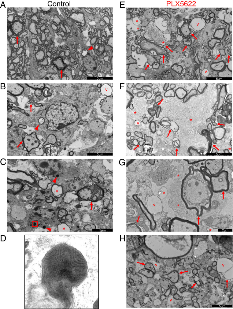
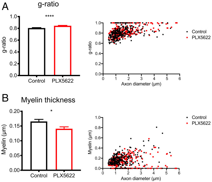
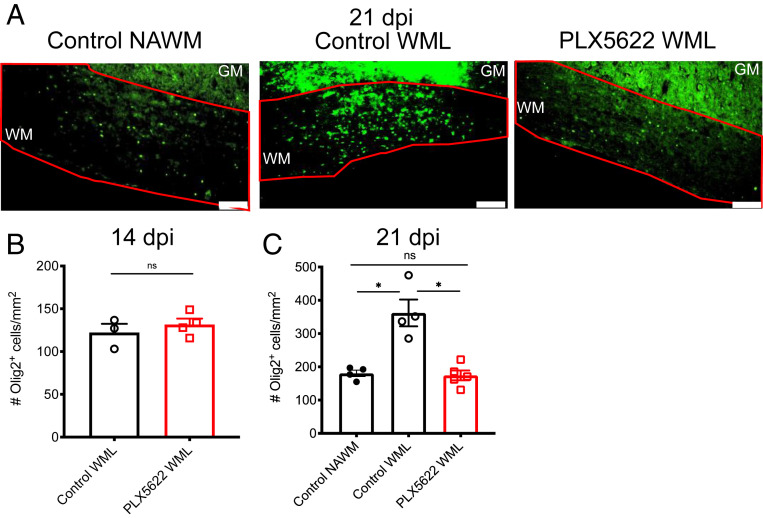
Microglia are considered both pathogenic and protective during recovery from demyelination, but their precise role remains ill defined. Here, using an inhibitor of colony stimulating factor 1 receptor (CSF1R), PLX5622, and mice infected with a neurotropic coronavirus (mouse hepatitis virus [MHV], strain JHMV), we show that depletion of microglia during the time of JHMV clearance resulted in impaired myelin repair and prolonged clinical disease without affecting the kinetics of virus clearance. Microglia were required only during the early stages of remyelination. Notably, large deposits of extracellular vesiculated myelin and cellular debris were detected in the spinal cords of PLX5622-treated and not control mice, which correlated with decreased numbers of oligodendrocytes in demyelinating lesions in drug-treated mice. Furthermore, gene expression analyses demonstrated differential expression of genes involved in myelin debris clearance, lipid and cholesterol recycling, and promotion of oligodendrocyte function. The results also demonstrate that microglial functions affected by depletion could not be compensated by infiltrating macrophages. Together, these results demonstrate that microglia play key roles in debris clearance and in the initiation of remyelination following infection with a neurotropic coronavirus but are not necessary during later stages of remyelination.

Keywords: coronavirus; microglia; neuroinflammation; remyelination; virus-induced demyelination.


Conflict of interest statement

The authors declare no competing interest.

Figures

Fig. 1.
Fig. 1.
Exacerbated disease in mice depleted of microglia. Mice were infected intracranially with 700-PFU JHMV, then fed at day 7 p.i. with PLX5622-containing or control chow (AF). Representative flow plots of spinal cords showing gating for lymphocytes (CD45+CD11b), monocytes/macrophages (CD45hiCD11b+, MΦ), and microglia (CD45intCD11b+) at day 14 p.i. (B). Summary of numbers of microglia in spinal cords at day 14 p.i. (C). Clinical scores at indicated days p.i. (D). (BD n = 10 mice/group). Expression levels of viral genomic RNA (gRNA) as assessed by qPCR (E) (n = 4–13 mice/group) and infectious virus titers as determined by plaque assay in the spinal cord (F) (n = 4–9 mice/group). Mice were infected and fed at day 10 (G and H) or day 15 (I and J) p.i. with PLX5622-containing or control chow. Mice were infected and fed PLX5622-containing or control chow at day 7, followed by replacement of PLX5622-containing chow with control chow at 14 dpi (K and L). Clinical scores at indicated days p.i. (H, J, and L) (n = 5–10 mice/group). Data represent the mean ± SEM, *P < 0.05, **P < 0.01, ***P < 0.001, ****P < 0.0001 by Mann–Whitney U test.
Fig. 2.
Fig. 2.
Prolonged and exacerbated demyelination following microglia depletion. Mice were infected and fed with PLX5622-containing or control chow starting at day 7 p.i. Demyelination was quantified at indicated times p.i. (A). Representative images of LFB and hematoxylin and eosin (H&E) stained thoracic spinal cords at day 21 (B) and cervical spinal cords at day 50 (C) p.i. Mice were infected and fed at day 15 p.i. with PLX5622-containing or control chow and demyelination quantified at day 28 p.i. (D). Mice were infected and fed PLX5622-containing or control chow at day 7, followed by replacement of PLX5622-containing chow with control chow at 14 dpi, and demyelination was quantified at day 28 p.i. (E). (Scale bars, 500 μm.) Except for two images on the right hand side of panel B where scale bar, 100 μm. Data shown in A are representative of two independent experiments with 5–7 mice per group. Data represent the mean ± SEM, *P < 0.05, **P < 0.01 by Mann–Whitney U test.
Fig. 3.
Fig. 3.
Decreased immune infiltration in PLX5622-treated mice. Mice were infected and treated at 7 dpi as described above, and flow cytometry was used to analyze cells from the spinal cord at 14 dpi in PLX5622-treated and control mice. Number and frequency of monocytes/macrophages (CD45hiCD11b+Ly6G) (A), CD4 T cells (C), Foxp3+ Tregs (F), and CD8 T cells (G). Normalized geometric MFI (gMFI) of Ly6C, MHCI, and MHCII staining in monocytes/macrophages (MΦ) (B). Virus-specific CD4+ T cell frequency and number were determined by IFN-γ intracellular cytokine staining (ICS) after stimulation with M133 peptide (D and E). Virus-specific CD8 T cell frequency and number were determined by IFN-γ ICS after stimulation with S510 peptide (H and I). Data represent combined results from two to three experiments with a combined total of 8–16 mice per group. Normalized gMFI (B) calculated as fold change over the mean of the control group for each experiment. Data represent the mean ± SEM, **P < 0.01, ***P < 0.001, ****P < 0.0001 by Mann–Whitney U test.
Fig. 4.
Fig. 4.
Microglia express proremyelination and myelin debris removal genes. Microglia from mice at 14 and 21 dpi as well as mock-infected mice were isolated by a fluorescence-activated cell sorter for RNA-seq analysis. Heat maps show the 1,818 unique differentially expressed genes across each pairwise comparison (A) and selected genes involved in debris clearance and remyelination (B). Isolated microglia at 14 dpi and after mock infection and isolated monocytes/macrophages (MΦ) at 14 dpi from control- and PLX5622-treated mice were analyzed for the indicated mRNA transcripts by qPCR (C). Following infection and treatment with PLX5622-containing or control feed at 7 dpi, flow cytometry was used to analyze the geometric gMFI of CD11c in monocytes/macrophages at 14 dpi (D). Data in A and B were derived from four mice per group and analyzed as described in Materials and Methods. Data in C represent 4–9 mice per group. Data in D represent combined results from two to three experiments with a combined total of 5–17 mice per group. n.d.: undetermined Ct values. Data represent the mean ± SEM, *P < 0.05, ***P < 0.001, ****P < 0.0001 by Mann–Whitney U test.
Fig. 5.
Fig. 5.
Increased vacuolization and cellular debris in PLX5622-treated mice. Mice were infected and fed with PLX5622-containing or control chow starting at 7 dpi. Representative images of toluidine blue-stained spinal cords at 21 dpi (arrows: vacuoles; arrowheads: remyelinating axons) (A). Representative images of LFB-stained spinal cords at 21 dpi with vacuolization (arrows) and pyknotic nuclear debris (arrowheads) (B). Graphs show quantification of vacuole numbers (C) and size (D) as well as quantification of cellular debris (E). Images in A are representative of three mice per group. Images and quantification in BE are representative of two fields per mouse and four mice per group (Scale bars, 20 μm.) Data represent the mean ± SEM, *P < 0.05 by Mann–Whitney U test.
Fig. 6.
Fig. 6.
Myelin debris clearance is impaired in PLX5622-treated mice. Mice were infected and fed with control (AD) or PLX5622-containing (EH) chow starting at 7 dpi. Shown are representative electron micrographs of spinal cord demyelinating lesions at 21 dpi with vacuolization (v), dystrophic, or degenerating axons (arrows), glial cells containing myelin or axonal inclusions (arrowheads), and vesiculated myelin debris (asterisks). Micrograph shown in D is a high magnification micrograph of the boxed region in C, demonstrating lamellar myelin debris within a glial cell. Micrographs are representative of three mice per group. Scale bars as indicated in the figure.
Fig. 7.
Fig. 7.
Remyelination is impaired in PLX5622-treated mice. Infected mice were fed PLX5622-containing or control chow starting at 7 dpi. EMs of spinal cord demyelinating lesions were analyzed at 21 dpi. Calculation of the g ratio and scatter plot depicting the g ratio as a function of inner axon diameter (A). Myelin thickness and scatter plot depicting myelin thickness relative to inner axon diameter (B). n = 265 axons analyzed per group. Data represent the mean ± SEM, *P < 0.05, ****P < 0.0001 by Mann–Whitney U test.
Fig. 8.
Fig. 8.
Diminished oligodendrocyte accumulation in demyelinating lesions of PLX5622-treated mice. Infected mice were treated at day 7 p.i. with PLX5622-containing or control feed. Representative images of Olig2 immunostaining in normal appearing WM (NAWM) and WMLs of control-treated mice and WMsL of PLX5622-treated mice at 21 dpi, Scale bars, 100 μm. (A). Quantification of Olig2 staining in WMLs at 14 dpi (B) and in NAWMs and WMLs at 21 dpi (C). Too little NAWM was detected in drug-treated mice at day 21 p.i. to allow quantification. GM: gray matter; WM: white matter. Data in B are representative of three to four mice per group. Data in C show individual mice with four to five mice per group and represent the mean ± SEM, *P < 0.05 by Mann–Whitney U test.

References

    1. Major E. O., Yousry T. A., Clifford D. B., Pathogenesis of progressive multifocal leukoencephalopathy and risks associated with treatments for multiple sclerosis: A decade of lessons learned. Lancet Neurol. 17, 467–480 (2018). - PubMed
    1. Soto-Hernandez J. L., Human herpesvirus 6 encephalomyelitis. Emerg. Infect. Dis. 10, 1700–1702 (2004). - PMC - PubMed
    1. Ozden S., Seilhean D., Gessain A., Hauw J.-J., Gout O., Severe demyelinating myelopathy with low human T cell lymphotropic virus type 1 expression after transfusion in an immunosuppressed patient. Clin. Infect. Dis. 34, 855–860 (2002). - PubMed
    1. Hall W. W., Choppin P. W., Measles-virus proteins in the brain tissue of patients with subacute sclerosing panencephalitis: Absence of the M protein. N. Engl. J. Med. 304, 1152–1155 (1981). - PubMed
    1. Sundqvist E. et al. ., Epstein-Barr virus and multiple sclerosis: Interaction with HLA. Genes Immun. 13, 14–20 (2012). - PubMed

Publication types

MeSH terms

Substances