. 2020 Sep 29;117(39):24545-24556.
doi: 10.1073/pnas.2004900117. Epub 2020 Sep 14.

Regulation of titin-based cardiac stiffness by unfolded domain oxidation (UnDOx)

Affiliations

Regulation of titin-based cardiac stiffness by unfolded domain oxidation (UnDOx)

Christine M Loescher et al. Proc Natl Acad Sci U S A. .

Abstract

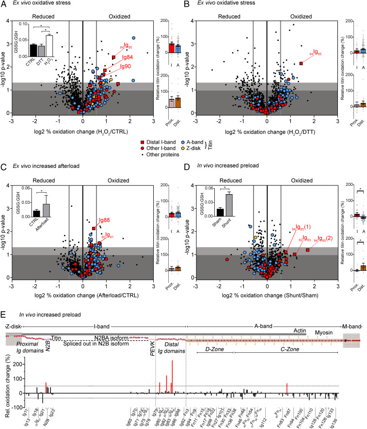
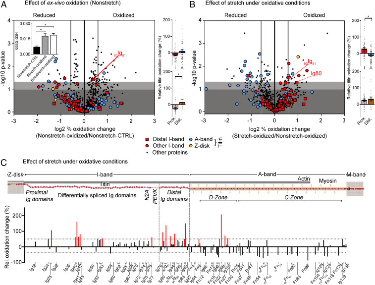
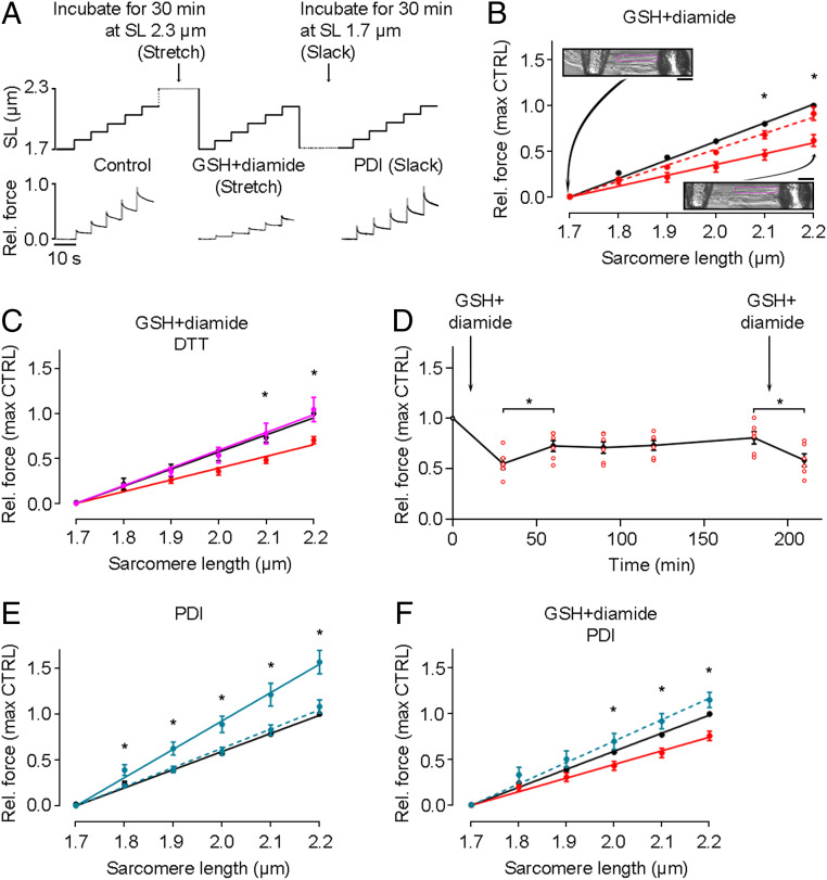
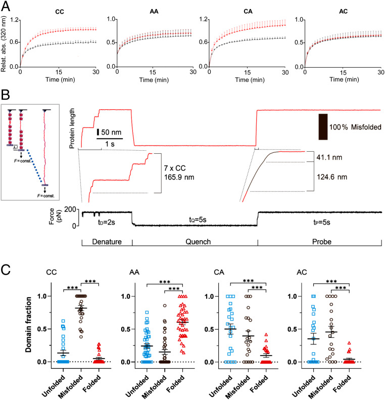
The relationship between oxidative stress and cardiac stiffness is thought to involve modifications to the giant muscle protein titin, which in turn can determine the progression of heart disease. In vitro studies have shown that S-glutathionylation and disulfide bonding of titin fragments could alter the elastic properties of titin; however, whether and where titin becomes oxidized in vivo is less certain. Here we demonstrate, using multiple models of oxidative stress in conjunction with mechanical loading, that immunoglobulin domains preferentially from the distal titin spring region become oxidized in vivo through the mechanism of unfolded domain oxidation (UnDOx). Via oxidation type-specific modification of titin, UnDOx modulates human cardiomyocyte passive force bidirectionally. UnDOx also enhances titin phosphorylation and, importantly, promotes nonconstitutive folding and aggregation of unfolded domains. We propose a mechanism whereby UnDOx enables the controlled homotypic interactions within the distal titin spring to stabilize this segment and regulate myocardial passive stiffness.

Keywords: mechanics; myocardial stiffness; oxidative stress; proteomics; single-molecule measurements.


Conflict of interest statement

The authors declare no competing interest.

Figures

Fig. 1.
Fig. 1.
Redox state of cardiac proteins in different mouse heart models. Volcano plots of peptides identified in the Langendorff H2O2 perfusion (n = 3) vs. control (n = 3) (A) or DTT (n = 3) (B), afterload-increase heart model (n = 4 for control and afterload increase) (C), and in vivo shunt (n = 4) vs. sham (n = 3) model (D). The difference in (log2) percentage oxidized was used. A threshold value of +0.58/−0.58 was chosen for redox classification (thick vertical gray line). Nonsignificant changes, dark gray background; sites trending toward significances between −log10 1.00 to 1.30, light gray background; and significantly altered sites above −log10 of 1.30, white background. Insets: GSSG:GSH ratio for H2O2 Langendorff perfusion (A; n = 6), afterload increase (C; n = 9) and shunt vs. sham (D; n = 4). Colored bar graphs on Right: percentage oxidation increase in I-band vs. A-band titin (Top) and proximal vs. distal I-band titin (Bottom). Data are means ± SEM *P < 0.05, two-tailed Mann–Whitney U test. For data in Insets, significance was tested by ANOVA, followed by Tukey’s test (A) or unpaired, two-tailed Student’s t test (C and D). (E) Schematic of mouse cardiac titin and bar graph showing the percentage oxidation change against the associated titin domains (UniProtKB ID A2ASS6). Increases by >50% are highlighted in red.
Fig. 2.
Fig. 2.
Redox state of the skeletal muscle. Volcano plots of peptides identified in nonstretched oxidized (n = 3) vs. nonstretched control muscle (n = 3) (A) and nonstretched oxidized (n = 3) vs. stretched oxidized muscle (n = 3) (B). Oxidized samples were incubated in relaxing buffer containing 2 mM GSH/0.5 mM diamide. The difference in (log2) percentage oxidized was used. A threshold value of +0.58/−0.58 was chosen (thick vertical gray line) for redox classification. Nonsignificant changes, dark gray background; sites trending toward significances between −log10 1.00 and 1.30, light gray background; and significantly altered sites above −log10 of 1.30, white background. Inset in A, GSSG:GSH ratio (n = 6). Colored bar graphs on Right: percentage oxidation increase in I-band vs. A-band titin (Top) and proximal vs. distal I-band titin (Bottom). Data are means ± SEM *P < 0.05, two-tailed Mann–Whitney U test. For data in Inset (A), significance was tested by ANOVA, followed by Tukey’s test. (C) Schematic of mouse skeletal muscle titin and bar graph showing the percentage oxidation change against the associated titin domains (UniProtKB ID A2ASS6). Increases by >50% are highlighted in red.
Fig. 3.
Fig. 3.
Effects of S-glutathionylation and disulfide bonding on passive force of human cardiomyocytes. (A) Stepwise stretch protocols used on skinned cardiomyocytes and example force traces. First, a control stretch protocol was conducted, followed by a 30-min incubation at either stretched or slack length in the absence or presence of 2 mM GSH/0.5 mM diamide or 35 µM PDI. (B) Passive force measured under control conditions (black), followed by incubation with 2 mM GSH/0.5 mM diamide at stretched length (solid red line, n = 5) or slack (dashed line, n = 3). Insets show images of a permeabilized cell attached to needles at slack (Top) and after stretch (Bottom). (Scale bars, 10 µm.) (C) Passive force measured under control conditions (black) followed by incubation with 2 mM GSH/0.5 mM diamide at stretched length (red), then another 30-min incubation with 5 mM DTT at stretched length (magenta) (n = 5). (D) Change of passive force with incubation of 2 mM GSH/0.5 mM diamide at stretched length and (after return to slack length) periodically remeasured before the oxidant was reintroduced (n = 6 cells; note that some data points overlay each other). (E) Passive force measured under control conditions (black), followed by incubation with 35 µM PDI at stretched length (solid blue line, n = 3) or slack (dashed line, n = 4). (F) Passive force measured under control conditions (black), followed by incubation with 2 mM GSH/0.5 mM diamide at stretched length (red), then another 30-min incubation with 35 µM PDI at slack length (blue) (n = 7). Cardiomyocytes were from a human donor heart. Peak force measured at the end of each stretch step was normalized to the control value at SL 2.2 µm. Data are means ± SEM; fits are linear regressions; *P < 0.05, by unpaired, two-tailed Student’s t test.
Fig. 4.
Fig. 4.
UnDOx in vitro and in vivo and unfolding-induced phosphorylation of Ig domains. (A) Schematic of mouse cardiac I-band titin and bar graphs showing the percentage oxidation change identified by MS against the associated titin domains (UniProtKB ID A2ASS6), for the Langendorff H2O2 vs. control (Top) or DTT (Middle) and increased afterload vs. control (Bottom) experiments. (B) Western blots (anti-GSH antibody) of recombinant Ig domains 82Ig83, Ig84, 84Ig85 and three-domain construct (Ig82-82Ig83-Ig83) thermally unfolded at 27 °C, 57 °C, or 77 °C and then oxidized with 2 mM GSH/0.5 mM diamide; 82Ig83 was also deglutathionylated with 5 mM DTT. (C) Representation of wild-type 82Ig83 and cysteine (C) mutant constructs (Top) and Western blots of recombinant 82Ig83 thermally unfolded at 27 °C or 57 °C and then oxidized with 2 mM GSH/0.5 mM diamide. CC, wild type; AA, both Cs replaced by alanine (A); CA, only C13601 replaced by A; AC, only C13585 replaced by A. (D) Double immunofluorescence staining of glutathione (GSH) and I-band titin in ischemic and remote heart tissue from myocardial ischemia mice (Top) and quantification of immunofluorescence intensity (Bottom). (Scale bars, 5 µm.) Data are mean ± SD; n = 103 (ischemic) and n = 93 (remote), from two hearts/group; ***P < 0.001 by ANOVA followed by Tukey’s test. n.s., non-significant. Care was taken to analyze cells of similar sarcomere length (∼2.1 µm). (E) Representation of 82Ig83 WT and phosphomutants (Top). P-0, all potential phosphosites replaced by A. P-1 to P-8, mutant constructs containing only one potential phosphosite. Bottom shows CaMKIIδ-mediated phosphorylation of 82Ig83 constructs unfolded at 57° and CaMKIIδ phosphorylation of unfolded 82Ig83 WT and P-7 mutant after unfolding and S-glutathionylation with 2.0 mM GSH/0.5 mM diamide, detected by autoradiography. Coomassie-stained gel bands are shown above the (γ-P32)ATP signal.
Fig. 5.
Fig. 5.
Molecular consequences of UnDOx of 82Ig83. (A) Aggregation of unfolded wild-type (CC) and mutant 82Ig83 (AA, CA, and AC) under control (black) and oxidative (2 mM GSH/0.5 mM diamide; red) conditions. Absorbance from partially refolded construct measured at 320 nm and 1-min intervals for 30 min. CC: control n = 4, oxi n = 5. Double mutant, no cysteines (AA): control n = 4, oxi n = 4. Single mutant C13601A (CA): control n = 6, oxi n = 5. Single mutant C13585A (AC): control n = 6, oxi n = 4. Data are normalized to highest aggregation value of 82Ig83 (CC) after oxidation. Data are means ± SD. Curves are exponential saturation function best fits. (B) Unfolding/refolding of recombinant polyprotein (82Ig83)8 measured by single-molecule AFM force spectroscopy (Left) and example trace with seven domains in the tether. In presence of 1 mM GSH/2 mM diamide, domains were first unfolded at 175 pN and held extended for 2 s (denature). Then, force was quenched to zero for 5 s to allow for refolding (quench), before it was increased back to 175 pN (probe). (C) Proportion of unfolded, misfolded, and folded events detected in S-glutathionylated 82Ig83 CC (n = 130), AA (n = 209), CA (n = 113), and AC (n = 98) polyproteins. Data are mean ± SEM; ***P < 0.001, by ANOVA followed by Tukey’s multiple comparisons test.
Fig. 6.
Fig. 6.
Proposed mechanism of UnDOx to control in-register aggregation of distal I-band titin. Several Ig domains from the distal I-band titin region (mainly the NH2-terminal half) are suggested to be in a partially unfolded state in vivo. S-glutathionylation of the unfolded domains stabilizes the unfolded state, which promotes aggregation of parallel titin molecules. Stretch under oxidant stress conditions recruits more Ig domains to the unfolded state and further promotes this mechanism. The result is an improved anchorage of the titin spring under mechanical and oxidant stress.

References

    1. Gao W. D. et al. ., Nitroxyl-mediated disulfide bond formation between cardiac myofilament cysteines enhances contractile function. Circ. Res. 111, 1002–1011 (2012). - PMC - PubMed
    1. Lovelock J. D. et al. ., Ranolazine improves cardiac diastolic dysfunction through modulation of myofilament calcium sensitivity. Circ. Res. 110, 841–850 (2012). - PMC - PubMed
    1. Beckendorf L., Linke W. A., Emerging importance of oxidative stress in regulating striated muscle elasticity. J. Muscle Res. Cell Motil. 36, 25–36 (2015). - PMC - PubMed
    1. Koser F., Loescher C., Linke W. A., Posttranslational modifications of titin from cardiac muscle: How, where, and what for? FEBS J. 286, 2240–2260 (2019). - PMC - PubMed
    1. Grützner A. et al. ., Modulation of titin-based stiffness by disulfide bonding in the cardiac titin N2-B unique sequence. Biophys. J. 97, 825–834 (2009). - PMC - PubMed

Publication types

LinkOut - more resources