. 2020 Oct 15;205(8):2231-2242.
doi: 10.4049/jimmunol.1901533. Epub 2020 Sep 14.

Small-Molecule Inhibitor of 8-Oxoguanine DNA Glycosylase 1 Regulates Inflammatory Responses during Pseudomonas aeruginosa Infection

Affiliations

Small-Molecule Inhibitor of 8-Oxoguanine DNA Glycosylase 1 Regulates Inflammatory Responses during Pseudomonas aeruginosa Infection

Shugang Qin et al. J Immunol. .

Abstract

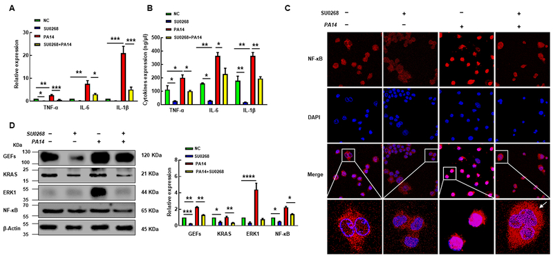
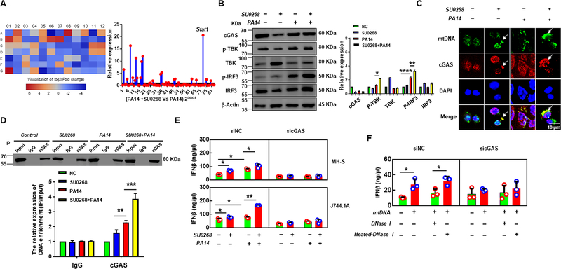
The DNA repair enzyme 8-oxoguanine DNA glycosylase 1 (OGG1), which excises 8-oxo-7,8-dihydroguanine lesions induced in DNA by reactive oxygen species, has been linked to the pathogenesis of lung diseases associated with bacterial infections. A recently developed small molecule, SU0268, has demonstrated selective inhibition of OGG1 activity; however, its role in attenuating inflammatory responses has not been tested. In this study, we report that SU0268 has a favorable effect on bacterial infection both in mouse alveolar macrophages (MH-S cells) and in C57BL/6 wild-type mice by suppressing inflammatory responses, particularly promoting type I IFN responses. SU0268 inhibited proinflammatory responses during Pseudomonas aeruginosa (PA14) infection, which is mediated by the KRAS-ERK1-NF-κB signaling pathway. Furthermore, SU0268 induces the release of type I IFN by the mitochondrial DNA-cGAS-STING-IRF3-IFN-β axis, which decreases bacterial loads and halts disease progression. Collectively, our results demonstrate that the small-molecule inhibitor of OGG1 (SU0268) can attenuate excessive inflammation and improve mouse survival rates during PA14 infection. This strong anti-inflammatory feature may render the inhibitor as an alternative treatment for controlling severe inflammatory responses to bacterial infection.


Conflict of interest statement

Footnotes

Disclosures

The authors have no financial conflicts of interest.

Figures

FIGURE 1.
FIGURE 1.
The chemical inhibitor SU0268 suppresses OGG1’s activity and inflammatory response in cell culture. (A) Cell viability detected by MTT assay. MH-S macrophages were treated with SU0268 (50, 25, 12.5, 6.25, 3.125, 1.5625, 0.78, 0.39 μM) for 24 hours and IC50 was calculated with GraphPad 7.0. (B) Cell viability evaluated after treating cells with SU0268 at 0, 2, 4, 8, 16 μM, respectively. (C) Cell viability was further evaluated by MTT at 2 μM for indicated times. (D) Endogenous OGG1 activity in lysates prepared from MH-S cells (0.1 mg/mL total protein) was measured using a previously described fluorescence assay (23). (E) MH-S Cells were cultured in the presence of SU0268 (1 μM) for 36 hours or treated with H2O2 (5 mM) and Fe2+ (100 μM) for 1 hour. Total DNA was then isolated and digested with nuclease P1 prior to measurement of 8-oxoG using a commercial 8-hydroxy 2-deoxyguanosine ELISA kit. The anti-inflammatory effect of SU0268 was evaluated by quantifying the expression level of TNF-α, IL-1β and IL-6 by using ELISA (F) and (G) western blotting after treatment with SU0268 (2 μM) for 0, 4, 8, 12 hours.
FIGURE 2.
FIGURE 2.
SU0268 inhibits inflammatory responses in MH-S cells via OGG1- GEFs/KRAS-NF-κB axis. MH-S cells were treated with SU0268 (2 μM) for 8 h (the control group received an equal amount of 100% ethanol), then infected with PA14 for 2 h. Anti-inflammatory activity of SU0268 was analyzed by measuring the expression of TNF-α, IL-1β and IL-6 by qPCR (A) and ELISA (B). The activation of NF-κB was detected by immunofluorescence (C) (arrows indicating cytoplasmic NF-κB staining, the typical cells were enlarged and placed on the bottom). (D) The proteins expression levels of OGG1-GEFs/KRAS-NF-κB axis were determined by western blotting.
FIGURE 3.
FIGURE 3.
SU0268 promotes mtDNA release into the cytoplasm after ROS-induced DNA damage in response to P. aeruginosa infection. MH-S cells were treated with SU0268 (2 μM) for 8 h (the control group received an equal amount of 100% ethanol), then infected with PA14 for 2 h. The ROS products were measured by NBT assay (A) and H2DCF assay (B) according to the manufacturer’s instructions. (C) DNA strand breaks damage were detected by comet assay, and the tail length of damaged DNA was measured with CLSM. (D) Mitochondrial membrane potential was assessed by JC-1 fluorescence assay. (E) Mitochondrial DNA was extracted and mtDNA copy numbers were analyzed by qPCR.
FIGURE 4.
FIGURE 4.
SU0268 promotes mtDNA release dependent on the BAK/BAX axis. MH-S cells were treated with SU0268 (2 μM) for 8 hours (the control group received an equal amount of 100% ethanol), subsequent infection with PA14 for 2 hours. (A) 60 genes involved in mtDNA release and mitochondrial function were quantified by using qPCR microarray, heat maps and red dots in scatter plot are used to indicate differential expression of genes (more than two-fold change and p<0.05 by Student’s t-test, respectively, each color of 1–60 represents the expression of one gene, primers are shown in Supplementary Table S2). (B) Cell apoptosis was measured by flow cytometry, ABT737, as a positive control, has been identified to cause apoptosis (77). (C) Released cytochrome c was detected by immunofluorescence (arrows indicating typical released cytochrome c) and western blotting (Right panel).
FIGURE 5.
FIGURE 5.
Increased release of mtDNA activates the cGAS pathway by directly binding cGAS protein to manipulate type I IFN responses. MH-S cells were treated with SU0268 (2 μM) for 8 hours (the control group received an equal amount of 100% ethanol), subsequent infection with PA14 for 2 hours. (A) RT2 Profiler PCR inflammation & immunity crosstalk array was used to identify key regulatory genes (330231 PAMM-181Z, QIAGEN, USA). Heat maps and red dots in scatter plots are used to indicate differential expression of genes (more than two-fold change and p<0.05 by Student’s t-test), respectively, each color of A01-G12 represents the expression of one gene. (B) The expression level of proteins (cGAS, p-TBK, TBK, p-IRF3, and IRF-3) were quantified through Western blotting. (C) Co-localization of mtDNA and cGAS was detected by immunofluorescence (arrows indicating typical co-localization of mtDNA and cGAS). (D) Mitochondria DNA that was bound to cGAS was analyzed by immunoprecipitation, then quantified by qPCR. (E) MH-S cells and J744.1A were transfected with cGAS siRNA for 48 h (negative siRNA as control) by using Lip RNAiMAX Reagent, then treated with SU0268 for 8 h (pure ethanol as control), followed by PA14 infection for 2 h. The expression level of IFNβ was determined by ELISA (42400–1, R&D Systems) according to the manufacturer’s instructions. (F) Mitochondria DNA deletion and replenishment assay were performed in J744.1A cells as previously described (26). J744.1A cells that deleted mtDNA were transfected with cGAS siRNA (Negative siRNA as control) for 36 hours and subsequently treated with PBS or DNAase I or heated-DNAase I for 4 hours, then mtDNA was replenished for 12 hours in pretreated PBS group by using Lip RNAiMAX Reagent, and the expression levels of IFNβ was analyzed by ELISA.
FIGURE 6.
FIGURE 6.
Small-molecule inhibitor of OGG1 significantly down-regulates pro-inflammatory responses and mitigates bacterial infection. Mice treated with SU0268 (10mg/kg) for 12 h and control mice without SU0268 treatment (n = 9) were intranasally challenged with PA14 at 6 × 10⁶ CFU (uninfected WT and SU0268 as a control) and observed up to 9 days. (A)The survival test is represented by Kaplan-Meier survival curves, and the results showed that pretreated with SU0268 increased survival rates compared with PA14 infection without SU0268 pretreatment (p = 0.0925; 95% confidence interval, n = 9 for each group). (B) Colony formation unit assay was used to count the number of bacteria in the lung, blood and BALF at 9 days after infection. (C) H&E staining was used to assess lung injury in mice (photograph is magnified 20 times). (D) ELISA was performed to determine the expression level of IFNβ in the mouse primary BMDM cells. (E) 10,000 cells that were stained by indicated antibodies were collected, the percentages of the myeloid progenitor cells (Stained CD45+/CD11b+ cells / total cells) and neutrophils (Stained CD11b+/Gr1+/MHCII+ cells / total cells) in mouse BALF, blood and lung were detected by flow cytometry.
FIGURE 7.
FIGURE 7.
Schematic illustration of the signaling pathways in which small-molecule inhibitor of OGG1 orchestrates inflammatory and mtDNA-mediated Type I IFN responses. Normally, PA14 infection triggers increased ROS production and DNA damage, which moderately activates OGG1’s damage repair function and induces an excessive inflammatory response by regulation of the KRAS-ERK1-NF-κB axis. Inhibiting the enzymatic activity of OGG1 decreases DNA inflammatory responses. Furthermore, a small-molecule inhibitor of OGG1 elicits massive mitochondrial DNA release into the cytoplasm, which then initiates IFNβ responses by the cGAS-TBK-IRF3-IFNβ circuit. Ultimately, elevated IFNβ mitigates P. aeruginosa infection and spread.

Similar articles

Cited by

References

    1. Hoggarth A, Weaver A, Pu Q, Huang T, Schettler J, Chen F, Yuan X, and Wu M. 2019. Mechanistic research holds promise for bacterial vaccines and phage therapies for Pseudomonas aeruginosa. Drug design, development and therapy 13: 909–924. - PMC - PubMed
    1. Zhang Y, Zhou CM, Pu Q, Wu Q, Tan S, Shao X, Zhang W, Xie Y, Li R, Yu XJ, Wang R, Zhang L, Wu M, and Deng X. 2019. Pseudomonas aeruginosa Regulatory Protein AnvM Controls Pathogenicity in Anaerobic Environments and Impacts Host Defense. mBio 10: e01362–01319. - PMC - PubMed
    1. Li R, Fang L, Pu Q, Lin P, Hoggarth A, Huang H, Li X, Li G, and Wu M. 2016. Lyn prevents aberrant inflammatory responses to Pseudomonas infection in mammalian systems by repressing a SHIP-1-associated signaling cluster. Signal transduction and targeted therapy 1: 16032. - PMC - PubMed
    1. Pu Q, Gan C, Li R, Li Y, Tan S, Li X, Wei Y, Lan L, Deng X, Liang H, Ma F, and Wu M. 2017. Atg7 Deficiency Intensifies Inflammasome Activation and Pyroptosis in Pseudomonas Sepsis. Journal of immunology 198: 3205–3213. - PMC - PubMed
    1. Pan L, Hao W, Zheng X, Zeng X, Ahmed Abbasi A, Boldogh I, and Ba X. 2017. OGG1-DNA interactions facilitate NF-kappaB binding to DNA targets. Scientific reports 7: 43297. - PMC - PubMed

Publication types

MeSH terms