. 2020 Sep 14;11(9):750.
doi: 10.1038/s41419-020-02964-2.

Activation of Wnt signaling by amniotic fluid stem cell-derived extracellular vesicles attenuates intestinal injury in experimental necrotizing enterocolitis

Affiliations

Activation of Wnt signaling by amniotic fluid stem cell-derived extracellular vesicles attenuates intestinal injury in experimental necrotizing enterocolitis

Bo Li et al. Cell Death Dis. .

Abstract

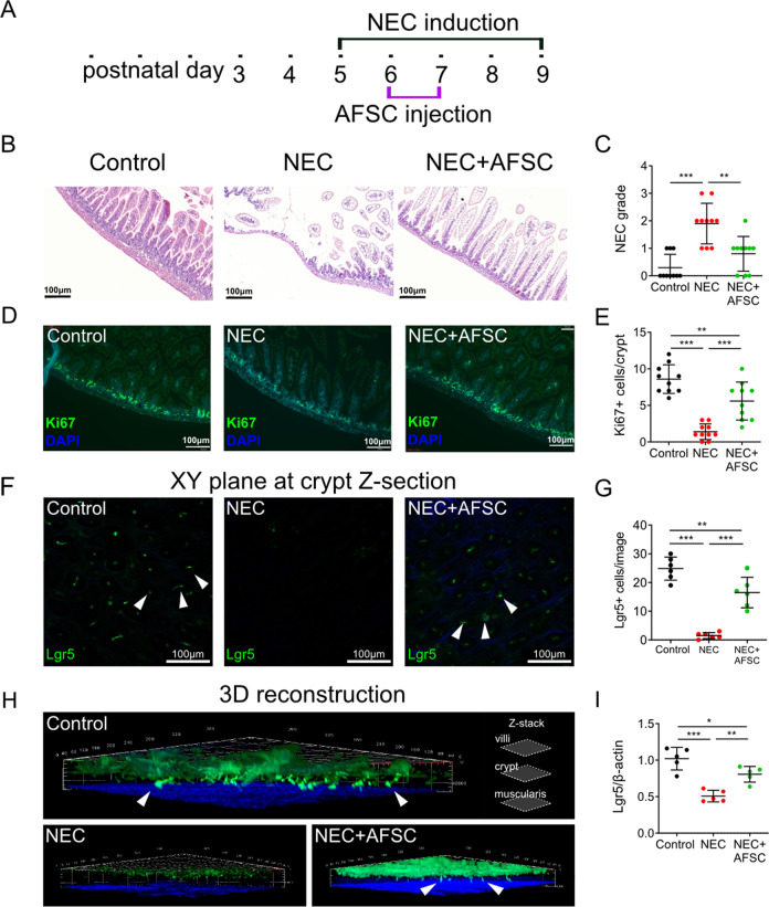
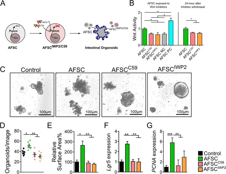
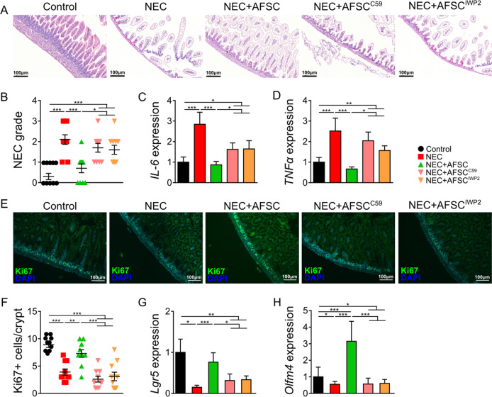
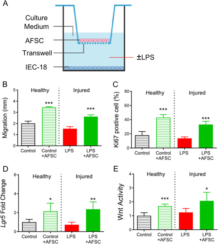
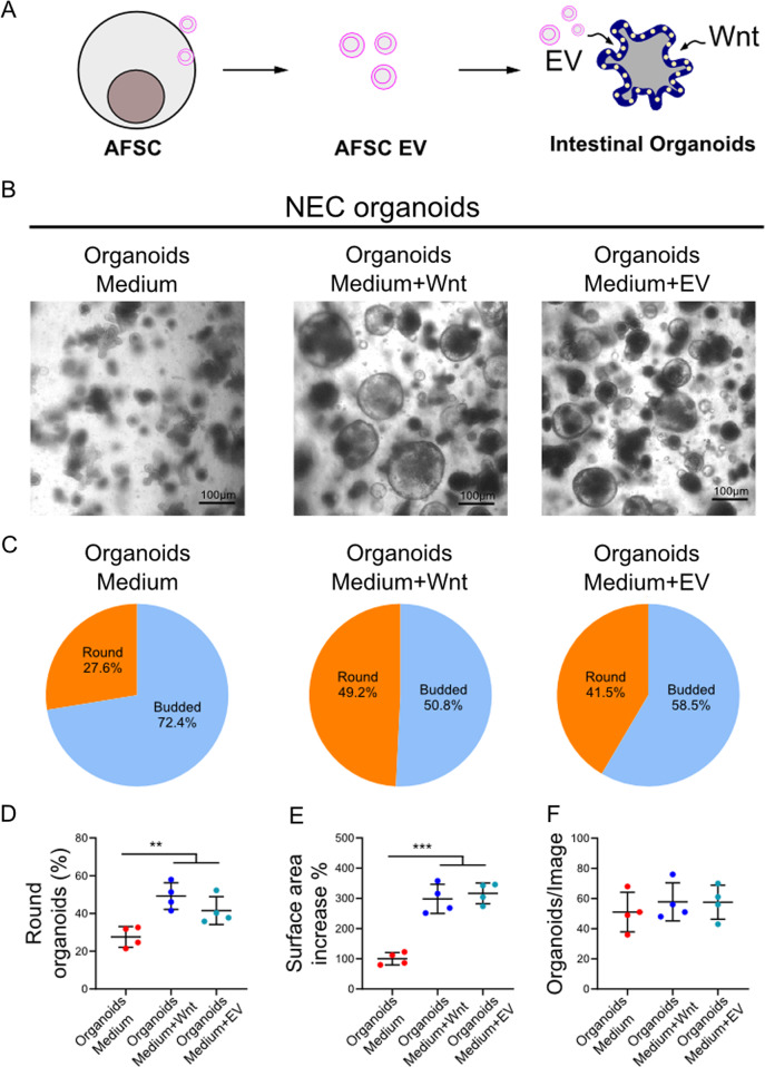
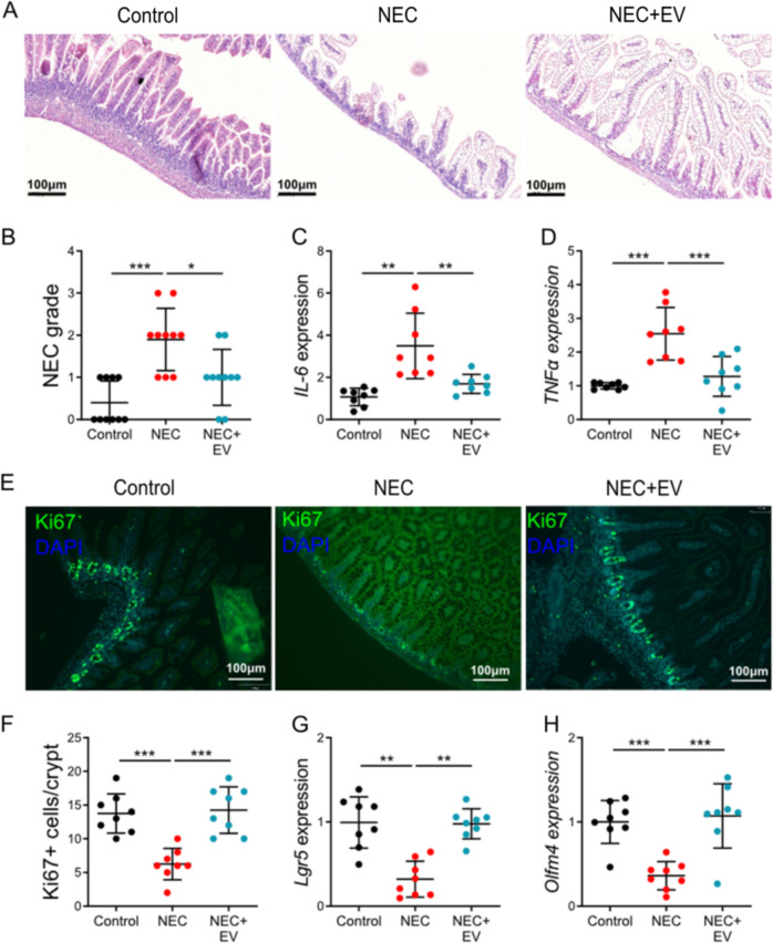
Necrotizing enterocolitis (NEC) is a devastating intestinal disease primarily affecting preterm neonates and causing high morbidity, high mortality, and huge costs for the family and society. The treatment and the outcome of the disease have not changed in recent decades. Emerging evidence has shown that stimulating the Wnt/β-catenin pathway and enhancing intestinal regeneration are beneficial in experimental NEC, and that they could potentially be used as a novel treatment. Amniotic fluid stem cells (AFSC) and AFSC-derived extracellular vesicles (EV) can be used to improve intestinal injury in experimental NEC. However, the mechanisms by which they affect the Wnt/β-catenin pathway and intestinal regeneration are unknown. In our current study, we demonstrated that AFSC and EV attenuate NEC intestinal injury by activating the Wnt signaling pathway. AFSC and EV stimulate intestinal recovery from NEC by increasing cellular proliferation, reducing inflammation and ultimately regenerating a normal intestinal epithelium. EV administration has a rescuing effect on intestinal injury when given during NEC induction; however, it failed to prevent injury when given prior to NEC induction. AFSC-derived EV administration is thus a potential emergent novel treatment strategy for NEC.


Conflict of interest statement

The authors declare that they have no conflict of interest.

Figures

Fig. 1
Fig. 1. Amniotic fluid stem cells (AFSC) rescued intestinal injury, restored epithelial regeneration, and increased active intestinal stem cells (ISC).
NEC induction was conducted on postnatal (p) days 5–9, with AFSC intraperitoneal injections given on p6 and p7 (a). Histopathology of ileal sections from mice administered with AFSC during necrotizing enterocolitis (NEC) induction showed normal villous structure when compared to NEC mice (b). Administration of AFSC during NEC significantly decreased histological scores (c). Intestinal epithelial proliferation (Ki67), which is reduced in NEC, was restored with AFSC administration (d, e). In vivo visualization of ISC after NEC induction showed a decrease in Lgr5+ ISC, some of which are denoted with white arrows, expression in NEC and restoration after AFSC treatment (fi). n = 10 for each group (be) and n = 6 for each group (fi). Data are presented as means ± SD. *p < 0.05; **p < 0.01; ***p < 0.001, using unpaired Student’s t test or one-way ANOVA with post-hoc tests as appropriate.
Fig. 2
Fig. 2. AFSC increased intestinal stem cells and epithelial proliferation via Wnt signaling in intestinal organoids.
AFSC treated with two known inhibitors of Porcn, Wnt-C59, and IWP2 that are involved in Wnt maturation and release, were added to intestinal organoids (a). Wnt activity was reduced in Wnt-C59 and IWP2-treated AFSC with effects lasting ≥24 h after removal of the inhibitors, and a negative control (NC) and positive control (PC) were also included (b). After 7 days in culture, AFSC induced organoid growth and increased both the number and surface area of organoids, while Wnt-deficient AFSC failed to promote organoid growth (ce). Organoids cocultured with AFSC had increased gene expression of the ISC marker Lgr5 and proliferation marker PCNA, and this was not observed in Wnt-deficient AFSC (f, g). Data are presented as means ± SD. n = 6 for each group. *p < 0.05; **p < 0.01; ***p < 0.001, using unpaired Student’s t test or one-way ANOVA with post-hoc tests as appropriate.
Fig. 3
Fig. 3. Wnt signaling was necessary to attenuate intestinal damage during experimental NEC.
Ileal sections from NEC mice administered with C59 and IWP2-treated AFSC demonstrated increased villus damage as compared to NEC+AFSC (a, b). AFSC reduced NEC-induced intestinal expression of pro-inflammatory cytokines IL-6 and TNFα (c, d), increased epithelial proliferation (Ki67) (e, f), and increased gene expression of ISC marker Lgr5 and Olfm4 (g, h). These changes were not evident with administration of Wnt-deficient AFSC (pretreated with C59 or IWP2). n = 10 for Control, NEC, NEC+AFSC, and n = 6 for NEC+AFSCC59, NEC+AFSCIWP2. Data are presented as means ± SD. *p < 0.05; **p < 0.01; ***p < 0.001, using unpaired Student’s t test or one-way ANOVA with post-hoc tests as appropriate.
Fig. 4
Fig. 4. AFSC-secreted factors that increased intestinal stem cell activity and Wnt pathway activation in both healthy and injured intestinal epithelial cells.
Transwells were used to coculture AFSC with intestinal epithelial cell line (IEC-18) cells (a). AFSC increased IEC migration to the apical compartment (b), intestinal cell proliferation (c), intestinal stem cell activity (d), and Wnt activity (e) during wound healing in both normal “healthy” and LPS-induced injury conditions. n = 6 for each group. Data are presented as means ± SD. *p < 0.05; **p < 0.01; ***p < 0.001, using unpaired Student’s t test or one-way ANOVA with post-hoc tests as appropriate.
Fig. 5
Fig. 5. AFSC-derived EV rescued organoids derived from NEC–injured intestinal tissue in a similar manner to Wnt.
EV derived from AFSC cultured media were harvested and used in NEC organoids (a). AFSC-derived EV added to culture medium increased organoid growth in a similar manner to exogenous Wnt enriched culture medium (b, c). NEC organoids treated with exogenous Wnt and EV also displayed more spherical shape, higher surface area, and increased numbers as compared to controls (df). n = 4 for each group. Data are presented as means ± SD. *p < 0.05; **p < 0.01; ***p < 0.001, using unpaired Student’s t test or one-way ANOVA with post-hoc tests as appropriate.
Fig. 6
Fig. 6. AFSC-derived EV improved intestinal growth in NEC-induced intestinal injury.
Administration of EV derived from AFSC-conditioned medium on P6-P7 during NEC progression improved intestinal histology (a, b), and reduced IL-6 (c) and TNFα expression (d). There was also increased Ki67 (e, f), Lgr5 (g), and Olfm4 (h) expression with EV administration relative to NEC alone. n = 8 for each group. Data are presented as means ± SD. *p < 0.05; **p < 0.01; ***p < 0.001, using unpaired Student’s t test or one-way ANOVA with post-hoc tests as appropriate.

Similar articles

Cited by

References

    1. Neu J, Walker WA. Necrotizing enterocolitis. N. Engl. J. Med. 2011;364:255–264. - PMC - PubMed
    1. Yee WH, et al. Incidence and timing of presentation of necrotizing enterocolitis in preterm infants. Pediatrics. 2012;129:e298–e304. - PubMed
    1. Pierro A. The surgical management of necrotising enterocolitis. Early Hum. Dev. 2005;81:79–85. - PubMed
    1. Thyoka M, et al. Advanced necrotizing enterocolitis part 1: mortality. Eur. J. Pediatr. Surg. 2012;22:8–12. - PubMed
    1. Nino DF, et al. Retinoic acid improves incidence and severity of necrotizing enterocolitis by lymphocyte balance restitution and repopulation of LGR5+ intestinal stem cells. Shock. 2017;47:22–32. - PMC - PubMed

Publication types