. 2020 Sep 14;10(1):312.
doi: 10.1038/s41398-020-00994-0.

Presynaptic dysfunction in CASK-related neurodevelopmental disorders

Affiliations

Presynaptic dysfunction in CASK-related neurodevelopmental disorders

Martin Becker et al. Transl Psychiatry. .

Abstract

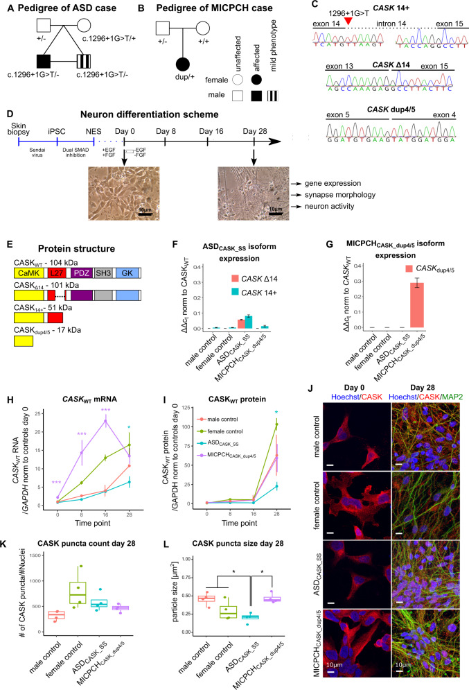
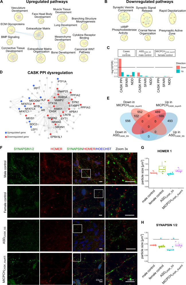
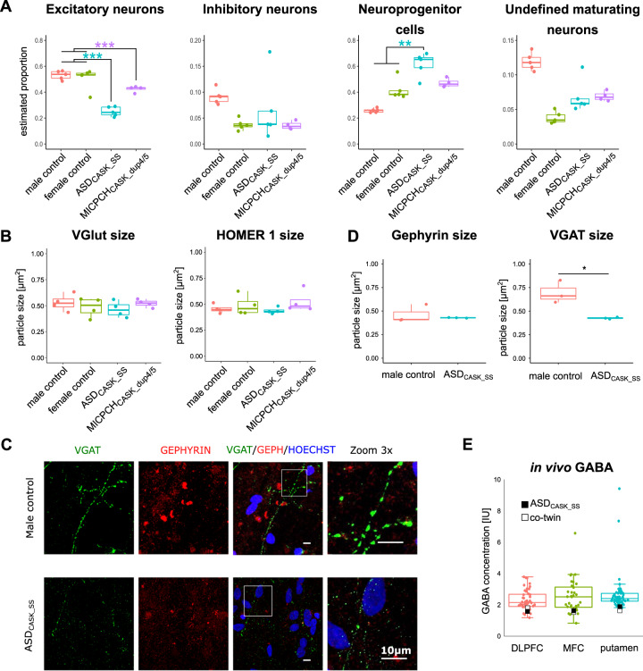
CASK-related disorders are genetically defined neurodevelopmental syndromes. There is limited information about the effects of CASK mutations in human neurons. Therefore, we sought to delineate CASK-mutation consequences and neuronal effects using induced pluripotent stem cell-derived neurons from two mutation carriers. One male case with autism spectrum disorder carried a novel splice-site mutation and a female case with intellectual disability carried an intragenic tandem duplication. We show reduction of CASK protein in maturing neurons from the mutation carriers, which leads to significant downregulation of genes involved in presynaptic development and of CASK protein interactors. Furthermore, CASK-deficient neurons showed decreased inhibitory presynapse size as indicated by VGAT staining, which may alter the excitatory-inhibitory (E/I) balance in developing neural circuitries. Using in vivo magnetic resonance spectroscopy quantification of GABA in the male mutation carrier, we further highlight the possibility to validate in vitro cellular data in the brain. Our data show that future pharmacological and clinical studies on targeting presynapses and E/I imbalance could lead to specific treatments for CASK-related disorders.


Conflict of interest statement

The authors declare no competing interests. Sven Bölte declares no direct conflict of interest related to this article. Bölte discloses that he has in the last 5 years acted as an author, consultant, or lecturer for Shire/Takeda, Medice, Roche, Eli Lilly, Prima Psychiatry, and SB Education and Psychological Consulting AB. He receives royalties for text books and diagnostic tools from Huber/Hogrefe, Kohlhammer, and UTB.

Figures

Fig. 1
Fig. 1. CASK mutations affect wild-type expression in carriers.
a Pedigree of a male diagnosed with ASD and his monozygotic cotwin who exhibits autistic traits, both carrying a splice-site mutation in the X-linked CASK gene (chrX: 41,586,906 C>A (hg38); NM_001126055: c.1296+1 G>T) inherited from a typically developed mother. b Pedigree of a female diagnosed with MICPCH, carrying a de novo duplication of 54.9 kb surrounding exons 4 and 5 (chrX:41710324–41765176 (hg38)) of the CASK gene. c Sanger chromatogram of CASK mRNA isoforms in cDNA of ASDCASK_SS fibroblasts and MICPCHCASK_dup4/5 NES cells. d Schematic summary of the differentiation protocol from skin biopsy to maturing neurons and representative phase-contrast microscopy images of male control NES cells and day 28 neurons. e Protein domain structure of CASKWT and in silico-predicted domain structure of CASKΔ14, CASK14+, and CASKdup4/5. f RT-qPCR quantification of CASKΔ14 and CASK14+ expression in three biological replicates of case and control NES cells. g RT-qPCR quantification of CASKdup4/5 in three biological replicates of case and control NES cells. h RT-qPCR quantification of CASKWT in three biological replicates of differentiating neurons. i Protein quantification of CASKWT protein in two biological replicates of differentiating neurons using capillary western blot quantification. j Representative confocal microscopy images of NES cells (day 0) and differentiated neurons (day 28) immunostained for CASK (red), MAP2 (green), and Hoechst (blue). Scale = 10 µm. k, l Quantification of CASK puncta k number and l size from confocal images (n = 4 per cell line). Statistical differences between cases and controls were calculated using ANOVA with post hoc Tukey HSD. *p < 0.05, ***p < 0.001. Asterisks in a and b are color-coded according to case cell lines.
Fig. 2
Fig. 2. Single-cell transcriptomics of MICPCHCASK_dup4/5.
ac t-Distributed stochastic neighbor embedding (tSNE) plot of 383 MICPCHCASK_dup4/5 cell transcriptomes after 28 days of differentiation colored by a the detection of mutant CASKdup4/5 expression, b the identified cell-type clusters, and c expression of SLC17A6, GAD2, NES, TOP2A, SEMA3D, and LHX1.
Fig. 3
Fig. 3. Consistent dysregulation of presynaptic and CASK-interacting genes in bulk RNA sequencing after 28 days of differentiation.
a Upregulated and b downregulated gene sets emerging from GSEA in bulk RNA-sequencing data from mutation carriers compared with controls. c GSEA for CASK-interacting proteins (CASK PPI), ASD risk genes (SFARI), and NDD genes (NDD) in bulk RNA-sequencing data from mutation carriers compared with controls. d Network of CASK-interacting proteins with upregulation marked in blue and downregulation in red. e Venn diagram illustrating the top upregulated and downregulated genes in ASDCASK_SS and MICPCHCASK_dup4/5 in comparison with sex-matched controls. f Representative confocal microscopy images of neurons differentiated for 28 days and immunostained for Synapsin-1/2 (green), Homer-1 (red), and Hoechst (blue). Scale = 10 µm. g Quantification of Homer-1 and h Synapsin-1/2 particle size (n = 4 per cell line). Statistical differences between cell lines were calculated using ANOVA with post hoc Tukey HSD. *p < 0.05, **p < 0.01, ***p < 0.001. Asterisks are color-coded according to case cell lines.
Fig. 4
Fig. 4. Presynaptic effect is limited to inhibitory VGAT presynaptic marker.
a Deconvolution of cell-type populations underlying bulk RNA-sequence samples from all cell lines. b Quantification of VGlut and Homer-1 particle size (n = 4 per cell line). c Representative confocal microscopy images of neurons differentiated for 28 days and immunostained for inhibitory VGAT (green), Homer-1 (red), and Hoechst (blue). Scale = 10 µm. d Quantification of VGAT and Homer-1 particle size (n = 3 per cell line). Statistical differences between cell lines were calculated using ANOVA with post hoc Tukey HSD. *p < 0.05, **p < 0.01, ***p < 0.001. Asterisks are color-coded according to case cell lines. e In vivo concentration of GABA in the DLPFC, MFC, and putamen of ASDCASK_SS and his cotwin in relation to a typical developed control group.
Fig. 5
Fig. 5. Neuronal activity measured by time-lapse calcium imaging.
a Number and b height of calcium spikes in active neurons of mutation carriers and controls differentiated for 4 and 5 weeks. Number of total neurons below the x-axis. Biological replicates at 4 weeks: female control (n = 2), male control (n = 2), ASDCASK_SS (n = 1), and MICPCHCASK_dup4/5 (n = 2). Five weeks: female control (n = 2), male control (n = 2), ASDCASK_SS (n = 5), and MICPCHCASK_dup4/5 (n = 2). Statistical differences between cell lines were calculated using pairwise Wilcoxon Rank Sum test followed by Bonferroni correction. *p < 0.05, **p < 0.01. c Representative 5-min traces of active neurons after 5-week differentiation with called peak locations (green).

References

    1. Deciphering Developmental Disorders S. Prevalence and architecture of de novo mutations in developmental disorders. Nature542, 433–438 (2017). - PMC - PubMed
    1. Satterstrom FK, et al. Large-scale exome sequencing study implicates both developmental and functional changes in the neurobiology of autism. Cell. 2020;180:568–584. - PMC - PubMed
    1. De Rubeis S, et al. Synaptic, transcriptional and chromatin genes disrupted in autism. Nature. 2014;515:209–U119. - PMC - PubMed
    1. Moog U, et al. Phenotypic and molecular insights into CASK-related disorders in males. Orphanet J. Rare Dis. 2015;10:44. - PMC - PubMed
    1. Moog U, et al. Phenotypic spectrum associated with CASK loss-of-function mutations. J. Med. Genet. 2011;48:741–751. - PubMed

Publication types

Substances