Review
. 2021 Jan;124(2):359-367.
doi: 10.1038/s41416-020-01048-4. Epub 2020 Sep 15.

Cytotoxic CD8+ T cells in cancer and cancer immunotherapy

Affiliations
Review

Cytotoxic CD8+ T cells in cancer and cancer immunotherapy

Hans Raskov et al. Br J Cancer. 2021 Jan.

Abstract

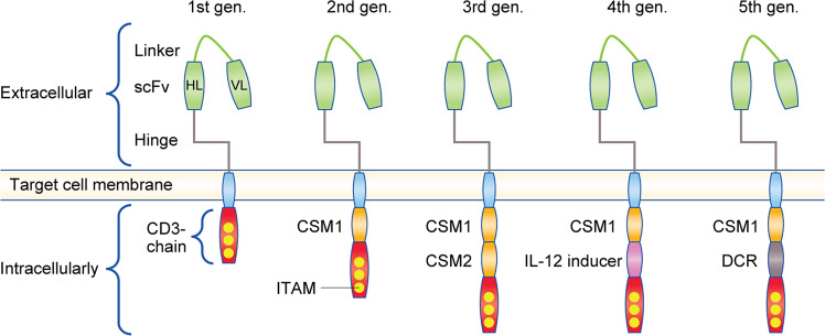
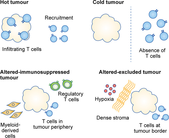
The functions of, and interactions between, the innate and adaptive immune systems are vital for anticancer immunity. Cytotoxic T cells expressing cell-surface CD8 are the most powerful effectors in the anticancer immune response and form the backbone of current successful cancer immunotherapies. Immune-checkpoint inhibitors are designed to target immune-inhibitory receptors that function to regulate the immune response, whereas adoptive cell-transfer therapies use CD8+ T cells with genetically modified receptors-chimaeric antigen receptors-to specify and enhance CD8+ T-cell functionality. New generations of cytotoxic T cells with genetically modified or synthetic receptors are being developed and evaluated in clinical trials. Furthermore, combinatory regimens might optimise treatment effects and reduce adverse events. This review summarises advances in research on the most prominent immune effectors in cancer and cancer immunotherapy, cytotoxic T cells, and discusses possible implications for future cancer treatment.


Conflict of interest statement

The authors declare no competing interests.

Figures

Fig. 1
Fig. 1. T-cell differentiation—an overview.
Common lymphoid progenitor cells giving rise to immature precursor T cells originate in the red bone marrow. Due to the production in the thymus of chemotactic agents/thymic factors (e.g., thymotaxin, thymosin and thymopoietin), immature precursor T cells (being TCR- and CD-negative [double negative]) enter the circulation and are directed to the thymus. Within the thymus, the same agents induce the production of TCR and CD proteins. Thymic cells present the now CD- and TCR-positive T cells for MHC-1 and MHC-2 molecules to identify T-cell reactivity and direct their maturation pathways. During the positive selection process, T cells being able to bind MHC class I or II molecules with at least a weak affinity are identified. By negative selection, T cells with a high affinity for self-peptides undergo apoptosis to minimise the risk of immune responses towards self-proteins in the periphery. T cells with TCR affinity for MHC-1 become CD8+ T cells and T cells with TCR affinity for MHC-2 become CD4+ T cells. Depending on cytokine and stromal cell signalling, they may also differentiate into T-helper and T-regulatory cells. MHC: major histocompatibility complex.
Fig. 2
Fig. 2. T-cell activation: the T-cell receptor (TCR) complex.
Extracellularly, the TCR consists of the α and β chains, both of which have a constant region (C) and variable region (V), with the latter determining antigen specificity. The TCRαβ antigen-binding subunit is non-covalently bound to three CD3 co-receptor signalling subunits (ζζ, CD3δε and CD3γε), all of which contain immunoreceptor tyrosine-based activation motifs (ITAMs) in their cytoplasmic domains. SS disulfide bridge.
Fig. 3
Fig. 3. T-cell activation.
The V domains of the α and β chains on T cells interact with an antigenic peptide presented by MHC-1 on the target cell, while the co-receptor CD8 associates with TCR–MHC-1 to tightly secure the TCR–CD3 complex to the major histocompatibility complex (MHC)–peptide complex. As CD8 binds to the MHC-1, Lck phosphorylates the intracellular portions of the CD3 ITAMs and positions ZAP-70 to phosphorylate the transmembrane proteins that allow the CD8+ T cell to secrete its cytokines. Lck: lymphocyte-specific protein tyrosine kinase, P: phosphorylation, CD45: receptor-linked protein tyrosine phosphatase, Zap70: ζ chain of T-cell-receptor-associated protein kinase 70.
Fig. 4
Fig. 4. CD8+ T-cell immune-checkpoint receptors and their ligands.
Receptors stimulating CD8+ T-cell functions include CD28, ICOS and B7.1. Receptors mediating inhibitory signals include CTLA-4, PD-1 and B7.1. CD28 and CTLA-4 compete for the ligands B7.1 and B7.2 during the early stages of the CD8+ T-cell response. pMHC: peptide-loaded major histocompatibility complex, ICOS: inducible T-cell co-stimulator.
Fig. 5
Fig. 5. Development of chimaeric antigen receptors (CARs).
The CAR construct comprises an antigen-binding scFv attached via a hinge to the CD3ζ signalling unit. Intracellularly, additional signalling units are attached to the CD3ζ chain. First-generation CARs contained only tyrosine-based activation motifs (ITAM) in the CD3 ζ-chain intracellular domain. Second-generation CARs include one co-stimulatory molecule, such as CD28 or 4-1BB, whereas third-generation CARs contain two co-stimulatory molecules, such as CD28 + 4-1BB (CD137). Fourth-generation CARs are based on second-generation CARs paired with a constitutively or inducibly expressed cytokine (e.g., IL-12) to further expand the T-cell population. Fifth-generation CARs are based on second-generation CARs with the addition of the intracellular domains of cytokine receptors (e.g., IL-2Rβ). CSM: co-stimulatory molecule, DCR: domain of cytokine receptors, IL-12: inducer activator of interleukin-12 transcription.
Fig. 6
Fig. 6. CD8+ T-cell distribution within tumours.
In solid cancers, tumours can be classified as hot, cold, altered–immunosuppressed or altered–excluded tumours based on the degree of tumour CD8+ T-cell infiltration and the composition of the tumour microenvironment (TME). Hot tumours are characterised by high infiltration of CD8+ T cells and respond better to immune-enhancing therapies, whereas cold tumours are less likely to benefit from such treatments. Cold tumours are characterised by the absence of CD8+ T-cell infiltration, while altered–immunosuppressed tumours have sparse CD8+ T-cell infiltration, localised in the tumour periphery, and the presence of myeloid-derived suppressor cells and regulatory CD8+ T cells in the tumour tissue. Altered–excluded tumours are dominated by an abnormal vasculature (and consequent hypoxia) and a dense stroma, while CD8+ T cells are located at the border of these tumours.

Similar articles

Cited by

References

    1. Wellenstein MD, de Visser KE. Cancer-cell-intrinsic mechanisms shaping the tumor immune landscape. Immunity. 2018;48:399–416. doi: 10.1016/j.immuni.2018.03.004. - DOI - PubMed
    1. Barnes TA, Amir E. HYPE or HOPE: the prognostic value of infiltrating immune cells in cancer. Br. J. Cancer. 2017;117:451–460. doi: 10.1038/bjc.2017.220. - DOI - PMC - PubMed
    1. Stadtmauer EA, Fraietta JA, Davis MM, Cohen AD, Weber KL, Lancaster E, et al. CRISPR-engineered T cells in patients with refractory cancer. Science. 2020;367:eaba7365. doi: 10.1126/science.aba7365. - DOI - PMC - PubMed
    1. Qin S, Xu L, Yi M, Yu S, Wu K, Luo S. Novel immune checkpoint targets: moving beyond PD-1 and CTLA-4. Mol. Cancer. 2019;18:155. doi: 10.1186/s12943-019-1091-2. - DOI - PMC - PubMed
    1. Overgaard NH, Jung J-W, Steptoe RJ, Wells JW. CD4+/CD8+ double-positive T cells: more than just a developmental stage? J. Leukoc. Biol. 2015;97:31–38. doi: 10.1189/jlb.1RU0814-382. - DOI - PubMed