Review
. 2020 Aug 8;10(22):10057-10074.
doi: 10.7150/thno.45990. eCollection 2020.

Palladium-based nanomaterials for cancer imaging and therapy

Affiliations
Review

Palladium-based nanomaterials for cancer imaging and therapy

Yongchun Liu et al. Theranostics. .

Abstract

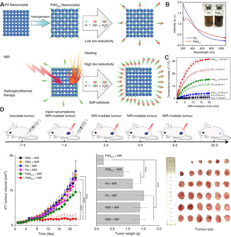
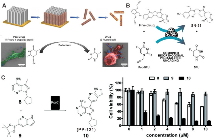
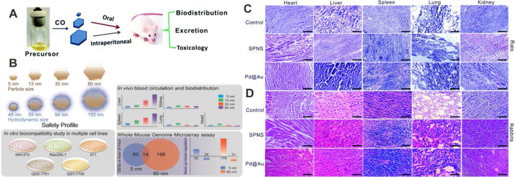
In recent decade, palladium-based (Pd-based) nanomaterials have shown significant potential for biomedical applications because of their unique optical properties, excellent biocompatibility and high stability in physiological environment. Compared with other intensively studied noble nanomaterials, such as gold (Au) and silver (Ag) nanomaterials, research on Pd-based nanomaterials started late, but the distinctive features, such as high photothermal conversion efficiency and high photothermal stability, have made them getting great attention in the field of nanomedicine. The goal of this review is to provide a comprehensive and critical perspective on the recent progress of Pd-based nanomaterials as imaging contrast agents and therapeutic agents. The imaging section focuses on applications in photoacoustic (PA) imaging, single-photon emission computed tomography (SPECT) imaging, computed tomography (CT) imaging and magnetic resonance (MR) imaging. For treatment of cancer, single photothermal therapy (PTT) and PTT combined with other therapeutic modalities will be discussed. Finally, the safety concerns, forthcoming challenges and perspective of Pd-based nanomaterials on biomedical applications will be presented.

Keywords: Palladium-based nanomaterials; cancer imaging; combined cancer therapy; photothermal therapy; safety profile.


Conflict of interest statement

Competing Interests: The authors have declared that no competing interest exists.

Figures

Figure 1
Figure 1
TEM images of (A) Pd NSs, (B) Pd collora, (C) Pd@Ag nanoplates, (D) Porous Pd NPs, (E) Pd NPs@PPy, (F) Pd NPs, (G) PdNS/ZIF-8 JNPs and (H) H-Pd nanospheres. Adapted with permission from ref. 10, Copyright from 2011, Springer Nature; adapted with permission from ref. 13. Copyright 2011, American Chemical Society; adapted with permission from ref. 14. Copyright 2011, Wiley-VCH; adapted with permission from ref. 18. Copyright 2014, Royal Society of Chemistry; adapted with permission from ref. 19. Copyright 2016, Royal Society of Chemistry; adapted with permission from ref. 20. Copyright 2018, Springer Nature; adapted with permission from ref. 21. Copyright 2018, Wiley-VCH; adapted with permission from ref. 22. Copyright 2018, American Chemical Society.
Figure 2
Figure 2
Optical spectrum and TEM images of (A) Pd NSs and (B) Au nanorods before and after laser irradiation. PA imaging application of different sized Pd NSs (C) in vitro and (D) in vivo. (E) Synthetic procedure for Pd@Au nanoplates and (F) their application in PA imaging of tumor. Adapted with permission from ref. 31. Copyright 2014, Royal Society of Chemistry; adapted with permission from ref. 11, Copyright from 2017, Springer Nature; adapted with permission from ref. 15. Copyright 2014, Wiley-VCH.
Figure 3
Figure 3
pH-sensitive radiolabeled Pd NSs for SPECT imaging in different tumor models, (A) radioactive iodide-labeled Pd NSs and (B) radioactive iodide and 99mTc-labeled Pd NSs as a multifunctional theranostic platform. Adapted with permission from ref. 47. Copyright 2018, Royal Society of Chemistry; adapted with permission from ref. 48. Copyright 2018, Elsevier.
Figure 4
Figure 4
(A) Pd NSs with different surface modification for MRI imaging. (B) UCNPs@Pd-PVP nanocomposites for T1-weighted MRI imaging. Adapted with permission from ref. 56. Copyright 2018, Elsevier; adapted with permission from ref. 57. Copyright 2019, Royal Society of Chemistry.
Figure 5
Figure 5
(A) Renal clearable ultra-small Pd NSs for PTT of cancer. (B) Optical spectra of porous Pd NPs and Pd nanocubes. (C) Chitosan oligosaccharide-coated Pd NPs for PPT of cancer. Adapted with permission from ref. 63. Copyright 2014, Wiley-VCH; adapted with permission from ref. 18. Copyright 2014, Royal Society of Chemistry; adapted with permission from ref. 20. Copyright 2018, Springer Nature.
Figure 6
Figure 6
(A) Schematic representation of the synthesis and application of Pd@Pt-PEG-Ce6 for combined cancer therapy. (B) Construction of Pd@Ag@mSiO2-Ce6 for combined cancer therapy. (C) Hollow Pd nanospheres for combined cancer therapy. (D) Schematic illustration of combined cancer therapy using H-Pd NSs. (E) Degradation behavior of H-Pd NSs in simulated body fluid. (F) Photothermal effect of Pd NSs and H-Pd NSs. (G) ESR spectra and (H) O2 concentration of Pd NSs and H-Pd NSs under different conditions. Adapted with permission from ref. 16. Copyright 2018, Wiley-VCH; adapted with permission from ref. 70. Copyright 2013, American Chemical Society; adapted with permission from ref. 22. Copyright 2018, American Chemical Society; adapted with permission from ref. 72. Copyright 2020, American Chemical Society.
Figure 7
Figure 7
(A) Direct adsorption of DOX on the surfaces of Pd NSs for combined cancer therapy. (B) Synthesis of prodrug conjugated Pd@Au nanoplates for combined cancer therapy. (C) Synergistic anticancer application of DOX/HCPT co-loaded UFO-like Pd-based Janus NPs. Adapted with permission from ref. 76. Copyright 2015, Springer Nature; adapted with permission from ref. 77. Copyright 2016, Royal Society of Chemistry; adapted with permission from ref. 21. Copyright 2018, Wiley-VCH.
Figure 8
Figure 8
(A) Pd@Au nanoplates with continuous production of O2 by catalyzing the decomposition of endogenous H2O2 to overcome tumor hypoxia. (B) pH-responsive radiolabeled Pd NSs for combined cancer therapy. Adapted with permission from ref. 32. Copyright 2019, Wiley-VCH; adapted with permission from ref. 47. Copyright 2018, Royal Society of Chemistry.
Figure 9
Figure 9
(A) Schematic illustration of the mechanism on the synergistic tumor inhibition of Pd-CpG. (B) In vivo combined cancer therapy of Pd-CpG. Adapted with permission from ref. 98. Copyright 2020, Royal Society of Chemistry.
Figure 10
Figure 10
(A) Schematic illustration showing the formation and laser-stimulated release of PdH0.2 nanocrystals for combined cancer therapy. (B) Optical spectra of Pd and PdH0.2 nanocrystals. (C) Photothermal effect of Pd and PdH0.2 nanocrystals. (D) In vivo combined cancer therapy of PdH0.2 nanocrystals. Adapted with permission from ref. 33. Copyright 2018, Springer Nature.
Figure 11
Figure 11
Illustration of Pd nanomaterials for prodrug activation and synthesis of toxic drugs in vivo for cancer chemotherapy. (A) The synthetic process of FePd NWs and activation of pro-5FU. (B) Schematic illustration of the activation of prodrug and pro-5FU by Pd devices. (C) Synthesis of PP-121 by Pd(0) catalysts and cytotoxicity tests. Adapted with permission from ref. 115. Copyright 2018, Wiley-VCH; adapted with permission from ref. 116. Copyright 2018, Wiley-VCH; adapted with permission from ref. 117. Copyright 2016, Royal Society of Chemistry.
Figure 12
Figure 12
(A) Effect of size and administration routes on the biobehaviors of Pd NSs. (B) Safety profile of different-sized Pd NSs in vitro and in vivo. H&E images of (C) rats and (D) rabbits after i.v. injection of Pd NSs and Pd@Au nanoplates. Adapted with permission from ref. 118. Copyright 2017, Royal Society of Chemistry; adapted with permission from ref. 11. Copyright 2017, Springer Nature; adapted with permission from ref. 120. Copyright 2019, Royal Society of Chemistry.

Similar articles

Cited by

References

    1. Siegel RL, Miller KD, Jemal A. Cancer statistics, 2020. CA Cancer J Clin. 2020;70:7–30. - PubMed
    1. Lammers T, Aime S, Hennink WE, Storm G, Kiessling F. Theranostic nanomedicine. Acc Chem Res. 2011;44:1029–38. - PubMed
    1. Shi J, Kantoff PW, Wooster R, Farokhzad OC. Cancer nanomedicine: progress, challenges and opportunities. Nat Rev Cancer. 2017;17:20. - PMC - PubMed
    1. Huo S, Ma H, Huang K, Liu J, Wei T, Jin S. et al. Superior penetration and retention behavior of 50 nm gold nanoparticles in tumors. Cancer Res. 2013;73:319–30. - PubMed
    1. Kim D, Shin K, Kwon SG, Hyeon T. Synthesis and biomedical applications of multifunctional nanoparticles. Adv Mater. 2018;30:1802309. - PubMed

Publication types