Review
. 2021 Jan 20;72(1):15-35.
doi: 10.1093/jxb/eraa414.

Unraveling the sugar code: the role of microbial extracellular glycans in plant-microbe interactions

Affiliations
Review

Unraveling the sugar code: the role of microbial extracellular glycans in plant-microbe interactions

Alan Wanke et al. J Exp Bot. .

Abstract

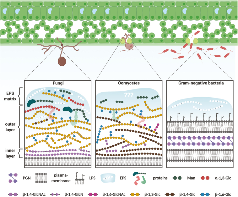
To defend against microbial invaders but also to establish symbiotic programs, plants need to detect the presence of microbes through the perception of molecular signatures characteristic of a whole class of microbes. Among these molecular signatures, extracellular glycans represent a structurally complex and diverse group of biomolecules that has a pivotal role in the molecular dialog between plants and microbes. Secreted glycans and glycoconjugates such as symbiotic lipochitooligosaccharides or immunosuppressive cyclic β-glucans act as microbial messengers that prepare the ground for host colonization. On the other hand, microbial cell surface glycans are important indicators of microbial presence. They are conserved structures normally exposed and thus accessible for plant hydrolytic enzymes and cell surface receptor proteins. While the immunogenic potential of bacterial cell surface glycoconjugates such as lipopolysaccharides and peptidoglycan has been intensively studied in the past years, perception of cell surface glycans from filamentous microbes such as fungi or oomycetes is still largely unexplored. To date, only few studies have focused on the role of fungal-derived cell surface glycans other than chitin, highlighting a knowledge gap that needs to be addressed. The objective of this review is to give an overview on the biological functions and perception of microbial extracellular glycans, primarily focusing on their recognition and their contribution to plant-microbe interactions.

Keywords: Cell wall; chitin; extracellular polysaccharides; glucan; immunity; matrix; microbes; symbiosis.


Figures

Fig. 1.
Fig. 1.
Schematic overview of microbial cell surface glycans. Recognition of microbe-derived cell wall polysaccharides represents an important mechanism by which plants surveil their microbial surrounding. Since cell wall structure and function are highly interlocked, core polysaccharides are conserved within different microbial groups. This scheme illustrates these core polysaccharides and their linkage types without representing exact quantitative proportions. Cell walls of filamentous fungi and oomycetes are network-like structures consisting of highly interconnected polysaccharide fibrils. In fungi, the inner cell wall layer consists of chitin (β-1,4-GlcNAc) and chitosan polymers (β-1,4-GlcN). It is covalently linked to the outer cell wall layer, which is mainly composed of β-1,3/1,6-glucans (β-1,3/1,6-Glc) with minor amounts of β-1,4-glucose (β-1,4-Glc). The outer layer is concealed by α-1,3-glucans (α-1,3-Glc) and mixed-linkage mannose (Man) polymers. Mannose polymers often occur as heterosaccharides with minor amounts of additional sugar types (e.g. rhamnose and galactose). A highly mobile, gel-like extracellular polysaccharide (EPS) matrix is loosely attached to the outer cell wall of many fungi. In contrast to fungi, no detailed studies on the cell wall architecture of oomycetes have been performed. Cross-linked cellulose (β-1,4-Glc) and β-1,3/1,6-glucans are major components of the inner part of oomycete cell walls. Chitin (β-1,4- and β-1,6-GlcNAc) only occurs in minute amounts; most of it is assumed to be connected to β-glucan polymers. The cellulose content is reduced in the external parts of the cell wall. Instead, branched β-glucans and mannose oligomers are present in that layer. To our knowledge, no detailed information on the architecture of an EPS matrix in oomycetes has been reported. Peptidoglycan (PGN) is a conserved part of bacterial cell walls present in Gram-positive and Gram-negative bacteria. The main heteropolysaccharides consist of alternating N-acetylglucosamine (GlcNAc) and N-acetylmuramic acid residues, which are interconnected through peptide chains. In Gram-negative bacteria, PGN is embedded between an inner (plasma membrane) and outer membrane layer. The outer membrane layer is decorated with lipid-linked polysaccharides, so-called lipopolysaccharides (LPSs). An amorphous matrix made of EPSs (e.g. xanthan and succinoglycan) encases many bacterial species. A more detailed overview on microbial cell surface glycans and their implications on plant–microbe interactions can be found in Supplementary Table S1.
Fig. 2.
Fig. 2.
Live cell images of root-associated bacteria embedded in the fungal β-glucan matrix. Confocal laser scanning microscopy live stain images of an A. thaliana Col-0 root segment, 12 d post-colonization with the fungus Serendipita indica and root-associated bacteria R11 (Bacillus sp.) (A) or R935 (Flavobacterium sp.) (B) (Bai et al., 2015). The fungal cell wall and bacteria (dots) were stainable with WGA-AF594 (green pseudo-color). β-1,3-glucan was visualized using the FITC488-labeled lectin WSC3 (magenta pseudo-color) (Wawra et al., 2019). Fungal chitin was only detected in hyphae growing in the extracellular space (yellow arrowheads), whereas the β-glucan matrix was also detectable after the hyphae entered the root cortical cells (white arrowheads). Interestingly, the β-glucan architecture was very distinct in the presence of the individual bacterial strains (structures indicated by the blue brackets). While a fine structure in the glucan network was visible for S. indica in combination with R11, the combination with R935 resulted in an apparently extended, more dense, and amorphous looking β-glucan layer. In addition, in the presence of R935, the fungal β-glucan matrix around the S. indica hyphae appeared more diffuse and less compact compared with the matrix detected around more isolated growing hyphae (B, yellow brackets). It is currently unclear whether the additionally deposited β-glucan is produced by the bacteria themselves or whether their presence triggers enhanced production and secretion by the fungus. Scale bars=25 µm.

References

    1. Acosta-Jurado S, Rodriguez-Navarro DN, Kawaharada Y, et al. 2016. Sinorhizobium fredii HH103 invades Lotus burttii by crack entry in a nod factor- and surface polysaccharide-dependent manner. Molecular Plant-Microbe Interactions 29, 925–937. - PubMed
    1. Adl SM, Simpson AG, Lane CE, et al. 2012. The revised classification of eukaryotes. Journal of Eukaryotic Microbiology 59, 429–493. - PMC - PubMed
    1. Albert I, Hua C, Nürnberger T, Pruitt RN, Zhang L. 2020. Surface sensor systems in plant immunity. Plant Physiology 182, 1582–1596. - PMC - PubMed
    1. Almendras K, Garcia J, Caru M, Orlando J. 2018. Nitrogen-fixing bacteria associated with Peltigera cyanolichens and Cladonia chlorolichens. Molecules 23. - PMC - PubMed
    1. Amor BB, Shaw SL, Oldroyd GE, Maillet F, Penmetsa RV, Cook D, Long SR, Denarie J, Gough C. 2003. The NFP locus of Medicago truncatula controls an early step of Nod factor signal transduction upstream of a rapid calcium flux and root hair deformation. The Plant Journal 34, 495–506. - PubMed

Publication types