Comparative Study
. 2020 Dec 16;37(12):3485-3506.
doi: 10.1093/molbev/msaa183.

The Molecular Machinery of Gametogenesis in Geodia Demosponges (Porifera): Evolutionary Origins of a Conserved Toolkit across Animals

Affiliations
Comparative Study

The Molecular Machinery of Gametogenesis in Geodia Demosponges (Porifera): Evolutionary Origins of a Conserved Toolkit across Animals

Vasiliki Koutsouveli et al. Mol Biol Evol. .

Abstract

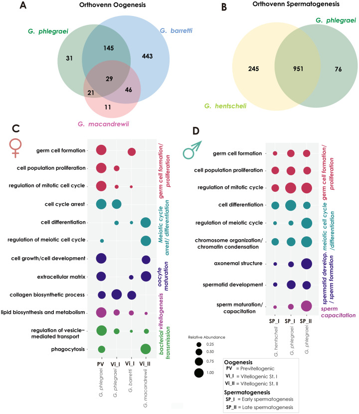
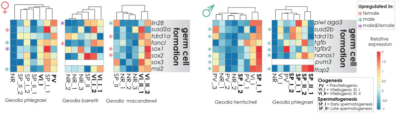
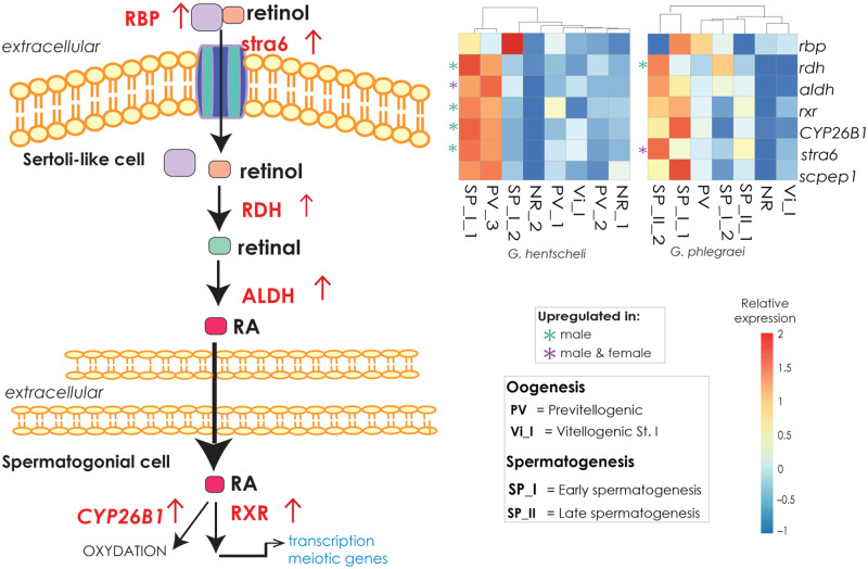
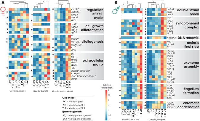
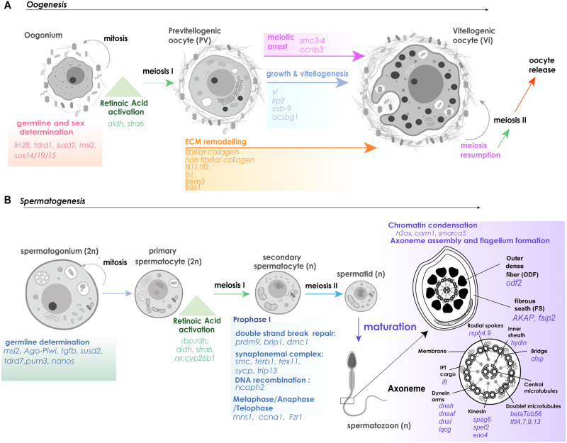
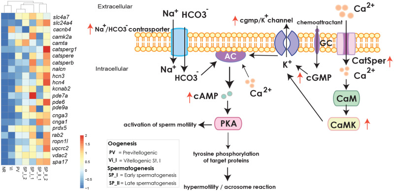
All animals are capable of undergoing gametogenesis. The ability of forming haploid cells from diploid cells through meiosis and recombination appeared early in eukaryotes, whereas further gamete differentiation is mostly a metazoan signature. Morphologically, the gametogenic process presents many similarities across animal taxa, but little is known about its conservation at the molecular level. Porifera are the earliest divergent animals and therefore are an ideal phylum to understand evolution of the gametogenic toolkits. Although sponge gametogenesis is well known at the histological level, the molecular toolkits for gamete production are largely unknown. Our goal was to identify the genes and their expression levels which regulate oogenesis and spermatogenesis in five gonochoristic and oviparous species of the genus Geodia, using both RNAseq and proteomic analyses. In the early stages of both female and male gametogenesis, genes involved in germ cell fate and cell-renewal were upregulated. Then, molecular signals involved in retinoic acid pathway could trigger the meiotic processes. During later stages of oogenesis, female sponges expressed genes involved in cell growth, vitellogenesis, and extracellular matrix reassembly, which are conserved elements of oocyte maturation in Metazoa. Likewise, in spermatogenesis, genes regulating the whole meiotic cycle, chromatin compaction, and flagellum axoneme formation, that are common across Metazoa were overexpressed in the sponges. Finally, molecular signals possibly related to sperm capacitation were identified during late stages of spermatogenesis for the first time in Porifera. In conclusion, the activated molecular toolkit during gametogenesis in sponges was remarkably similar to that deployed during gametogenesis in vertebrates.

Keywords: Porifera; evolution; oogenesis; proteomics; spermatogenesis; transcriptomics.


Figures

Fig. 1.
Fig. 1.
Morphological features of oocytes in sponge tissues of Geodia species. (A) Light microscopy image showing a PV oocyte (oc) of Geodia phlegraei (≤20 μm in diameter) (insert in the upper right) and a vitellogenic stage I (Vi_I) oocyte in the tissue of Geodia barretti (∼30μm in diameter). (B) Light microscopy image of several vitellogenic oocytes of maturation stage III (Vi_III) in Geodia atlantica close to the canals (ca), ready to be released. A thick layer of collagen (c) surrounds the mature oocytes. (C) Transmission Electron Micrograph (TEM) of a PV oocyte of Geodia hentscheli. The nucleolated (nu) nucleus (n) occupies most of the oocyte. Many vesicles (vs), few lipid droplets (li) as well as mitochondrial clouds (mi) can be seen in the ooplasm. A thin layer of collagen is deposited around the oocyte. Many bacterial (b) symbionts were observed in the mesohyl, none inside the oocyte. (D) TEM image of an ameboid shape PV oocyte of G. hentscheli with a large nucleolated nucleus and the ooplasm full of vesicles but no yolk or lipid droplet. (E) TEM image of a vitellogenic oocyte (Vi_II) of Geodia macandrewii full of lipid yolk (li) and bacterial symbionts in vesicles. The oocyte has pseudopodia (ps) to acquire further bacteria and nutrients for the maternal tissue. A thick layer of collagen surrounds the oocyte. (F) Close-up of (E) of G. macadrewii. Dense accumulations of mitochondria are observed in the ooplasm, close to the nucleus, and among those the nuage (ng) granules. The Golgi apparatus (go) can be seen in the periphery of the nucleus.
Fig. 2.
Fig. 2.
Morphological features of spermatic cysts in sponge tissues of Geodia species. (A) Light microscopy image indicating spermatic cyst (sc) in developmental stage I (SP_I) in the mesohyl of Geodia hentscheli. (B) Light microscopy image depicting several spermatic cysts (sc) of developmental stage II (SP_II) accumulated around the canals (ca) in the tissue of Geodia phlegraei. (C) TEM micrograph of a SP_I spermatic cyst from G. hentscheli in which cells at the beginning of gametic development; spermatogonia (s) and primary spermatocytes (ps) were observed. Internalization of the flagellum (f) from the choanocyte occurs at the early stages. (D) TEM micrograph of a SP_II spermatic cyst from G. phlegraei with asynchronous development: spermatogonia (s) in the outer part of the cyst, and primary (ps) to secondary spermatocytes (ss) and mature spermatid/preliminary sperm (ms) in the inner part of the spermatic cyst.
Fig. 3.
Fig. 3.
Shared upregulated genes during gametogenesis and GO enrichment. Venn diagram depicting the shared ortholog genes which were upregulated during (A) oogenesis across the species Geodia phlegraei, G. barretti, and G. macandrewii and (B) spermatogenesis across the species Geodia hentscheli and G. phlegraei. (C) Bubble graph depicting selected GO-enriched categories related to oogenesis, extracted from the upregulated genes among female individuals with oocytes in different developmental stages, PV (G. phlegraei); Vi_I (G. phlegraei, G. barretti) and Vi_II (G. macandrewii). (D) Bubble graph depicting selected GO-enriched categories related to spermatogenesis, extracted from the upregulated genes among male individuals with spermatic cysts in SP_I (G. hentscheli, G. phlegraei) and SP_II developmental stages (G. phlegraei). GO enrichment analysis was conducted selecting a P value ≤0.05. Each color represents a different group of gene categories according to their function. The size of the circle in each case is relative to the expression level of each gene category. Each gene category was analysed seperately .
Fig. 4.
Fig. 4.
Heatmaps depicting the expression level of genes related to germ cell formation during oogenesis and spermatogenesis in individuals of the studied species. The scale for relative expression values increases from blue to red. The asterisks indicate the upregulated genes, pink for upregulated genes found in females, green asterisk for upregulated genes found in males, and purple in both female and male specimens.
Fig. 5.
Fig. 5.
The RA pathway. (A) Schematic representation of the RA pathway (modified from Griswold [2016]), which permits a balance between germ cell maintenance and activation of meiosis. Red arrows indicate upregulation of genes in our data. (B) Heatmap of depicting the expression level of the main genes in the RA pathway mainly focused on individuals with gametes in early stages among the studied species (Geodia hentscheli and G. phlegraei). The scale for relative expression values increases from blue to red. The asterisks indicate the upregulated genes in female and or male specimens; the color green was used for upregulated genes in male and purple when found in both female and male individuals. There was no gene exlusively upregulated in female individuals.
Fig. 6.
Fig. 6.
(A) Heatmaps depicting the expression level of selected genes related to oogenesis, across individuals with oocytes in different developmental stages: PV, V_I, and V_II (in Geodia phlegraei, G. barretti, and G. macandrewii, respectively). (B) Heatmaps depicting the expression level of selected genes related to spermatogenesis in individuals with spermatid cysts in different developmental stages: SP_I (Geodia hentscheli and G. phlegraei) and SP_II (G. phlegraei). The genes are separated into few big categories according to their function. The scale for relative expression values increases from blue to red. The asterisks indicate the differentially expressed genes in female and male specimens, respectively.
Fig. 7.
Fig. 7.
A schematic representation of the molecular toolkits recruited and physiological changes during the progress of (A) oogenesis and (B) spermatogenesis in Geodia spp., based on our histological and transcriptomic results and the theoretical progress of gametogenesis in other Metazoa. The main overexpressed regulatory pathways and genes involved in each developmental stage and transition phase are reported.
Fig. 8.
Fig. 8.
Illustration of Ca+2 driven regulation cascades related to sperm motility and capacitation. Bicarbonate, HCO3 is transported via a Na+/HCO3 cotransporter and Ca+2 via Ca+2 channels including the sperm-specific Ca+2 channel, CatSper. Those activate adenylyl cyclase (AC) which increases the levels of cAMP and leads to PKA activation. CaM-dependent increase of Ca+2 as also activation of guanylyl cyclase receptor (GC) and further increase of cGMP into the cell also induce activation of AC and final activation of PKA. Activation of PKA causes flagellar movement. Further phosphorylation of PKA substrates leads to tyrosine phosphorylation and activation of all the final sperm capacitation steps (from sperm motility to axonemal reaction before fertilization) in animals. The illustration is a modified version according to Morisawa and Yoshida (2005), Ickowicz et al. (2012), and Rahman et al. (2017). The red arrows indicate the overexpression of the relevant genes in male specimens of the species Geodia phlegraei as it was the only one with several individuals containing SP_II spermatic cysts. The graph is accompanied by the expression level of the relevant genes in all the studied specimens of G. phlegraei and presented through a heatmap. All the genes indicated were upregulated in males of G. phlegraei. The expression level increases from blue to red color.

Similar articles

Cited by

References

    1. Abdo DA, Fromont J, McDonald JI.. 2008. Strategies, patterns and environmental cues for reproduction in two temperate haliclonid sponges. Aquat Biol. 1:291–302.
    1. Amiel A, Leclère L, Robert L, Chevalier S, Houliston E.. 2009. Conserved functions for Mos in eumetazoan oocyte maturation revealed by studies in a cnidarian. Curr Biol. 19(4):305–311. - PubMed
    1. Anderson EL, Baltus AE, Roepers-Gajadien HL, Hassold TJ, de Rooij DG, van Pelt AMM, Page DC.. 2008. Stra8 and its inducer, retinoic acid, regulate meiotic initiation in both spermatogenesis and oogenesis in mice. Proc Natl Acad Sci U S A. 105(39):14976–14980. - PMC - PubMed
    1. Andrews S. 2010. FastQC: A Quality Control Tool for High Throughput Sequence Data [Online]. Available from:http://www.bioinformatics.babraham.ac.uk/projects/fastqc/.
    1. Aouacheria A, Geourjon C, Aghajari N, Navratil V, Deléage G, Lethias C, Exposito JY.. 2006. Insights into early extracellular matrix evolution: spongin short chain collagen-related proteins are homologous to basement membrane type IV collagens and form a novel family widely distributed in invertebrates. Mol Biol Evol. 23(12):2288–2302. - PubMed

Publication types