. 2020 Oct;19(10):e13229.
doi: 10.1111/acel.13229. Epub 2020 Sep 15.

Underlying features of epigenetic aging clocks in vivo and in vitro

Affiliations

Underlying features of epigenetic aging clocks in vivo and in vitro

Zuyun Liu et al. Aging Cell. 2020 Oct.

Abstract

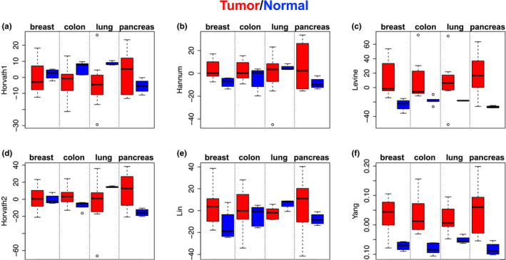
Epigenetic clocks, developed using DNA methylation data, have been widely used to quantify biological aging in multiple tissues/cells. However, many existing epigenetic clocks are weakly correlated with each other, suggesting they may capture different biological processes. We utilize multi-omics data from diverse human tissue/cells to identify shared features across eleven existing epigenetic clocks. Despite the striking lack of overlap in CpGs, multi-omics analysis suggested five clocks (Horvath1, Horvath2, Levine, Hannum, and Lin) share transcriptional associations conserved across purified CD14+ monocytes and dorsolateral prefrontal cortex. The pathways enriched in the shared transcriptional association suggested links between epigenetic aging and metabolism, immunity, and autophagy. Results from in vitro experiments showed that two clocks (Levine and Lin) were accelerated in accordance with two hallmarks of aging-cellular senescence and mitochondrial dysfunction. Finally, using multi-tissue data to deconstruct the epigenetic clock signals, we developed a meta-clock that demonstrated improved prediction for mortality and robustly related to hallmarks of aging in vitro than single clocks.

Keywords: DNA methylation; biological aging; cellular senescence; epigenetic clock; mitochondria.


Conflict of interest statement

The authors declare no competing interests.

Figures

Figure 1
Figure 1
Roadmap of this comparative analysis. To simplify the description, we used the last name of the first author to refer to each clock. The upper part shows the timeline of eleven epigenetic clocks included in this study. The next two parts include hypothesis development and testing. In hypothesis development part, we did comparative analysis for eleven epigenetic clocks mainly in four aspects. In hypothesis test part, we deconstructed the core signals across them, and developed and validated a novel meta‐clock
Figure 2
Figure 2
Age correlations for the clock scores across tissue and cell types. Pearson's correlations were used to assess associations between chronological age (x‐axis) and DNAmAge (y‐axis) by pooling 16 distinct tissue and cell types. Epigenetic clocks are denoted using the last name of the first author. DLPFX, dorsolateral prefrontal cortex
Figure 3
Figure 3
Transcriptomic pattern of 11 existing epigenetic clocks. Hieratical clustering of epigenetic clocks was performed based on the log2FC values for age‐adjusted associations with 8589 genes in monocytes (a) and DLPFC (b). (c) Comparisons of the strength of differential expression associations between clocks, for five clocks (reference clock = Horvath1) in monocytes. The x‐axis represents the log2FC for the association between Horvath1 and 8589 genes. The y‐axis represents the log2FC for the association between five epigenetic clocks (Yang, Hannum, Lin, Levine, and Horvath2) and 8589 genes, with clocks distinguished by colors. The slope represents the fitted line of the association between the log2FC for Horvath1 and the log2FC for each of the other five clocks, respectively. Thus, slope > 1 suggests the respective clock has stronger gene expression signals compared to Horvath1; a slope < 1 suggests Horvath1 has stronger gene expression signals compared to the respective clock; and a slope = 1 suggests comparable gene expression associations between Horvath1 and the respective clock. (d) Comparisons of the strength of differential methylation for six clocks (Levine, Weidner, VidalBralo, Hannum, Lin, and Horvath2) in DLPFC relative to Horvath1. Selected GO terms (e) and KEGG pathways (f) by the enrichment analysis for co‐expression modules (identified via WGCNA) that were shown to be associated with multiple epigenetic clocks in monocytes and/or DLPFC. Modules are denoted by color (turquoise, yellow, green, and red). For each module, the five most enriched biological processes are shown, based on q value (FDR). DLPFX, dorsolateral prefrontal cortex
Figure 4
Figure 4
Epigenetic clocks distinguish cancer vs. normal tissues. DNAmAge (adjusted for tissue type and age of the donor) was compared between tumor (red color) and normal tissue (blue color) for breast, colon, lung, and pancreas. Bars indicate standard errors
Figure 5
Figure 5
Epigenetic clocks, cellular senescence, and mitochondrial DNA depletion. (a–f) DNAmAge was estimated in BJ fibroblasts using early passage (EP) control samples, near senescent cells (NS), terminally passaged replicative senescent cells (RS), and oncogene‐induced senescent (OIS) cells via HRAS. (g–l) DNAmAge was estimated and compared between control (rho+) and mitochondrial DNA depleted (rho−) 143B cells. Bars indicate standard errors
Figure 6
Figure 6
Meta‐clock validation. (a) Results for mortality prediction using the validation sample from the Framingham Heart Study (FHS), in comparison with two robust mortality prediction clocks. Bars represent 95% confidence intervals for hazard ratios (green point). (b) Biweight midcorrelations between the meta‐clock and chronological age when pooling four tissues, denoted by color. Tissue‐specific biweight midcorrelations and p‐values are shown in the legend. (c) Meta‐clock estimates comparing tumor (red) versus normal (blue) tissue across four cancer/tissue types (breast, colon, lung, and pancreas). Bars indicate standard errors. (d) Meta‐clock estimates in BJ fibroblasts, comparing early passage (EP) control samples, near senescent cells (NS), terminally passaged replicative senescent cells (RS), and oncogene‐induced senescent (OIS) cells via HRAS. Bars indicate standard errors. DLPFC, dorsolateral prefrontal cortex

References

    1. Ambatipudi, S. , Horvath, S. , Perrier, F. , Cuenin, C. , Hernandez‐Vargas, H. , Le Calvez‐Kelm, F. , … Herceg, Z. (2020). DNA methylome analysis identifies accelerated epigenetic ageing associated with postmenopausal breast cancer susceptibility. European Journal of Cancer, 75, 299–307. 10.1016/j.ejca.2017.01.014 - DOI - PMC - PubMed
    1. Brodin, P. , & Davis, M. M. (2017). Human immune system variation. Nature Reviews Immunology, 17(1), 21–29. 10.1038/nri.2016.125 - DOI - PMC - PubMed
    1. Chen, B. H. , Marioni, R. E. , Colicino, E. , Peters, M. J. , Ward‐Caviness, C. K. , Tsai, P.‐C. , … Horvath, S. (2016). DNA methylation‐based measures of biological age: meta‐analysis predicting time to death. Aging (Albany NY), 8(9), 1844–1865. 10.18632/aging.101020 - DOI - PMC - PubMed
    1. Field, A. E. , Robertson, N. A. , Wang, T. , Havas, A. , Ideker, T. , & Adams, P. D. (2018). DNA methylation clocks in aging: Categories, causes, and consequences. Molecular Cell, 71(6), 882–895. 10.1016/j.molcel.2018.08.008 - DOI - PMC - PubMed
    1. Florath, I. , Butterbach, K. , Muller, H. , Bewerunge‐Hudler, M. , & Brenner, H. (2014). Cross‐sectional and longitudinal changes in DNA methylation with age: an epigenome‐wide analysis revealing over 60 novel age‐associated CpG sites. Human Molecular Genetics, 23(5), 1186–1201. 10.1093/hmg/ddt531 - DOI - PMC - PubMed

Publication types