. 2020 Oct 1;42(2):1093-1099.
doi: 10.1093/jnen/nlaa082.

Mechanisms of Cerebral Microbleeds

Affiliations

Mechanisms of Cerebral Microbleeds

Lara C Wadi et al. J Neuropathol Exp Neurol. .

Erratum in

  • Erratum to: Mechanisms of Cerebral Microbleeds.
    [No authors listed] [No authors listed] J Neuropathol Exp Neurol. 2021 Mar 22;80(4):388. doi: 10.1093/jnen/nlaa145. J Neuropathol Exp Neurol. 2021. PMID: 33463693 Free PMC article. No abstract available.

Abstract

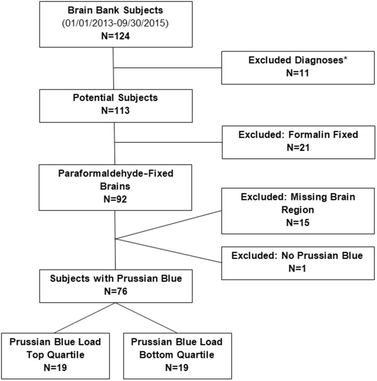
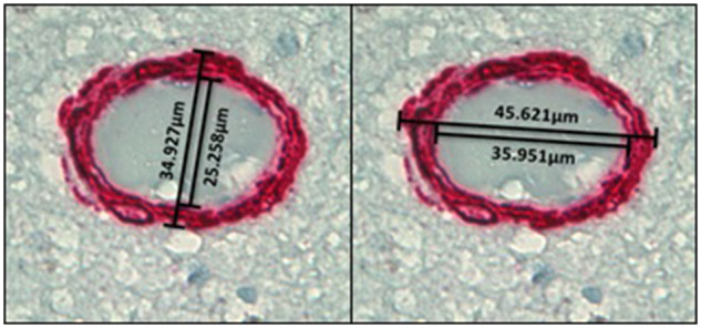
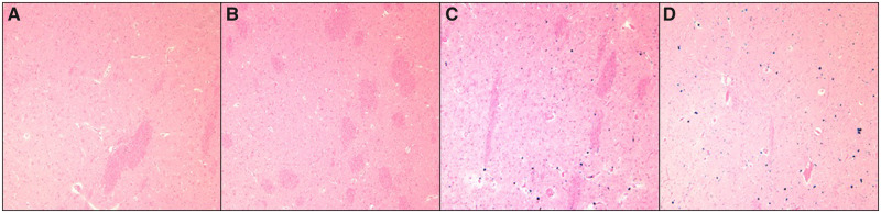
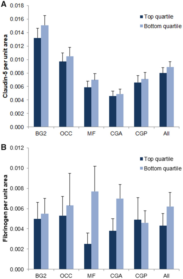
Cerebral microbleeds (CMB) are a common MRI finding, representing underlying cerebral microhemorrhages (CMH). The etiology of CMB and microhemorrhages is obscure. We conducted a pathological investigation of CMH, combining standard and immunohistological analyses of postmortem human brains. We analyzed 5 brain regions (middle frontal gyrus, occipital pole, rostral cingulate cortex, caudal cingulate cortex, and basal ganglia) of 76 brain bank subjects (mean age ± SE 90 ± 1.4 years). Prussian blue positivity, used as an index of CMH, was subjected to quantitative analysis for all 5 brain regions. Brains from the top and bottom quartiles (n = 19 each) were compared for quantitative immunohistological findings of smooth muscle actin, claudin-5, and fibrinogen, and for Sclerosis Index (SI) (a measure of arteriolar remodeling). Brains in the top quartile (i.e. with most extensive CMH) had significantly higher SI in the 5 brain regions combined (0.379 ± 0.007 vs 0.355 ± 0.008; p < 0.05). These findings indicate significant coexistence of arteriolar remodeling with CMH. While these findings provide clues to mechanisms of microhemorrhage development, further studies of experimental neuropathology are needed to determine causal relationships.

Keywords: Arteriosclerosis; Autopsy; Blood-brain barrier; Cerebral microhemorrhage; Histology; Immunohistochemistry.


Figures

FIGURE 1.
FIGURE 1.
Study population. *Excluded diagnoses: Trisomy 21, hereditary diffuse leukoencephalopathy with axonal spheroids, corticobasal degeneration, and frontotemporal lobar degeneration.
FIGURE 2.
FIGURE 2.
Illustrative images of Sclerosis Index (SI) measurement at 60× magnification. The final SI value per vessel is the average of the SI of the widest and narrowest axes of the arteriole.
FIGURE 3.
FIGURE 3.
Prussian blue stains of cerebral microhemorrhages. (A, B) From bottom quartile of Prussian blue load positivity. (C, D) From top quartile of Prussian blue load positivity.
FIGURE 4.
FIGURE 4.
Top and bottom Prussian blue-positivity quartile differences in claudin-5 and fibrinogen immunostaining. Top and bottom Prussian blue-positivity quartile differences in claudin-5 (A) and fibrinogen (B) immunostaining in the following brain regions: Striatum at the level of the mammillary bodies (BG2), calcarine/pericalcarine cortex (OCC), middle frontal gyrus (MF), rostral cingulate cortex (CGA), caudal cingulate cortex (CGP). No significant differences in claudin-5 or fibrinogen immunoreactivity are noted between top and bottom quartiles.
FIGURE 5.
FIGURE 5.
Top and bottom Prussian blue-positivity quartile differences in Sclerosis Index (SI). Top and bottom Prussian blue positivity quartile differences in SI in the following brain regions taken separately and collectively: Striatum at the level of the mammillary bodies (BG2), calcarine/pericalcarine cortex (OCC), middle frontal gyrus (MF), rostral cingulate cortex (CGA), caudal cingulate cortex (CGP). (A) SI was significantly higher in the top quartile versus bottom quartile in the MF brain region (0.410 ± 0.015 vs 0.358 ± 0.018; p < 0.05), in the CGA region (0.376 ± 0.010 vs 0.341 ± 0.013; p < 0.05), and in all 5 brain regions taken collectively (0.379 ± 0.007 vs 0.355 ± 0.008; p < 0.05). (B, C) SI in the bottom and top quartiles, respectively, under 60× magnification. *p < 0.05.

References

    1. Charidimou A, Krishnan A, Werring DJ, et al. Cerebral microbleeds: A guide to detection and clinical relevance in different disease settings. Neuroradiology 2013;55:655–74 - PubMed
    1. Fisher M, French S, Ji P, et al. Cerebral microbleeds in the elderly: A pathological analysis. Stroke 2010;41:2782–5 - PMC - PubMed
    1. Fazekas F, Kleinert R, Roob G, et al. Histopathologic analysis of foci of signal loss on gradient-echo T2*-weighted MR images in patients with spontaneous intracerebral hemorrhage: Evidence of microangiopathy-related microbleeds. Am J Neuroradiol 1999;20:637–42 - PMC - PubMed
    1. Kim TW, Lee SJ, Koo J, et al. Cerebral microbleeds and functional outcomes after ischemic stroke. Can J Neurol Sci 2014;41:577–82 - PubMed
    1. Bokura H, Saika R, Yamaguchi T, et al. Microbleeds are associated with subsequent hemorrhagic and ischemic stroke in healthy elderly individuals. Stroke 2011;42:1867–71 - PubMed

Publication types