. 2020 Oct 15;5(20):e142446.
doi: 10.1172/jci.insight.142446.

Modulating the tension-time integral of the cardiac twitch prevents dilated cardiomyopathy in murine hearts

Affiliations

Modulating the tension-time integral of the cardiac twitch prevents dilated cardiomyopathy in murine hearts

Joseph D Powers et al. JCI Insight. .

Abstract

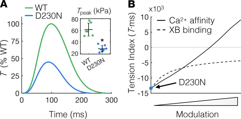
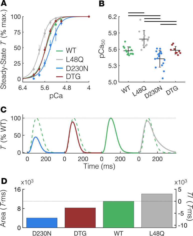
Dilated cardiomyopathy (DCM) is often associated with sarcomere protein mutations that confer reduced myofilament tension-generating capacity. We demonstrated that cardiac twitch tension-time integrals can be targeted and tuned to prevent DCM remodeling in hearts with contractile dysfunction. We employed a transgenic murine model of DCM caused by the D230N-tropomyosin (Tm) mutation and designed a sarcomere-based intervention specifically targeting the twitch tension-time integral of D230N-Tm hearts using multiscale computational models of intramolecular and intermolecular interactions in the thin filament and cell-level contractile simulations. Our models predicted that increasing the calcium sensitivity of thin filament activation using the cardiac troponin C (cTnC) variant L48Q can sufficiently augment twitch tension-time integrals of D230N-Tm hearts. Indeed, cardiac muscle isolated from double-transgenic hearts expressing D230N-Tm and L48Q cTnC had increased calcium sensitivity of tension development and increased twitch tension-time integrals compared with preparations from hearts with D230N-Tm alone. Longitudinal echocardiographic measurements revealed that DTG hearts retained normal cardiac morphology and function, whereas D230N-Tm hearts developed progressive DCM. We present a computational and experimental framework for targeting molecular mechanisms governing the twitch tension of cardiomyopathic hearts to counteract putative mechanical drivers of adverse remodeling and open possibilities for tension-based treatments of genetic cardiomyopathies.

Keywords: Cardiology; Cardiovascular disease; Molecular pathology.


Conflict of interest statement

Conflict of interest: ADM is a cofounder of and has an equity interest in Insilicomed Inc. and an equity interest in Vektor Medical Inc. He serves on the scientific advisory board of Insilicomed and as scientific advisor to both companies. Some of his research grants have been identified for conflict-of-interest management based on the overall scope of the project and its potential benefit to these companies. The author is required to disclose this relationship in publications acknowledging the grant support; however, the research subject and findings reported in this study did not involve the companies in any way. The terms of this arrangement have been reviewed and approved by the University of California San Diego in accordance with its conflict-of-interest policies.

Figures

Figure 1
Figure 1. A TI based on cardiac twitch T-time integrals correlates with the type and severity of myocardial growth.
(A) Depiction of the relationship between the TI and the degree and severity of cardiac growth (modified from ref. 7). The TI is determined by subtracting the area under a twitch T curve of a cardiomyocyte with perturbed contractility from that of a “normal” or “WT” cardiomyocyte. A positive TI correlates with concentric hypertrophy, whereas a negative TI correlates with eccentric hypertrophy (7). (B) Top panel: Theoretical 300 ms twitch T traces of a WT cardiomyocyte (black) and 4 potential variants with altered twitch T magnitude and kinetics. All twitches are represented as a percentage of the Tpeak of the WT twitch, which is shown as a dashed black trace with each variant twitch for comparison. Twitches were generated using a simple exponential model, as in ref. . Bottom panel: The area under the T-time trace (left ordinate) for each corresponding twitch above and the resulting TI (right ordinate) calculated as the difference in the area under the twitch T-time curve between WT and each variant twitch. Blue and gray correspond to twitches with decreased and increased (respectively) twitch-time integrals compared with WT (black). We note that because the TI encompasses contraction and relaxation kinetics, the absolute value of the TI does not depend on the peak twitch tension. T, tension; TI, tension index.
Figure 2
Figure 2. Measuring and modulating the tension index of D230N hearts.
(A) Average twitch T traces of intact trabeculae from WT and D230N hearts as a percentage of WT T. The Tpeak of trabeculae from D230N hearts is approximately half that of WT (inset). Sample sizes of n = 6 and n = 7 for WT and D230N trabeculae, respectively. The error bars of the inset represent SD and *P < 0.01 using a 2-tailed unpaired Student’s t test. (B) Dependence of the T index of simulated D230N twitches on modulation of XB or Ca2+ binding. The simulated T index for D230N twitches without any modulation is indicated by the blue circle. The rate of XB transition from a weak to a strong (T-generating) state was independently increased to simulate D230N twitches with augmented XB binding (dashed line). The affinity of Ca2+ for cTnC was also independently increased to simulate twitches of D230N cardiomyocytes with augmented Ca2+ sensitivity (solid line). T, tension; Tpeak, peak twitch tension; XB, cross-bridge; Ca2+, calcium; cTnC, cardiac troponin C.
Figure 3
Figure 3. Computational structural analysis of an atomically detailed thin filament with RUs containing D230N-Tm and L48Q cTnC.
(A) Distances (in Å) between the Ca2+ ion and each Ca2+-coordinating oxygen atom in site II of cTnC for a WT RU (green), a RU with D230N-Tm (blue) and a RU with both D230N-Tm and L48Q cTnC (red). (B) Distances (in Å) between the inhibitory peptide of cTnI and the center of mass of the closest actin monomer for each RU. The color scheme is the same as described for panel A. (C and D) Structural analysis of cTnC and cTnI subunits in the 3 different RUs. The cTnI subunit is shown as green when in the WT RU, blue when in the RU with D230N-Tm, and red when in the RU with both D230N-Tm and L48Q cTnC. cTnC is shown in gray, and Ca2+ ions are indicated by the yellow spheres. The switch and inhibitory peptides of cTnI are indicated by the arrows in the close-up insets. (E and F) Changes in the interactions between cTnC and the inhibitory and switch peptides of cTnI, relative to the WT RU, for the RU containing D230N-Tm (E) and the RU containing both D230N-Tm and L48Q cTnC (F). The color bar denotes the change in distances (in Å) between cTnC and cTnI residues in each variant RU relative to those in the WT RU. Thus, magenta indicates movement of cTnC–cTnI residues away from one another and black indicates movement of cTnC–cTnI residues toward one another (relative to WT). Residues corresponding to the inhibitory and switch peptides of cTnI are on either side of the vertical dashed line. Tm, tropomyosin; cTnC, cardiac troponin C; RU, regulatory unit; Ca2+, calcium; cTnI, cardiac troponin I.
Figure 4
Figure 4. L48Q cTnC prevents contractile abnormalities in cardiac tissue isolated from hearts containing D230N tropomyosin.
(A) Steady-state T as a percentage of the maximum value (at pCa 4.0) of demembranated cardiac muscle measured over a range of extracellular Ca2+ concentrations (pCa = –log([Ca2+]). The data are fit with the Hill equation (see Methods section) shown by the solid lines. (B) The pCa at half-maximal T (pCa50) of cardiac preparations from D230N hearts is significantly less than all other groups, whereas the pCa50 of preparations from L48Q plus D230N DTG hearts is not different from WT. Error bars represent SD. Black lines above the bars indicate P < 0.05 between groups using a 1-way ANOVA and a Tukey’s post hoc test of significance. (C) Average twitch T-time traces (in % WT Tpeak) of intact trabeculae for each genotype (same color scheme as panel A). The WT T-time trace is shown as a dashed green trace against each variant twitch for comparison. (D) The area under the T-time trace (left ordinate) for each genotype and the resulting TI (right ordinate). See Supplemental Table 2 for numerical values. T, tension; cTnC, cardiac troponin C; Ca2+, calcium; Tpeak, peak twitch tension; DTG, double-transgenic.
Figure 5
Figure 5. Ventricular remodeling and dysfunction in D230N hearts is prevented by expression of L48Q cTnC.
Echocardiographic measurements from mice of 2 to 5 months of age reveal that the left ventricular diastolic (A) and systolic (B) inner diameter (LVIDD and LVIDS, respectively) of D230N hearts (blue) progressively increase with age, whereas those of DTG hearts (red) do not change with age and are not significantly different from WT (green) at any age. The fractional shortening (C) and ejection fraction (D) also progressively worsen with age in D230N hearts, whereas those in DTG hearts remain approximately constant and do not significantly differ from WT. * indicates P < 0.05 for D230N vs. WT and + indicates P < 0.05 for D230N vs. DTG using a 1-way ANOVA and a Tukey’s post hoc test of significance. Error bars represent the SEM. See Supplemental Table 3 for all values and sample sizes. cTnC, cardiac troponin C; DTG, double-transgenic.

References

    1. McNally EM, Mestroni L. Dilated cardiomyopathy: genetic determinants and mechanisms. Circ Res. 2017;121(7):731–748. doi: 10.1161/CIRCRESAHA.116.309396. - DOI - PMC - PubMed
    1. Hershberger RE, Morales A, Siegfried JD. Clinical and genetic issues in dilated cardiomyopathy: a review for genetics professionals. Genet Med. 2010;12(11):655–667. doi: 10.1097/GIM.0b013e3181f2481f. - DOI - PMC - PubMed
    1. Hershberger RE, Siegfried JD. Update 2011: clinical and genetic issues in familial dilated cardiomyopathy. J Am Coll Cardiol. 2011;57(16):1641–1649. doi: 10.1016/j.jacc.2011.01.015. - DOI - PMC - PubMed
    1. Herman DS, et al. Truncations of titin causing dilated cardiomyopathy. N Engl J Med. 2012;366(7):619–628. doi: 10.1056/NEJMoa1110186. - DOI - PMC - PubMed
    1. McNally EM, Golbus JR, Puckelwartz MJ. Genetic mutations and mechanisms in dilated cardiomyopathy. J Clin Invest. 2013;123(1):19–26. doi: 10.1172/JCI62862. - DOI - PMC - PubMed

Publication types

MeSH terms