

Manganese-enhanced MRI (MEMRI) in breast and prostate cancers: Preliminary results exploring the potential role of calcium receptors
- PMID: 32931488
- PMCID: PMC7491733
- DOI: 10.1371/journal.pone.0224414
Manganese-enhanced MRI (MEMRI) in breast and prostate cancers: Preliminary results exploring the potential role of calcium receptors
Abstract
Procedures: To preliminary assess the relationship between Manganese Enhanced Magnetic Resonance Imaging (MEMRI) and the expression of calcium receptors in human prostate and breast cancer animal models.
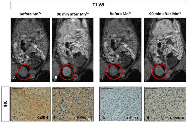
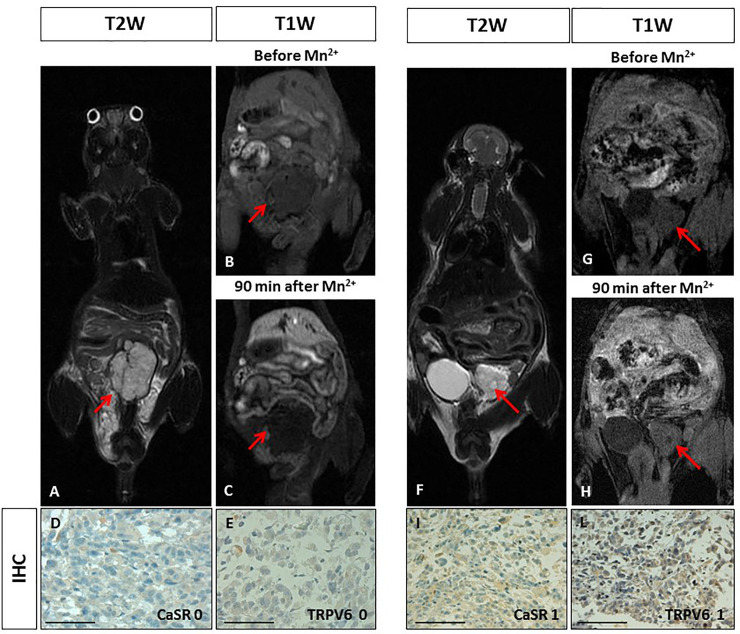
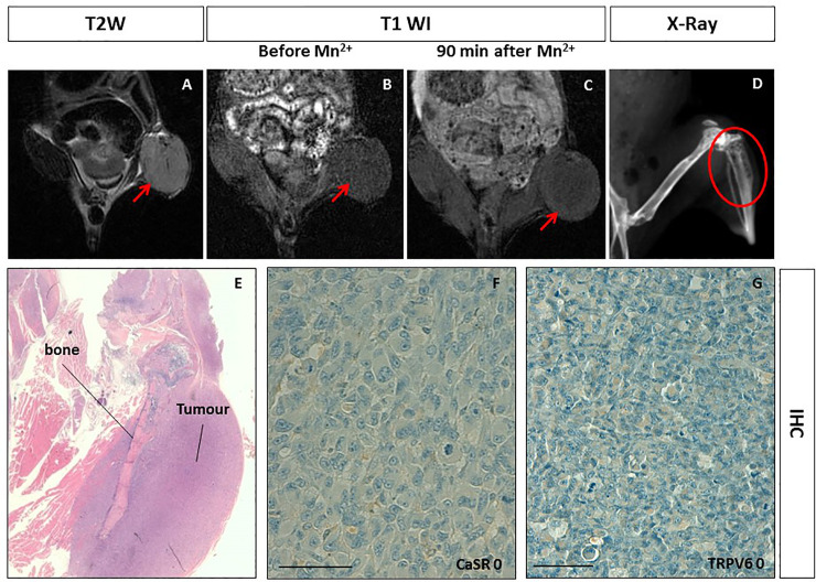
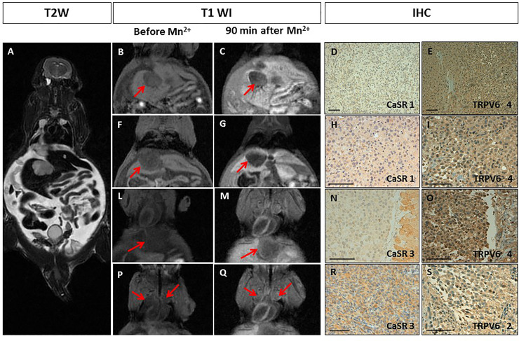
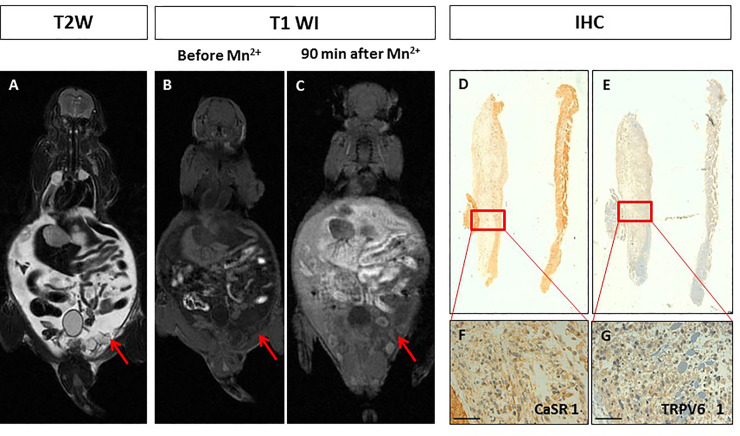
Methods: NOD/SCID mice were inoculated with MDA-MB-231 breast cancer cells and prostate PC3 cancer cells to develop orthotopic or pseudometastatic cancer animal models. Mice were studied on a clinical 3T scanner by using a prototype birdcage coil before and after intravenous injection of MnCl2. Assessment of receptor's status was carried out after the MR images acquisition by immunohistochemistry on excised tumours.
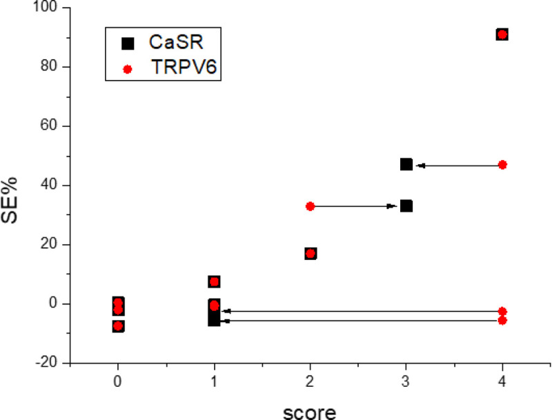
Results: Manganese contrast enhancement in breast or prostate cancer animal models well correlated with CaSR expression (p<0.01), whereas TRPV6 expression levels appeared not relevant to the Mn uptake.
Conclusion: Our preliminary results suggest that MEMRI appears an efficient tool to characterize human breast and prostate cancer animal models in the presence of different expression level of calcium receptors.
Conflict of interest statement
The author(s) declare no competing interests. All of the authors are aware of and agree to the content of the paper, as well as to their being listed as authors. None of the material has been published or is under consideration elsewhere, including the Internet. We also declare that none of the authors had any prior discussions with a Plos One Editorial Board Member about the work here described in the manuscript.
Figures
Similar articles
-
In vivo imaging of human breast cancer mouse model with high level expression of calcium sensing receptor at 3T.Eur Radiol. 2012 Mar;22(3):551-8. doi: 10.1007/s00330-011-2285-1. Epub 2011 Sep 24. Eur Radiol. 2012. PMID: 21947485
-
Comparison of manganese biodistribution and MR contrast enhancement in rats after intravenous injection of MnDPDP and MnCl2.Acta Radiol. 1997 Jul;38(4 Pt 2):700-7. doi: 10.1080/02841859709172402. Acta Radiol. 1997. PMID: 9245965
-
Transcranial manganese delivery for neuronal tract tracing using MEMRI.Neuroimage. 2017 Aug 1;156:146-154. doi: 10.1016/j.neuroimage.2017.05.025. Epub 2017 May 13. Neuroimage. 2017. PMID: 28506873 Free PMC article.
-
Mn2+ dynamics in manganese-enhanced MRI (MEMRI): Cav1.2 channel-mediated uptake and preferential accumulation in projection terminals.Neuroimage. 2018 Apr 1;169:374-382. doi: 10.1016/j.neuroimage.2017.12.054. Epub 2017 Dec 19. Neuroimage. 2018. PMID: 29277401
-
Manganese-enhanced MRI: an exceptional tool in translational neuroimaging.Schizophr Bull. 2008 Jul;34(4):595-604. doi: 10.1093/schbul/sbn056. Epub 2008 Jun 11. Schizophr Bull. 2008. PMID: 18550591 Free PMC article. Review.
Cited by
-
Two in One: Use of Divalent Manganese Ions as Both Cross-Linking and MRI Contrast Agent for Intrathecal Injection of Hydrogel-Embedded Stem Cells.Pharmaceutics. 2021 Jul 13;13(7):1076. doi: 10.3390/pharmaceutics13071076. Pharmaceutics. 2021. PMID: 34371767 Free PMC article.
-
Surveillance Value of Apparent Diffusion Coefficient Maps: Multiparametric MRI in Active Surveillance of Prostate Cancer.Cancers (Basel). 2023 Feb 10;15(4):1128. doi: 10.3390/cancers15041128. Cancers (Basel). 2023. PMID: 36831471 Free PMC article.
-
Biocompatible and bioactivable terpolymer-lipid-MnO2 Nanoparticle-based MRI contrast agent for improving tumor detection and delineation.Mater Today Bio. 2024 Jan 17;25:100954. doi: 10.1016/j.mtbio.2024.100954. eCollection 2024 Apr. Mater Today Bio. 2024. PMID: 38304342 Free PMC article.
-
Longitudinal manganese-enhanced magnetic resonance imaging of neural projections and activity.NMR Biomed. 2022 Jun;35(6):e4675. doi: 10.1002/nbm.4675. Epub 2022 Mar 6. NMR Biomed. 2022. PMID: 35253280 Free PMC article. Review.
References
-
- Berridge M.J, Bootman M.D, Roderick H.L. Calcium signalling: dynamics, homeostasis and remodelling, Nat Rev Mol. Cell Biol. 2003; 4: 517–529. - PubMed
-
- Berridge M.J, Lipp P. Bootman M.D. The versatility and universality of calcium signalling, Nat. Rev. Mol. Cell Biol. 2000; 1: 11–21. - PubMed
-
- Clapham D.E. Calcium signaling, Cell 2007; 131: 1047–1058. - PubMed
-
- Machaca K. Ca (2+) signaling, genes and the cell cycle, Cell Calcium 2011; 49: 323–330. - PubMed
Publication types
MeSH terms
Substances
LinkOut - more resources
Full Text Sources
Medical
Miscellaneous
