Review
. 2020 Sep;10(9):200186.
doi: 10.1098/rsob.200186. Epub 2020 Sep 16.

Looking back and looking forward: contributions of electron microscopy to the structural cell biology of gametes and fertilization

Affiliations
Review

Looking back and looking forward: contributions of electron microscopy to the structural cell biology of gametes and fertilization

Ravi Teja Ravi et al. Open Biol. 2020 Sep.

Abstract

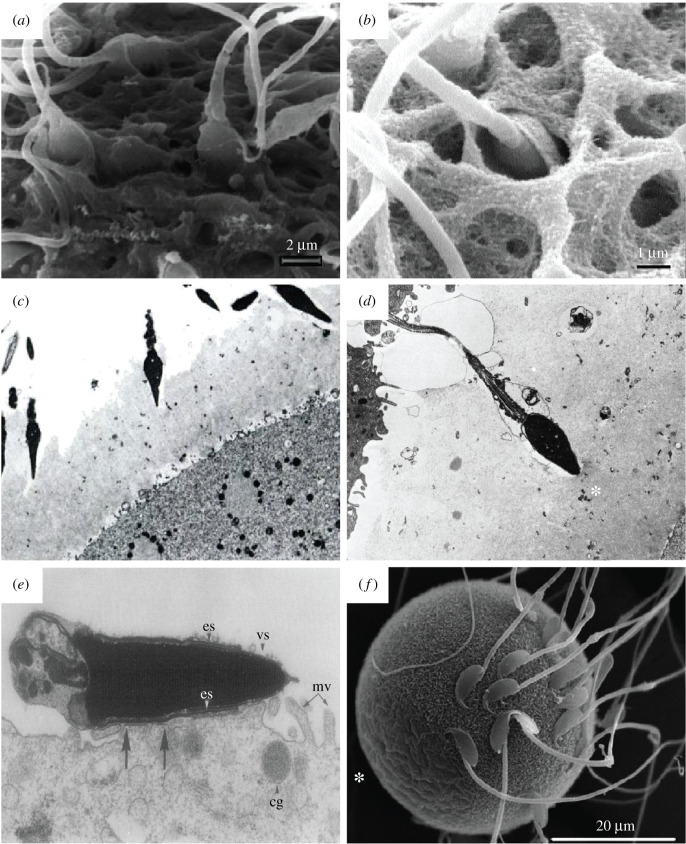
Mammalian gametes-the sperm and the egg-represent opposite extremes of cellular organization and scale. Studying the ultrastructure of gametes is crucial to understanding their interactions, and how to manipulate them in order to either encourage or prevent their union. Here, we survey the prominent electron microscopy (EM) techniques, with an emphasis on considerations for applying them to study mammalian gametes. We review how conventional EM has provided significant insight into gamete ultrastructure, but also how the harsh sample preparation methods required preclude understanding at a truly molecular level. We present recent advancements in cryo-electron tomography that provide an opportunity to image cells in a near-native state and at unprecedented levels of detail. New and emerging cellular EM techniques are poised to rekindle exploration of fundamental questions in mammalian reproduction, especially phenomena that involve complex membrane remodelling and protein reorganization. These methods will also allow novel lines of enquiry into problems of practical significance, such as investigating unexplained causes of human infertility and improving assisted reproductive technologies for biodiversity conservation.

Keywords: cryo-electron tomography; cryo-focused ion beam milling; egg; electron microscopy; fertilization; sperm.


Conflict of interest statement

We declare we have no competing interests.

Figures

Figure 1.
Figure 1.
Conventional EM of mammalian spermatozoa. (a) TEM micrograph of a thin section of human sperm head (×30 500): ac, acrosomal cap region; sas, subacrosomal space; nuc, nucleus; es, equatorial segment; pas, postacrosomal sheath; rne, redundant nuclear envelope; bp, basal plate; scp, striated connecting piece [27]. (b) TEM micrograph of a longitudinal thin section through the neck and proximal tail regions of a human spermatozoon (×64 000): bp, basal plate; rne, redundant nuclear envelope (nuclear pores indicated by the triangle); pc, proximal centriole; scp, striated connecting piece; m, mitochondria; afc, axial filament complex (comprising the axoneme and dense fibres) [27]. (c) SEM micrograph of a human spermatozoon, showing the surface morphologies in the anterior and posterior regions of the head (×15 000) [28]. (d) Freeze etching electron micrograph of a human spermatozoon head, depicting a face of the acrosomal membrane beneath a portion of the overlying plasma membrane (×50 000) [29].
Figure 2.
Figure 2.
Conventional EM of mammalian oocytes. (a) Thin-section TEM micrograph through the cortex of a mouse oocyte, showing the extent of the zona pellucida (ZP) and numerous transzonal projections (*), which are thin cytoplasmic extensions of the cumulus granulosa cells connecting them to the oocyte, penetrating through it [30]. (b) SEM micrograph of a mature human oocyte, showing the porous nature of the outer surface of the ZP (×1200) [31]. (c) SEM micrograph of the outer surface of the ZP of a mature human ooctye at a higher magnification, showing the filamentous-like arrangement of globule-bearing structures (×50 000) [31]. (d) SEM micrograph at a very high magnification of an unfertilized mouse oocyte, showing a branch of the filamentous structure of the ZP (×50 000) [32].
Figure 3.
Figure 3.
Conventional EM of mammalian sperm–oocyte interactions. (a) SEM micrograph of a mature human oocyte, showing the vertical binding of the sperm with a penetration of the apical tip of the sperm head into the ZP [33]. (b) SEM micrograph of a human sperm–oocyte interaction, showing the vertical binding of a sperm head vanishing into the the ZP [14]. (c) TEM micrograph of human sperm–oocyte interaction in vitro, showing acrosome-reacted sperm invading the ZP of a polyspermic ovum at different angles (×3330) [34]. (d) TEM micrograph of human sperm–oocyte interaction in vitro, showing an acrosome-reacted sperm that has penetrated about half the thickness of the ZP, just blocked outside the inner surface of the ZP (*) which is denser and more compact than the outer surface, thus depicting the block to polyspermy (×4330) [34]. (e) TEM micrograph showing the fusion of a tubal mouse egg incubated with capacitated epididymal sperm for 60 min. It shows the fusion of the sperm at the postacrosomal cap of the equatorial segment (es); cg, cortical granules; mv, microvilli; vs, vesiculated plasma and outer acrosomal membranes (×31 200) [35]. (f) SEM micrograph of a wild-type mouse egg incubated with sperm for 25 min, clearly showing sperm are not bound to microvilli-free region (*) [36].
Figure 4.
Figure 4.
Cryo-ET of pig sperm. (ac) The general cryo-ET workflow involves transferring live sperm (a) to an EM grid (shown under the light microscope, b), followed by plunge-freezing and imaging under a TEM (c). (d) A montage of cryo-EM projection images tracing a whole pig spermatozoon. A projection image of the midpiece is shown enlarged in the centre. (e,f) Computational slices through cryo-tomograms of the midpiece (e) and the principal piece (f). (e) In thicker regions of the cell, such as the midpiece, it is possible to resolve fine suborganellar features, such as membranes (inset: green, outer mitochondrial membrane; yellow, inner mitohondrial membrane). (f) In thinner regions of the cell, such as the distal part of the principal piece, it is possible to resolve individual protein complexes (inset). In (e,f), pm, plasma membrane; mito, mitochondrion; odf, outer dense fibre; fs, fibrous sheath; mtd, microtubule doublet; cpa, central pair apparatus; rs, radial spoke.
Figure 5.
Figure 5.
Computational image processing by subtomogram averaging and segmentation aid the analysis and interpretation of cryo-ET data. (a) Subtomogram averaging resolved the molecular architecture of doublet microtubules in the axoneme of sea urchin sperm, including positions and conformations of the dynein arms that drive flagellar motility [101]. Scale bar: 10 nm. (b) Combined with genetic perturbation, subtomogram averaging defined the roles of individual dynein assembly factors in the organization of the zebrafish sperm axoneme [102]. (c) Three-dimensional segmentation unveiled the complex organization of the isolated bovine sperm connecting piece, revealing novel fibrous linkers between plates of the segmented columns [103]. Scale bars: upper left panel, 250 nm; lower left panel, 50 nm.
Figure 6.
Figure 6.
Cryo-FIB milling and use of the VPP improve cryo-ET imaging. Slices through cryo-tomograms of pig sperm midpieces, acquired (a,b) on whole cells with an accelerating voltage of 200 kV and without the VPP; (c,d) on whole cells with an accelerating voltage of 200 kV and with the VPP; (e,f) on whole cells with an accelerating voltage of 300 kV and with the VPP; (g,h) on lamellae (approx. 300 nm thick) with an accelerating voltage of 300 kV and with the VPP. Left panels (a,c,e,g) show central longitudinal slices, while right panels (b,d,f,h) show tangential slices. Scale bars: left panels, 250 nm; right panels (digital zooms), 100 nm.

References

    1. van Leeuwenhoek A. 1677. Observationes D. Anthonii Lewenhoeck, de natis'e semine genitali animalculis. Phil. Trans. R. Soc. 12, 1040–1046. ( 10.1098/rstl.1677.0068) - DOI
    1. von Baer KE. 1827. De ovi mammalium et hominis genesi (On the genesis of the ovum of mammals and of man). Leipzig, Germany: Leopold Voss.
    1. Hertwig O. 1876. Beiträge zur kenntniss der bildung, befruchtung und theilung des thierischen eies. Morph. Jahrb. 1, 347–434.
    1. Fishman EL, et al. 2018. A novel atypical sperm centriole is functional during human fertilization. Nat. Commun. 9, 2210 ( 10.1038/s41467-018-04678-8) - DOI - PMC - PubMed
    1. Le Guennec M, et al. 2020. A helical inner scaffold provides a structural basis for centriole cohesion. Sci. Adv. 6, eaaz4137 ( 10.1126/sciadv.aaz4137) - DOI - PMC - PubMed

Publication types