. 2020 Nov 5;27(5):798-812.e6.
doi: 10.1016/j.stem.2020.08.005. Epub 2020 Sep 14.

Hydrogel Network Dynamics Regulate Vascular Morphogenesis

Affiliations

Hydrogel Network Dynamics Regulate Vascular Morphogenesis

Zhao Wei et al. Cell Stem Cell. .

Abstract

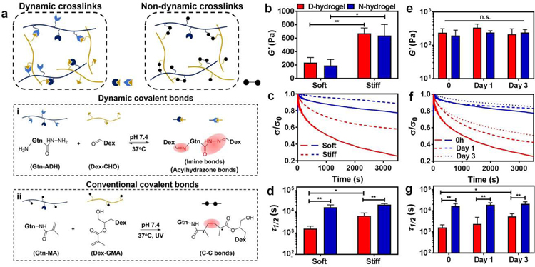
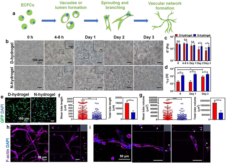
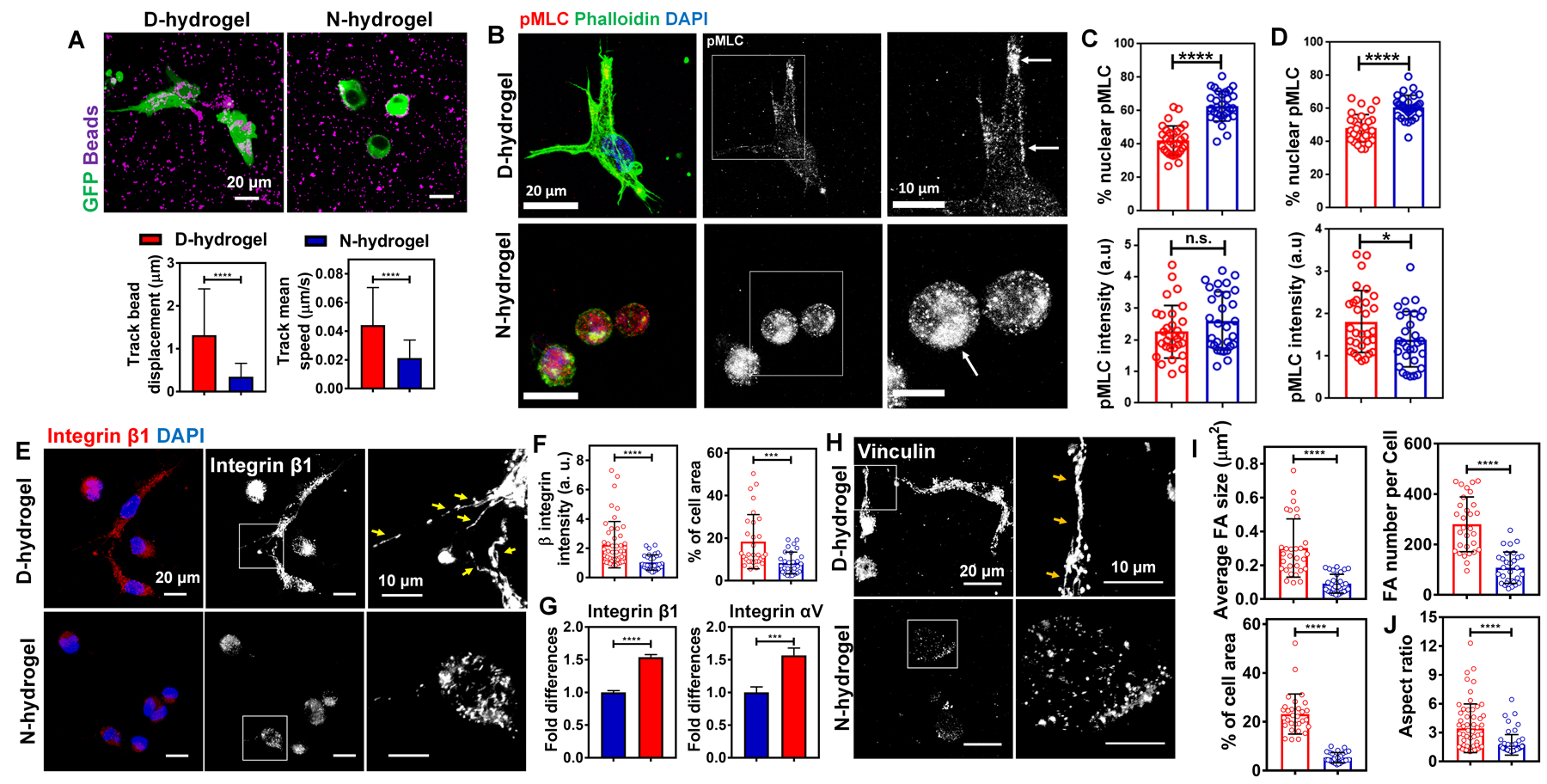
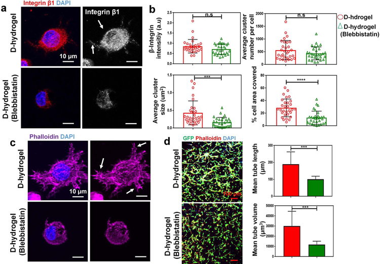
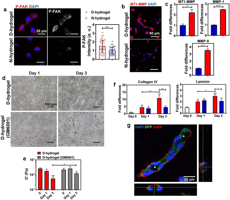
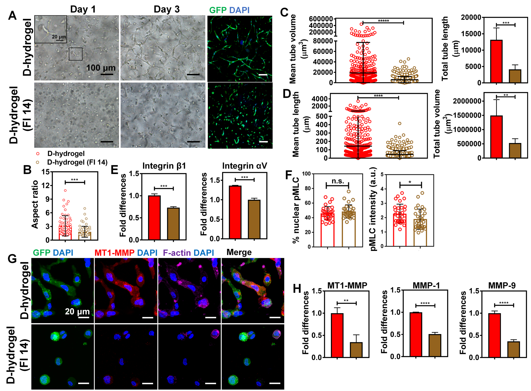
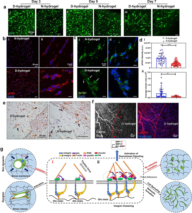
Matrix dynamics influence how individual cells develop into complex multicellular tissues. Here, we develop hydrogels with identical polymer components but different crosslinking capacities to enable the investigation of mechanisms underlying vascular morphogenesis. We show that dynamic (D) hydrogels increase the contractility of human endothelial colony-forming cells (hECFCs), promote the clustering of integrin β1, and promote the recruitment of vinculin, leading to the activation of focal adhesion kinase (FAK) and metalloproteinase expression. This leads to the robust assembly of vasculature and the deposition of new basement membrane. We also show that non-dynamic (N) hydrogels do not promote FAK signaling and that stiff D- and N-hydrogels are constrained for vascular morphogenesis. Furthermore, D-hydrogels promote hECFC microvessel formation and angiogenesis in vivo. Our results indicate that cell contractility mediates integrin signaling via inside-out signaling and emphasizes the importance of matrix dynamics in vascular tissue formation, thus informing future studies of vascularization and tissue engineering applications.

Keywords: cell contractility; integrin clustering; stress-relaxation; vasculogenesis.


Conflict of interest statement

Declaration of Interests The authors declare no competing interests.

Figures

Figure. 1.
Figure. 1.. Design and characterization of Dynamic-network hydrogels (D-hydrogels) and Non-dynamic-network hydrogels (N-hydrogels).
See also Figure S1. (a) Schematic depicting networks of D-hydrogel cross-linked by the dynamic covalent bonds of imine and acylhydrazone bonds between Gtn-ADH and Dex-CHO, and N-hydrogel cross-linked by the covalent bonds between Gtn-MA and Dex-GMA. (b) Storage moduli (G’) of D-hydrogels and N-hydrogels with different stiffness. (D-hydrogels are in red and N-hydrogels are in blue) (c) Stress relaxation curves of D-hydrogels and N-hydrogels with different stiffness. Stress is normalized to the initial stress. (d) Quantification of timescale at which the stress is relaxed to half its original value, Τ1/2, from stress relaxation tests of D-hydrogels and N-hydrogels with differing stiffness. (e) No significant change in stiffness, G’, of D-hydrogels and N-hydrogel along 3 days of incubation in endothelial growth medium-2 (EGM-2). (f) Significant increase in stress relaxation time over 3 days in culture in EGM-2 but (g) half stress relax time, Τ1/2, is maintained lower than in N-hydrogel along the 3 days culture period. Significance levels were set at n.s. p > 0.05, *p ≤ 0.05, and **p ≤ 0.01.
Figure. 2.
Figure. 2.. Hydrogels with dynamic and non-dynamic networks differently modulate morphogenesis of encapsulated endothelial colony forming cells (ECFCs).
See also Figures S2 and S3. (a) Schematic displaying the vasculogenesis of ECFCs encapsulated in hydrogels: from initial step of vacuole or lumen formation, then to sprouting and branching, to final tubulogenesis of complex vascular bed. (b) Light micrographic images showing phenotype changes of encapsulated ECFCs in D-hydrogels and N-hydrogels along 3 days in culture in EGM-2 media. Scale bars are 100 µm for the larger images and 20 µm for the insets. (c) The stiffness, G’, and (d) Τ1/2 of D-hydrogels and N-hydrogels encapsulated with ECFCs along the 3 days culture period. D-hydrogels are in red and N-hydrogels are in blue) (e) Confocal maximum intensity projection images of vascular phenotypes in D-hydrogels and N-hydrogels after 3 days in culture (GFP-ECFC in green, nuclei in blue) showing extensive vascular bed formed in D-hydrogels. Scale bars are 100 µm. (f-g) Quantitative analysis of vascular tube formation after 3 days in culture shows higher mean and total tube length (f), and higher mean and total tube volume (g) of ECFCs encapsulated in D-hydrogels compared with N-hydrogels (analysis using Imaris Filament Tracer; N = 3 biological replicates with 5–6 images per replicate). (h) Representative confocal maximum intensity projection with orthogonal views (on the upper and right side of the images) of luminal structures (indicated with asterisks) in D-hydrogels after 3 days in culture (F-actin in purple and nuclei in blue). Scale bars are 50 µm. (i) Magnified confocal maximum intensity projection images of vessels with orthogonal views (on the upper and right side of the two images of the left) of luminal structures (indicated with asterisks) in D-hydrogels after 3 days in culture (F-actin in purple and nuclei in blue). Scale bars are 50 µm. Significance levels were set at n.s. p > 0.05, *p ≤ 0.05, **p ≤ 0.01, and ****p ≤ 0.0001.
Fig. 3.
Fig. 3.. Hydrogels with dynamic networks promote focal adhesion (FA) formation in encapsulated ECFCs.
See also Figure S4. (a) Representative IF images of ECFCs (in green) with fluorescent beads (in purple) in D-hydrogels and N-hydrogels, and quantification of displacement and speed of the beads over time lapse (n = 15 cells from biological triplicates). (D-hydrogels are in red and N-hydrogels are in blue). Scale bars are 20 µm (b) Maximum intensity projections of confocal images of cells 12h after embedding into N- and D-hydrogels show protrusion formation and localization of pMLC to actin in D-hydrogels. Cells embedded in N-hydrogels have round shape without any protrusions and pMLC localized to the nucleus (phalloidin in green, pMLC in red, DAPI in blue). Scale bars are 20 µm. (c) corresponding quantification (n=30 cells from biological triplicates) of normalized intensities (lower graph) and % nuclear protein to overall protein levels (upper graph). (d) Quantification of normalized intensities (lower graph) and % nuclear protein to overall protein levels (upper graph) of cells embedded into D- and N-hydrogels after 24 h in culture. (e) IF images of integrin β1 staining and (f) corresponding quantifications (n = 30 cells from biological triplicates) of the normalized intensities (left graph) and FA areas (calculated as a percentage of the total cell area; right graph) showing more FA in ECFCs encapsulated within D-hydrogels compared to N-hydrogels, after day 1 in culture. Scale bars are 20 µm and 10 µm for higher magnification of FA. (g) Real-time RT-PCR analysis show higher integrin β1 and integrin αV mRNA expression in ECFCs encapsulated in D-hydrogels compared with N-hydrogels after day 1 in culture. (h) IF images of GFP-ECFCs encapsulated in D-hydrogels and N-hydrogels stained for vinculin after day 1 in culture. Scale bar is 20 µm and 10 µm for higher magnification of FA. (i) Quantifications (n = 30 cells from biological triplicates) of FA size, number and areas using vinculin staining showing more FA in ECFCs encapsulated in D-hydrogels compared with N-hydrogels after day 1 in culture. (j) Quantifications (n = 60 cells from biological triplicates) of aspect ratio using F-actin staining, demonstrating ECFCs spreading to a higher degree when encapsulated in D-hydrogels compared with N-hydrogels after 1 day in culture. Significance levels were set at ***p ≤ 0.001 and ****p ≤ 0.0001.
Fig. 4.
Fig. 4.. Cell contraction mediates integrin clustering and subsequent vessel formation.
Inhibition of cell contractility with Blebbistatin leads to reduction in integrin cluster size. (a) Maximum intensity projections of confocal images show reduced integrin cluster size and area coverage in cells treated with 60 µm blebbistatin after 24 hrs in culture. (integrin β1 in red, nuclei in blue). Scale bars are 10 µm. (b) analysis of integrin cluster size, number per cell, are covered and relative intensity (n = 30 cells from biological triplicates) of the normalized intensities (top left graph) and integrin area (calculated as a percentage of the total cell area; bottom right graph) showing smaller integrin clusters in ECFCs encapsulated within D-hydrogels treated with blebbistatin for 24 hrs. (c) Maximum intensity projections of representative confocal image of reduced lamellipodial extension in ECFCs in D-hydrogels treated with blebbistatin for 24 hrs (nuclei blue, phalloidin magenta). Scale bare 10 µm. (d) Confocal projection images of day 3 (GFP-ECFC in green, nuclei in blue and phalloidin in red), showing inhibition of vasculature formation in blebbistatin treated cells compared to untreated controls. Scale bars are 100 µm. Quantitative analysis of vascular tube formation, in D-hydrogels and D-hydrogels treated with blebbistatin after 3 days showing a decrease in mean tube length, as well as mean and tube volume (analysis using Imaris Filament Tracer; N = 3 biological replicates with 4 images per replicate). Significance levels were set at ***p ≤ 0.001 and ****p ≤ 0.0001.
Fig. 5
Fig. 5. Dynamic networks lead to the activation of FAK, matrix degradation via increased MMP expression and ECM deposition.
See also Figure S5. (a) Representative IF images and quantifications of the normalized intensities of P-FAK showing increased activation in ECFCs encapsulated in D-hydrogels compared to N-hydrogels (P-FAK in red, nuclei in blue) (n = 30 cells from biological triplicates). Scale bars are 20 µm. (b) Representative IF images of MT1-MMP stains showing higher expression of MT1-MMP in ECFCs encapsulated in D-hydrogels compared to N-hydrogels after 24 hrs in culture (MT1-MMP in red, nuclei in blue). Scale bars are 50 µm. (c) Real-time RT-PCR analysis show that ECFCs encapsulated in D-hydrogels highly express MT1-MMP, MMP-1 and MMP-9 mRNA compared to N-hydrogels. (D-hydrogels are in red and N-hydrogels are in blue) (d) Light micrographic images of ECFCs encapsulated in D-hydrogels treated with MMP inhibitor GM6001, at a concentration of 0.1 mM after days 1 and 3 of culture, showing inhibition of sprouting and vasculature formation compared to untreated controls. Scale bars are 100 µm (e) The G’ of ECFC-loaded D-hydrogel controls and D-hydrogels treated with GM6001 along with 3 days culture period. (f) Real-time RT-PCR analysis show that ECFCs encapsulated in D-hydrogels highly express Collagen IV and laminin on day 3 of culture compared to N-hydrogels. (g) Representative confocal maximum intensity projection with orthogonal views (on the bottom and right side of the image) of ColIV stains (in white/red; cells in green; nuclei in blue) after 3 days in D-hydrogels show strong localization of ColIV at the basement membrane of the lumenized vessels. Lumens are indicated with an asterisk. Scale bars are 20 µm. For graphs: D-hydrogels are in red, D-hydrogels treated with GM6001 or cells before encapsulation are in grey. Significance levels were set at *p ≤ 0.05, **p ≤ 0.01, ***p ≤ 0.001 and ****p ≤ 0.0001.
Fig. 6.
Fig. 6.. FAK activation in ECFCs is required for D-hydrogel remodeling and vascular morphogenesis.
See also Figure S5. (a) Light micrographic images of ECFCs encapsulated within D-hydrogels treated with FAK inhibitor 14 (FI 14) at a concentration of 10 µM after days 1 and 3 of culture and corresponding confocal projection images of day 3 (GFP-ECFC in green, nuclei in blue), showing inhibition of sprouting and vessel formation compared to untreated controls. Scale bars are 100 µm and 20 µm for the inset. (b) Quantifications of ECFC aspect ratio showing cell spreading is reduced when encapsulated in D-hydrogels treated with FI 14 compared with control D-hydrogels, after 24 hrs in culture (n = 60 cells from biological triplicates). (D-hydrogels are in red and D-hydrogels treated with FI 14 are in brown). (c) Quantitative analysis of vascular tube formation in D-hydrogels and D-hydrogels treated with FI 14 after 3 days showing a decrease in mean and total tube length as well as (d) mean and total tube volume (analysis using Imaris Filament Tracer; N = 3 biological replicates with 5–6 images per replicate). (e) RT-PCR analysis shows downregulation of integrin β1 and integrin αV mRNA expression in ECFCs in D-hydrogels treated with FI 14 compared to D-hydrogel controls after 24 hrs in culture. Significance levels were set at **p ≤ 0.01, ***p ≤ 0.001 and ****p ≤ 0.0001. (f) Quantification of normalized intensities (right graph) and % nuclear protein to overall protein levels (left graph) of cells embedded into D-hydrogels and treated with 10 µm FAK inhibitor. (g) Representative IF images show lower expression for MT1-MMP in ECFCs encapsulated in D-hydrogels treated with FI 14 compared to control D-hydrogels after 24 hrs in culture (GFP in green, MT1-MMP in red, phalloidin in purple and nuclei in blue). Scale bar are 20 µm. (h) RT-PCR analysis shows downregulation of MT1-MMP, MMP-1 and MMP-9 mRNA expression in ECFCs encapsulated in D-hydrogels treated with FI 14 compared to D-hydrogel controls after 24 hrs in culture.
Fig. 7.
Fig. 7.. Dynamic networks accelerate vasculogenesis in vivo and proposed molecular pathway of ECFCs in response to dynamic networks.
See also Figures S6 and S7. (a) GFP-ECFC-loaded D- and N-hydrogels were directly implanted subcutaneously in nude mice and retrieved after (i) day 3, (ii) day 5, and (iii) day 7 (n = 3). Representative confocal images show GFP-ECFC (in green) of the corresponding extracted hydrogels. (b) FAK activation is increased in ECFC-loaded D-hydrogels compared with N-hydrogels in vivo on day 5 indicated by pFAK signal intensity (pFAK in red, nuclei in blue) scale bars are 20 µm (i) and 5 µm (ii) (c) integrin cluster size is larger in ECFC-loaded D-hydrogels (β1-integrin in green some indicated by arrows, nuclei in blue) in vivo on day 5 scale bars are 20 µm (i) and 5 µm (ii). (d) quantification of pFAK signal intensity (i) and β1-integrin cluster size. (e) Representative histological images of CD31+ vessels infiltrating into acellular hydrogels in D-hydrogels compared to individual cells invading into the edge of N-hydrogels (indicated by arrows). Scale bars are 100 µm (f) Vessels, labeled with lectin, infiltrating into D-hydrogels were perfused (indicated by arrows) with Evans blue dye injected intravenously. Scale bars are 100 µm. Significance levels were set at **p ≤ 0.01, ***p ≤ 0.001 and ****p ≤ 0.0001. (g) Hydrogels with dynamic networks enable the rapid formation of FA in a stiffness-independent manner. Contrarily, static covalent hydrogels do not facilitate the formation of FA, leading to an abrogation of vascular morphogenesis. In both systems, ECFCs interact with the hydrogel binding sites, leading to vacuole and lumen formation. (i) The rigidity of the non dynamic matrix prevents the formation of integrin clusters via cell contractility inhibition; (ii) In the dynamic matrix, integrin β1 interaction with RGD binding sites of the Gtn leads to the recruitment of FAK and other FA proteins. In a second step, pMLC mediated actin contractility leads to the formation of larger integrin clusters. Integrin clustering and the recruitment of vinculin to the FAs leads to the formation of larger, stable FAs. These FAs allow for robust downstream signaling and further FAK activation. Activated FAK then further contributes to cell contraction and integrin expression promoting the formation of larger integrin clusters and taking in part in robust downstream signaling. The activation of FAK leads to the upregulation of the MT1-MMP and MMP-1, MMP-9, resulting in matrix degradation and remodeling, allowing the progression of ECFC vasculogenesis.

Comment in

Similar articles

Cited by

References

    1. Bayless KJ, Salazar R, and Davis GE (2000). RGD-dependent vacuolation and lumen formation observed during endothelial cell morphogenesis in three-dimensional fibrin matrices involves the αvβ3 and α5β1 integrins. The American journal of pathology 156, 1673–1683. - PMC - PubMed
    1. Beamish JA, Juliar BA, Cleveland DS, Busch ME, Nimmagadda L, and Putnam AJ (2019). Deciphering the relative roles of matrix metalloproteinase-and plasmin-mediated matrix degradation during capillary morphogenesis using engineered hydrogels. Journal of Biomedical Materials Research Part B: Applied Biomaterials. - PMC - PubMed
    1. Blatchley M, Park KM, and Gerecht S (2015). Designer hydrogels for precision control of oxygen tension and mechanical properties. Journal of Materials Chemistry B 3, 7939–7949. - PMC - PubMed
    1. Blatchley MR, Hall F, Wang S, Pruitt HC, and Gerecht S (2019). Hypoxia and matrix viscoelasticity sequentially regulate endothelial progenitor cluster-based vasculogenesis. Science advances 5, eaau7518. - PMC - PubMed
    1. Bloom RJ, George JP, Celedon A, Sun SX, and Wirtz D (2008). Mapping local matrix remodeling induced by a migrating tumor cell using three-dimensional multiple-particle tracking. Biophysical journal 95, 4077–4088. - PMC - PubMed

Publication types

LinkOut - more resources