. 2020 Sep 23;107(6):1071-1079.e2.
doi: 10.1016/j.neuron.2020.08.005. Epub 2020 Sep 14.

A Systematic Nomenclature for the Drosophila Ventral Nerve Cord

Affiliations

A Systematic Nomenclature for the Drosophila Ventral Nerve Cord

Robert Court et al. Neuron. .

Abstract

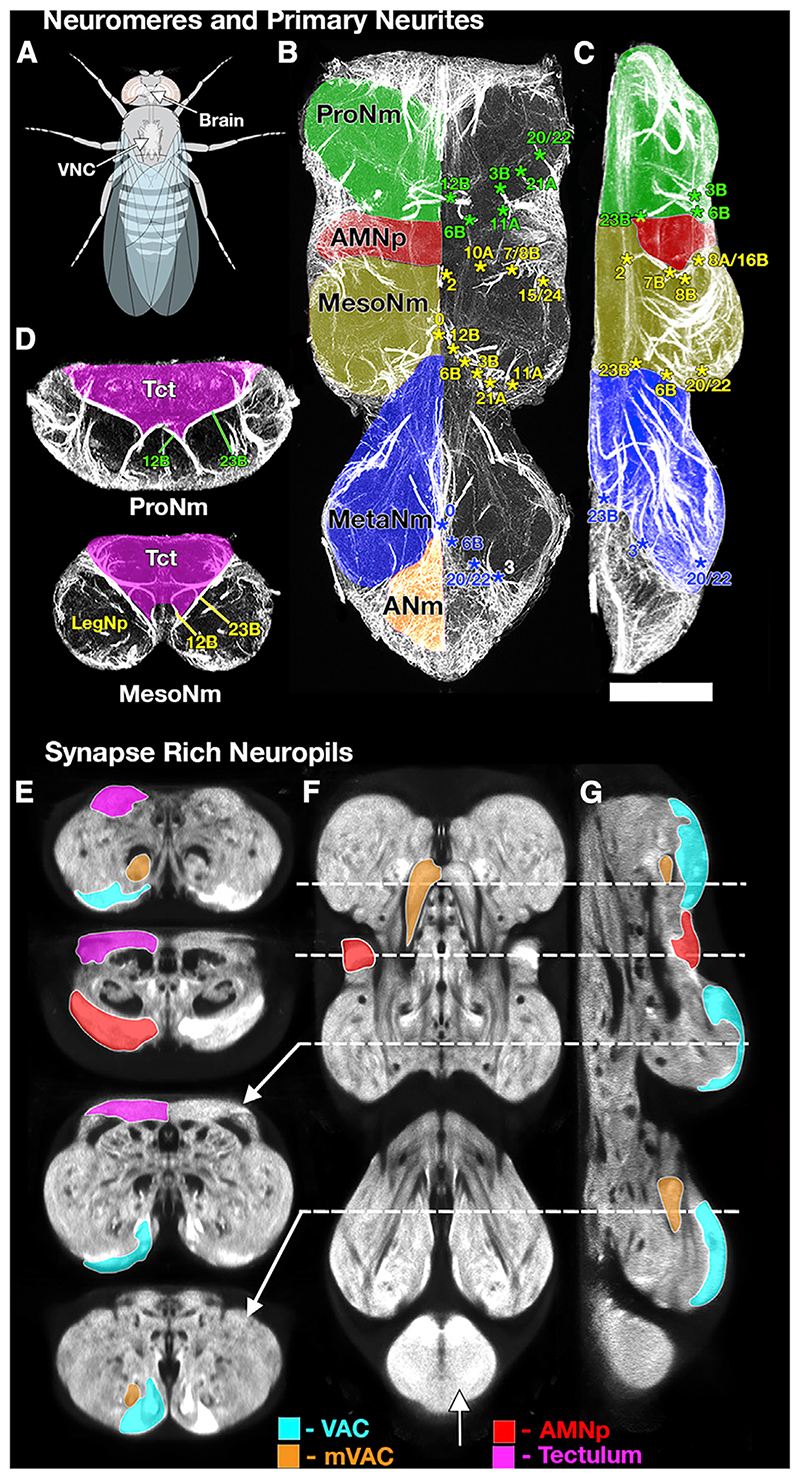
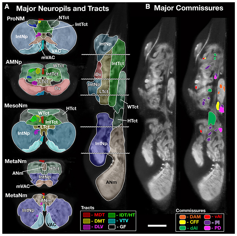
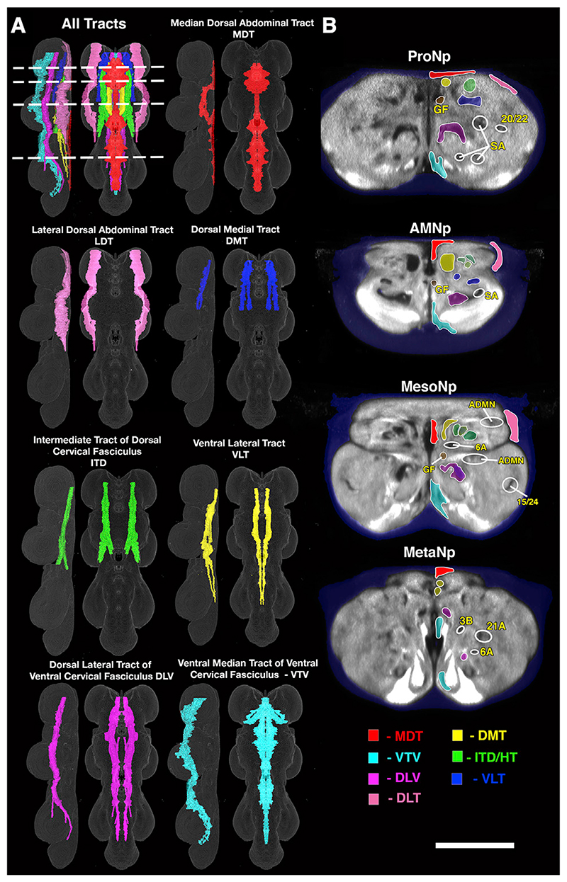
Drosophila melanogaster is an established model for neuroscience research with relevance in biology and medicine. Until recently, research on the Drosophila brain was hindered by the lack of a complete and uniform nomenclature. Recognizing this, Ito et al. (2014) produced an authoritative nomenclature for the adult insect brain, using Drosophila as the reference. Here, we extend this nomenclature to the adult thoracic and abdominal neuromeres, the ventral nerve cord (VNC), to provide an anatomical description of this major component of the Drosophila nervous system. The VNC is the locus for the reception and integration of sensory information and involved in generating most of the locomotor actions that underlie fly behaviors. The aim is to create a nomenclature, definitions, and spatial boundaries for the Drosophila VNC that are consistent with other insects. The work establishes an anatomical framework that provides a powerful tool for analyzing the functional organization of the VNC.

Keywords: anatomy; commissure; hemilineage; insect; motorneuron; neuromere; neuropil; ontology; tectulum; tract.


Conflict of interest statement

Declaration of Interests The authors declare no competing interests.

Figures

Figure 1
Figure 1. Selected Sections through an Adult VNC Illustrating the Tools Used to Define the Major Structures of the VNC
(A) Schematic of Drosophila illustrating the position of the VNC with respect to the body and brain. (B–D) Neuroglian immunostaining showing neuromeres and Primary Neurite bundles in horizontal (B), lateral (C), and transverse (D) sections to reveal the tracts of the primary neurites of the postembryonic neuronal lineages. The pattern of labeled pathways is highly stereotyped; each pathway corresponds to the primary neurites of neurons derived from a single neuroblast. These tracts provide a robust basis for identifying the key structures of the VNC such as the following: (B and C) the neuromere boundaries (ProNm [green], MesoNm [yellow], MetaNm [blue], and ANm [red]) and (D) the tectulum (magenta— Tct). The numbers refer to specific hemilineage primary neurite bundles, with the color indicating their neuromere of origin. (E–G) Brp-SNAP labeling (Bogovic et al., 2019) revealing the fine structure of the neuropil shown in transverse (E), horizontal (F), and lateral (G) sections. The bruchpilot (Brp) staining reveals characteristic regions of neuropil with high-density staining indicating synapse-rich neuropils. These synapse-rich neuropils can be used to define and segment specific neuropils such as the VAC (cyan), mVAC (orange), AMNp (red), and those of the tectulum (magenta, neck neuropil, wing neuropil, and haltere neuropil). The planes of the sections are indicated by the dotted lines. See also Video S1. A list ofthe abbreviations is given in Table 1. Scale 50 μm.
Figure 2
Figure 2. Major Neuropils, Tracts, and Commissures of the VNC
(A) Major Neuropils and Tracts—segmented VNC shown in transverse and lateral sections illustrating the outlines of the major neuropils and longitudinal tracts described in this study. The tectulum domains are shown in different shades of green, and the leg neuropil domains are shown in shades of blue. To further aid visualization, labeled tracts are only shown in the left half of the transverse sections. The plane of the transverse sections is indicated by dotted lines. (B) The position of the major commissural pathways shown on a lateral section at the midline of the VNC. Tracts derived from the same larval commissure are shown in the same colors. An unlabeled section is provided to show the detail unhindered by labeling. See also Figure S1 and Video S2. A list of the abbreviations is given in Table 1. Scale 50 μm.
Figure 3
Figure 3. Major Longitudinal Tracts of the VNC
(A) The major tracts of the VNC shown as rendered volumes from lateral and dorsal perspectives. (B) Transverse section views of the tracts at selected points in the VNC. The areas outlined by white circles identify other key structures (GF, giant fiber; ADMN, sensory afferents entering from the ADMN; SA, sensory afferents entering from the leg nerve; the numbers refer to hemilineage-derived axon fascicles). The planes of section are indicated by dotted lines in (A). See also Video S3. A list of the abbreviations is given in Table 1. Scale (A), 100 μm; (B), 50 μm.

References

    1. Bacon JP, Strausfeld NJ. The dipteran ‘Giant fibre’ pathway: neurons and signals. J Comp Physiol A. 1986;158:529–548.
    1. Baek M, Mann RS. Lineage and birth date specify motor neuron targeting and dendritic architecture in adult Drosophila. J Neurosci. 2009;29:6904–6916. - PMC - PubMed
    1. Bellen HJ, Tong C, Tsuda H. 100 years of Drosophila research and its impact on vertebrate neuroscience: a history lesson for the future. Nat Rev Neurosci. 2010;11:514–522. - PMC - PubMed
    1. Bidaye SS, Machacek C, Wu Y, Dickson BJ. Neuronal control of Drosophila walking direction. Science. 2014;344:97–101. - PubMed
    1. Bodenstein D. The Postembryonic Development of Drosophila. Wiley; 1950. pp. 275–367.

Publication types