. 2021 Jan;110(1):412-421.
doi: 10.1016/j.xphs.2020.09.013. Epub 2020 Sep 12.

Pregnancy-Related Hormones Increase Nifedipine Metabolism in Human Hepatocytes by Inducing CYP3A4 Expression

Affiliations

Pregnancy-Related Hormones Increase Nifedipine Metabolism in Human Hepatocytes by Inducing CYP3A4 Expression

Raju Khatri et al. J Pharm Sci. 2021 Jan.

Abstract

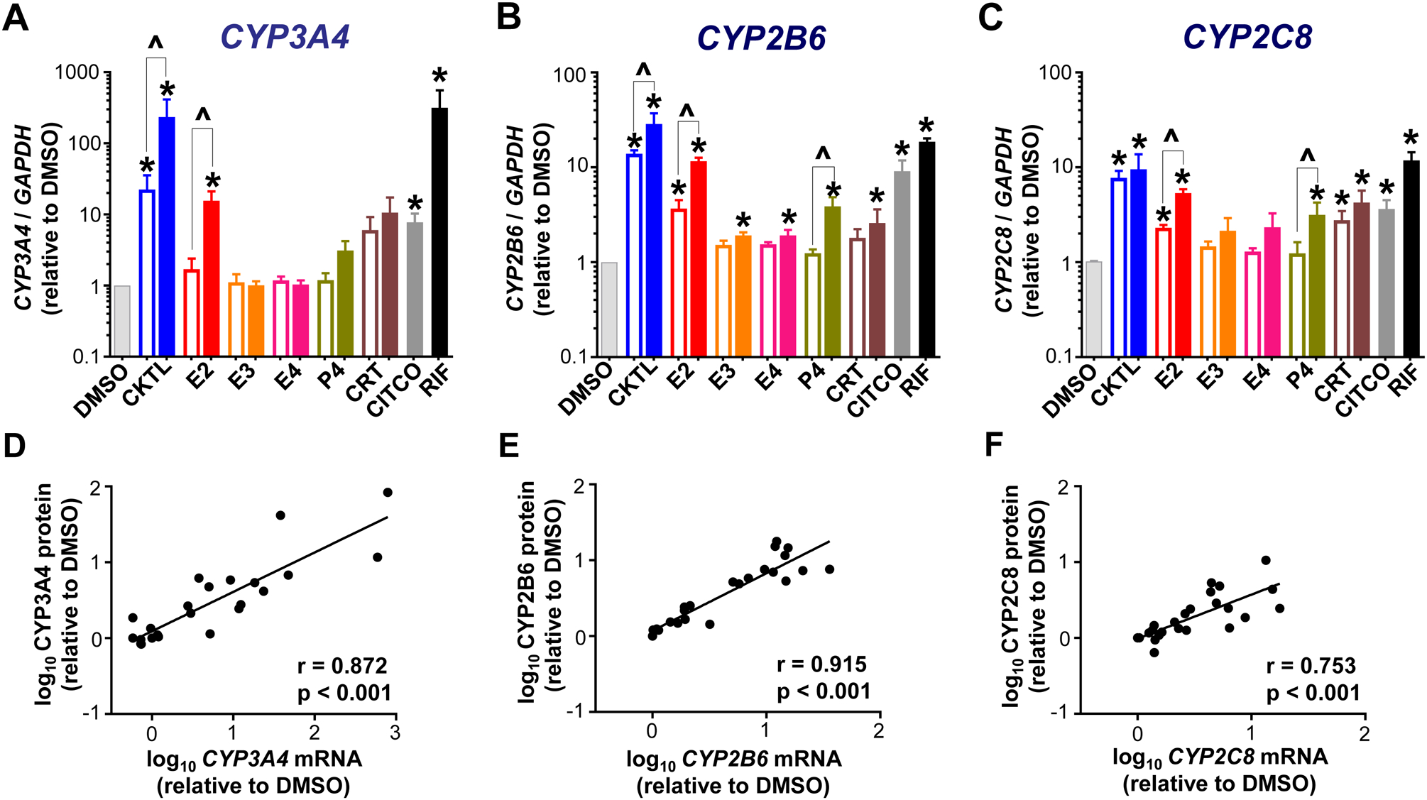
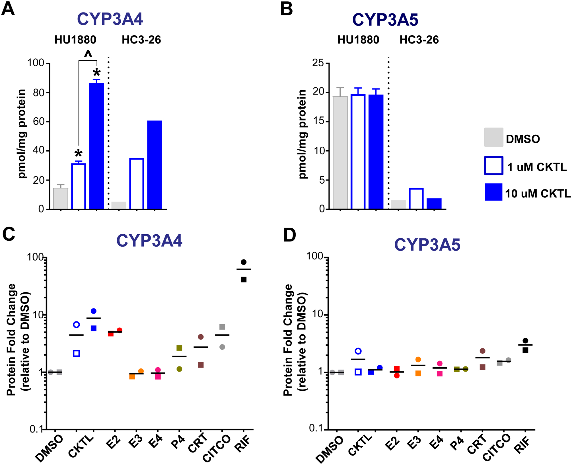
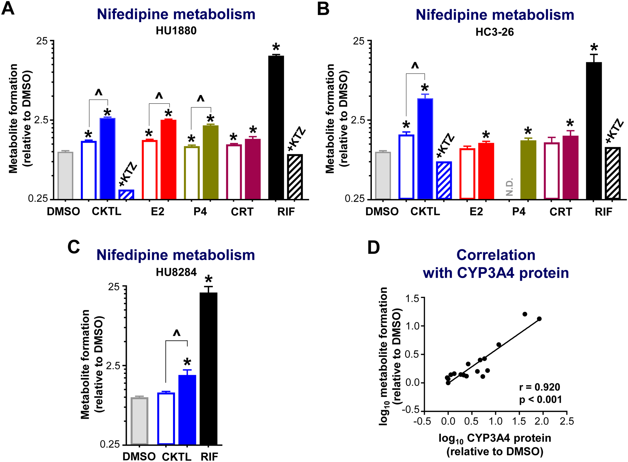
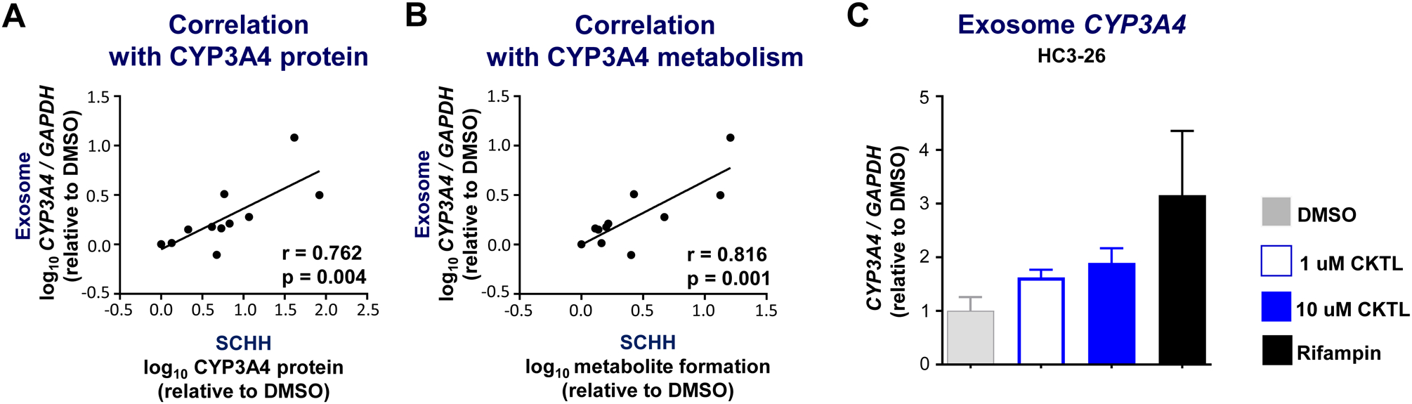
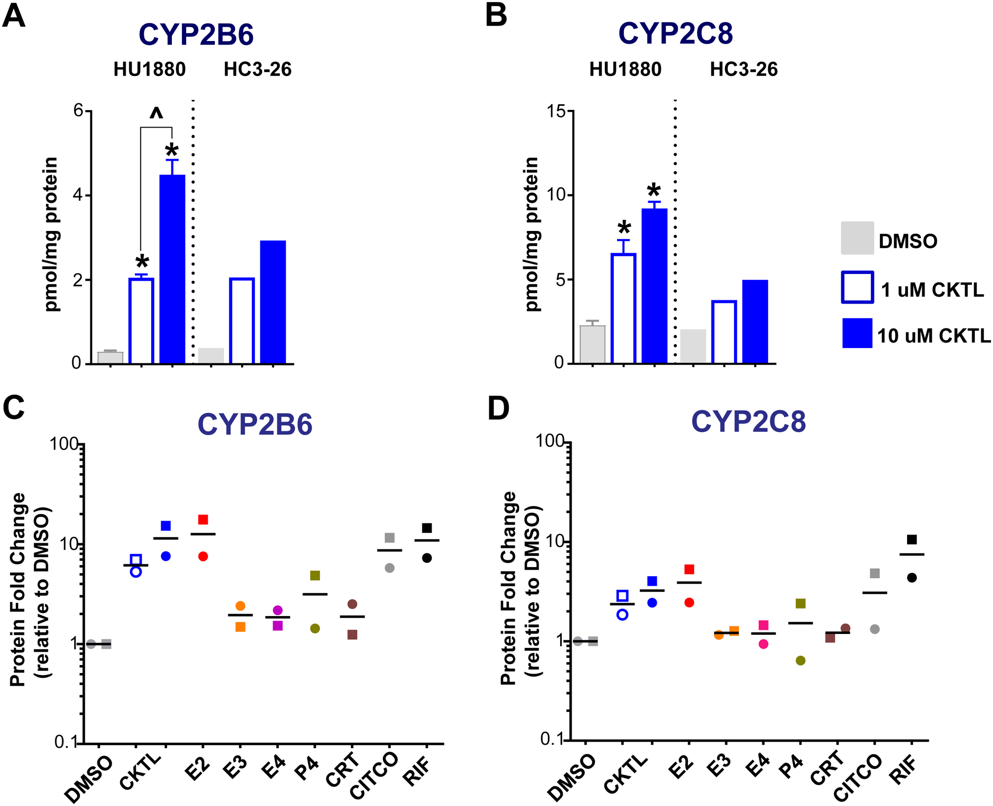
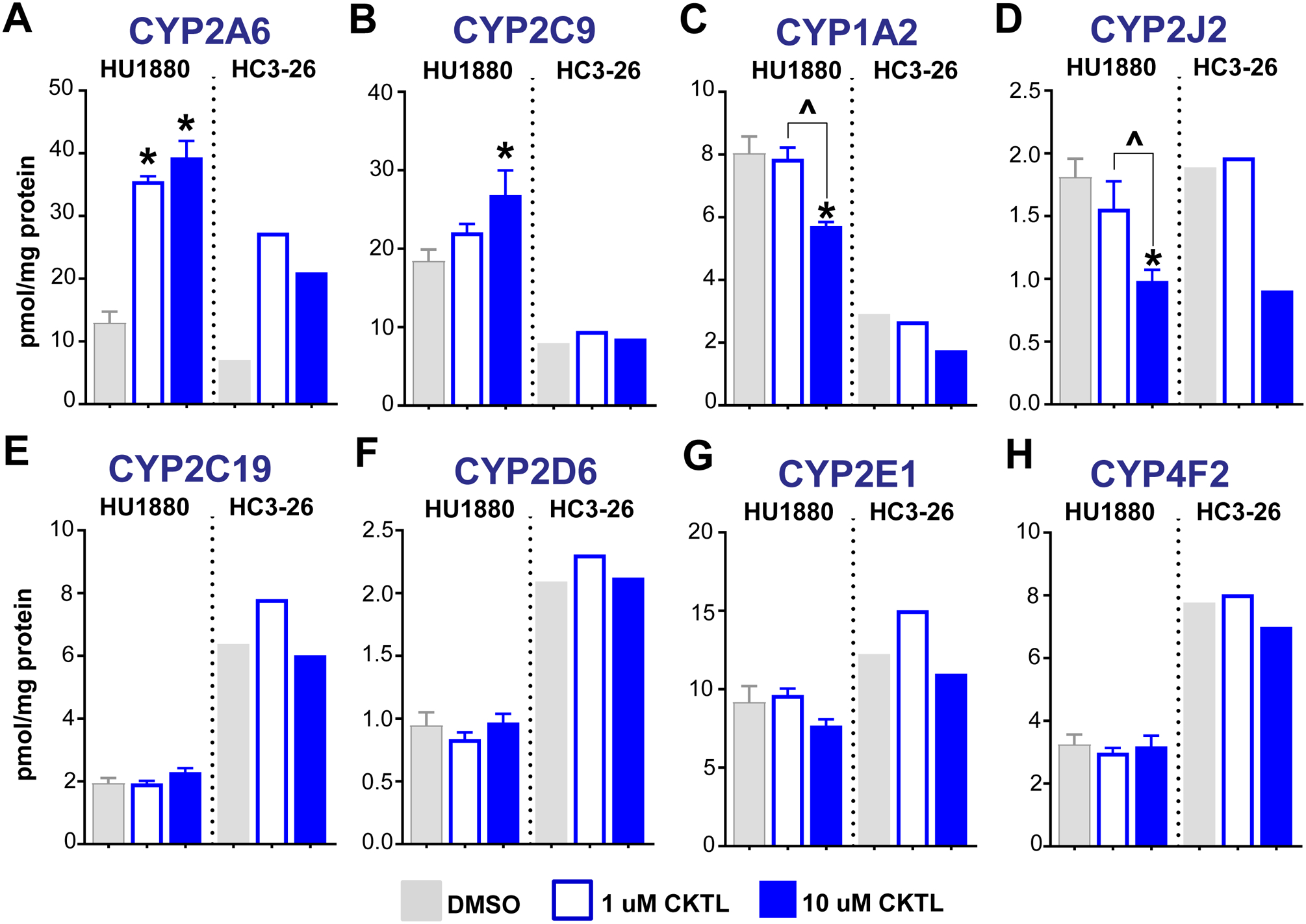
Pregnancy-related hormones (PRH) have emerged as key regulators of hepatic cytochrome P450 (CYP) enzyme expression and function. The impact of PRH on protein levels of CYP3A4 and other key CYP enzymes, and the metabolism of nifedipine (a CYP3A4 substrate commonly prescribed during pregnancy), was evaluated in primary human hepatocytes. Sandwich-cultured human hepatocytes (SCHH) from female donors were exposed to PRH (estradiol, estriol, estetrol, progesterone, and cortisol), individually or in combination as a cocktail. Absolute protein concentrations of twelve CYP isoforms in SCHH membrane fractions were quantified by nanoLC-MS/MS, and metabolism of nifedipine to dehydronifedipine in SCHH was evaluated. PRH significantly increased CYP3A4 protein concentrations and nifedipine metabolism to dehydronifedipine in a concentration-dependent manner. CYP3A4 mRNA levels in hepatocyte-derived exosomes positively correlated with CYP3A4 protein levels and dehydronifedipine formation in SCHH. PRH also increased CYP2B6, CYP2C8 and CYP2A6 levels. Our findings demonstrate that PRH increase nifedipine metabolism in SCHH by inducing CYP3A4 expression and alter expression of other key CYP proteins in an isoform-specific manner, and suggest that hepatocyte-derived exosomes warrant further investigation as biomarkers of hepatic CYP3A4 metabolism. Together, these results offer mechanistic insight into the increases in nifedipine metabolism and clearance observed in pregnant women.

Keywords: Cytochrome P450; Estradiol; Exosomes; Hepatic metabolism; Hypertension; Nifedipine; Pregnancy; Progesterone; Targeted proteomics.


Conflict of interest statement

Conflict of interest

Dr. Kim Brouwer is a co-inventor of the sandwich-cultured hepatocyte technology for quantification of biliary excretion (B-CLEAR®) and related technologies, which have been licensed exclusively to Qualyst Transporter Solutions, recently acquired by BioIVT. The other authors have no conflicts of interest to disclose.

Figures

Figure 1.
Figure 1.. Effect of pregnancy-related hormones on mRNA levels of key CYP isoforms in SCHH.
Human hepatocytes from three female donors (HU1880, HC3–26 and HC5–40) were exposed to hormones (E2, E3, E4, P4, CRT), either individually or in combination as a cocktail [CKTL] of all hormones, or controls (DMSO, CITCO, Rifampin [RIF]) for 72 h (n=2/group within each donor). (A) CYP3A4, (B) CYP2B6 and (C) CYP2C8 mRNA levels were quantified, normalized to GAPDH, expressed relative to vehicle (DMSO) control within each donor, and then combined for comparison across experimental groups (n=3 donors/group; mean ± SEM; *p<0.05 vs. DMSO). Concentration-dependent effects were evaluated (open bar: 1 μM, solid bar: 10 μM; ^p<0.05 1 vs. 10 μM). (D-F) Correlation between CYP mRNA levels and protein concentrations quantified by QTAP in SCHH membrane-associated protein in donors HU1880 and HC3–26. Data points represent the mean fold-changes in expression for each treatment group, relative to DMSO, within each donor. The Pearson correlation coefficient (r) and corresponding p-values are provided.
Figure 2.
Figure 2.. Effect of pregnancy-related hormones on protein concentrations of CYP3A4 and CYP3A5 in SCHH.
Following 72 h of hormone exposure, CYP3A4 and CYP3A5 protein concentrations were quantified by QTAP in SCHH membrane-associated protein isolated from two donors (HU1880, HC3–26). (A) CYP3A4 and (B) CYP3A5 absolute protein concentrations in the DMSO and hormone cocktail (CKTL) groups were compared separately in donor HU1880 (mean ± SEM: n=3–4/group; *p<0.05 vs. DMSO, ^p<0.05 1 vs. 10 μM) and donor HC3–26 (mean: n=2/group). (C) CYP3A4 and (D) CYP3A5 protein levels were expressed relative to vehicle (DMSO) within each donor, and then combined for comparison across experimental groups. The effect within donor HC3–26 (circles) and donor HU1880 (squares) is represented by the individual data points. Open circles and squares represent 1 μM CKTL. Solid circles and squares represent 10 μM CKTL and 10 μM for the individual hormones. Comparison of PRH effects within each donor are provided in Supplemental Figure 2.
Figure 3.
Figure 3.. Effect of pregnancy-related hormones on nifedipine metabolism in SCHH.
Following 72 h of hormone exposure, SCHH from three donors (HU1880, HC3–26, HU8284) were incubated with nifedipine (100 μM) for 60 min. Dehydronifedipine formation was quantified and compared across treatment groups in (A) donor HU1880 (n=4–7/group), (B) donor HC3–26 (n=3–8/group), and (C) donor HU8284 (n=3/group). Data are calculated as fold-change in metabolite formation relative to DMSO (*p<0.05 vs. DMSO). Concentration-dependent effects were evaluated (open bar: 1 μM, solid bar: 10 μM; ^p<0.05 1 vs. 10 μM). Ketoconazole co-administration (+KTZ, 5 μM) was included in the hormone cocktail (CKTL) and rifampin (RIF) treated groups (n=1/group in each donor) to confirm CYP3A-mediated effects. Since the KTZ rescue experiment was conducted in a single well (n=1), the observed value is presented without error bars. N.D., experimental group not studied. (D) Correlation between dehydronifedipine formation and CYP3A4 protein levels in donors HU1880 and HC3–26. Data points represent the mean fold-change of each treatment group, relative to DMSO, within each donor. The Pearson correlation coefficient (r) and corresponding p-value are provided.
Figure 4.
Figure 4.. Correlation between hepatocyte-derived exosome CYP3A4 mRNA levels and CYP3A4 expression and metabolism in SCHH.
Hepatocyte-derived exosomes were enriched from SCHH culture media collected during a subset of hormone exposure experiments conducted in donors HC3–26 and HU1880. CYP3A4 mRNA levels in enriched exosomes were quantified by RT-PCR, normalized to GAPDH, and expressed relative to vehicle (DMSO) control. Correlation between exosome CYP3A4 mRNA levels and (A) CYP3A4 protein expression in SCHH membrane fractions or (B) dehydronifedipine formation in SCHH media. Data points represent the mean fold-change of each treatment group, relative to DMSO, within each donor. The Pearson correlation coefficient (r) and corresponding p-value are provided. (C) CYP3A4 mRNA levels in enriched exosomes from DMSO, CKTL (1 μM [open bar] and 10 μM [closed bar]), and RIF (10 μM) treated SCHH in donor HC3–26 (n=4–7/group).
Figure 5.
Figure 5.. Effect of pregnancy-related hormones on protein concentrations of CYP2B6 and CYP2C8 in SCHH.
Following 72 h of hormone exposure, CYP2B6 and CYP2C8 protein concentrations were quantified by QTAP in SCHH membrane-associated protein isolated from two donors (HU1880, HC3–26). (A) CYP2B6 and (B) CYP2C8 absolute protein concentrations in the DMSO and hormone cocktail (CKTL) groups were compared separately in donor HU1880 (mean ± SEM: n=3–4/group; *p<0.05 vs. DMSO, ^p<0.05 1 vs. 10 μM) and donor HC3–26 (mean: n=2/group). (C) CYP2B6 and (D) CYP2C8 protein levels were expressed relative to vehicle (DMSO) within each donor, and then combined for comparison across experimental groups. The effect within donor HC3–26 (circles) and donor HU1880 (squares) is represented by the individual data points. Open circles and squares represent 1 μM CKTL. Solid circles and squares represent 10 μM CKTL and 10 μM for the individual hormones. Comparison of PRH effects within each donor are provided in Supplemental Figure 2.
Figure 6.
Figure 6.. Effect of pregnancy-related hormones on protein concentrations of other key CYP isoforms in SCHH.
Following 72 h of hormone exposure, protein concentrations of eight additional CYP isoforms (A: CYP2A6, B: CYP2C9, C: CYP1A2, D: CYP2J2, E: CYP2C19, F: CYP2D6, G: CYP2E1, H: CYP4F2) were quantified by QTAP in SCHH membrane-associated proteins isolated from two donors (HU1880, HC3–26). Absolute protein concentrations in the DMSO and hormone CKTL groups were compared separately in donor HU1880 (mean ± SEM: n=3–4/group; *p<0.05 vs. DMSO, ^p<0.05 1 vs. 10 μM) and donor HC3–26 (mean: n=2/group).

Similar articles

Cited by

References

    1. Ayad M, Costantine MM. Epidemiology of medications use in pregnancy. Semin Perinatol 2015;39(7):508–511. - PMC - PubMed
    1. Tasnif Y, Morado J, Hebert MF. Pregnancy-Related Pharmacokinetic Changes. Clin Pharmacol Ther 2016;100(1):53–62. - PubMed
    1. Gonzalez D, Boggess KA, Cohen-Wolkowiez M. Lessons learned in pediatric clinical research to evaluate safe and effective use of drugs in pregnancy. Obstet Gynecol 2015;125(4):953–958. - PMC - PubMed
    1. Dallmann A, Pfister M, van den Anker J, Eissing T. Physiologically Based Pharmacokinetic Modeling in Pregnancy: A Systematic Review of Published Models. Clin Pharmacol Ther 2018;104(6):1110–1124. - PubMed
    1. Shuster DL, Bammler TK, Beyer RP et al. Gestational age-dependent changes in gene expression of metabolic enzymes and transporters in pregnant mice. Drug Metab Dispos 2013;41(2):332–342. - PMC - PubMed

Publication types

LinkOut - more resources