. 2020 Nov 1:406:115242.
doi: 10.1016/j.taap.2020.115242. Epub 2020 Sep 12.

Biological effects of inhaled hydraulic fracturing sand dust. VI. Cardiovascular effects

Affiliations

Biological effects of inhaled hydraulic fracturing sand dust. VI. Cardiovascular effects

Kristine Krajnak et al. Toxicol Appl Pharmacol. .

Abstract

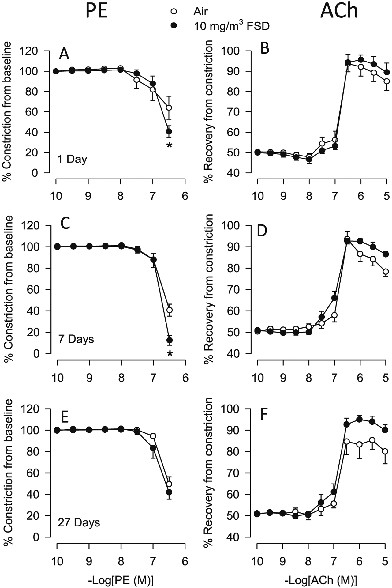
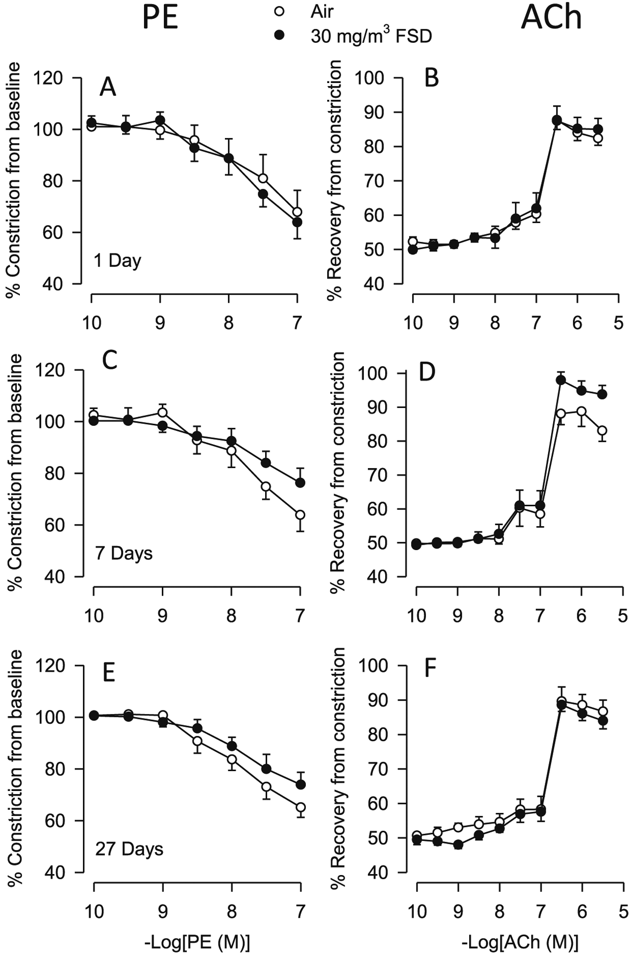
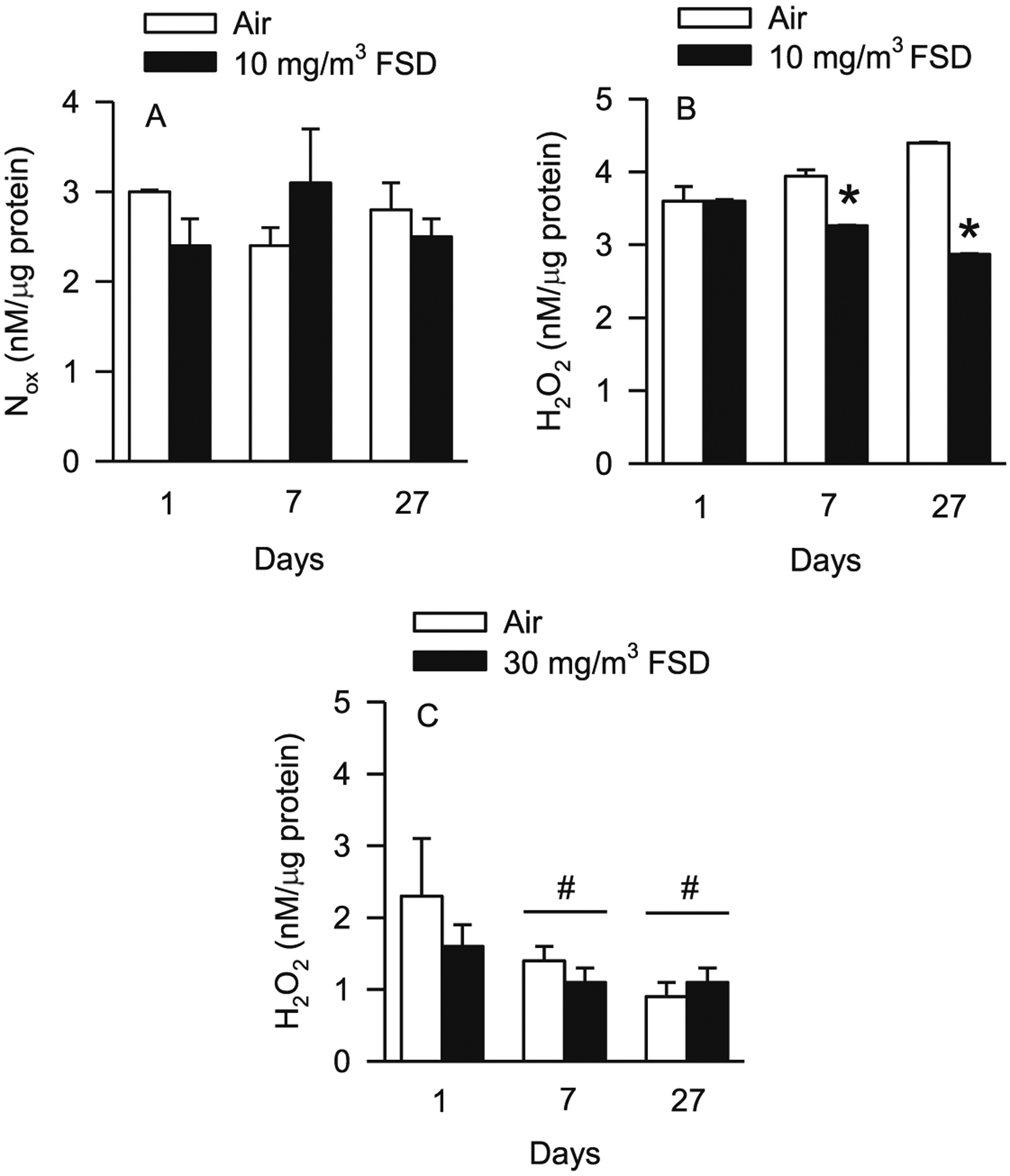
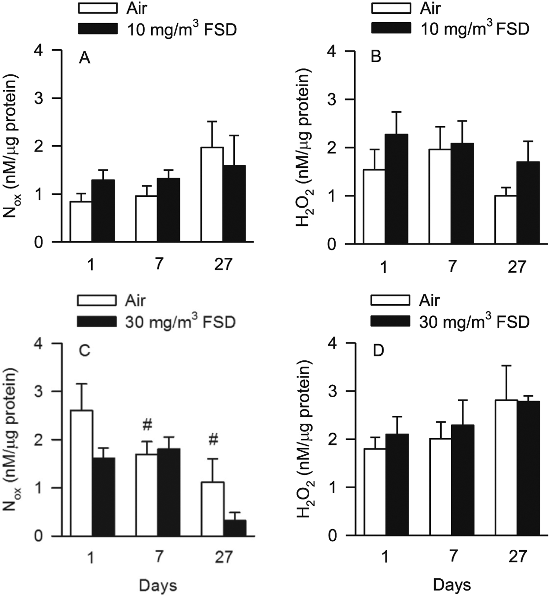
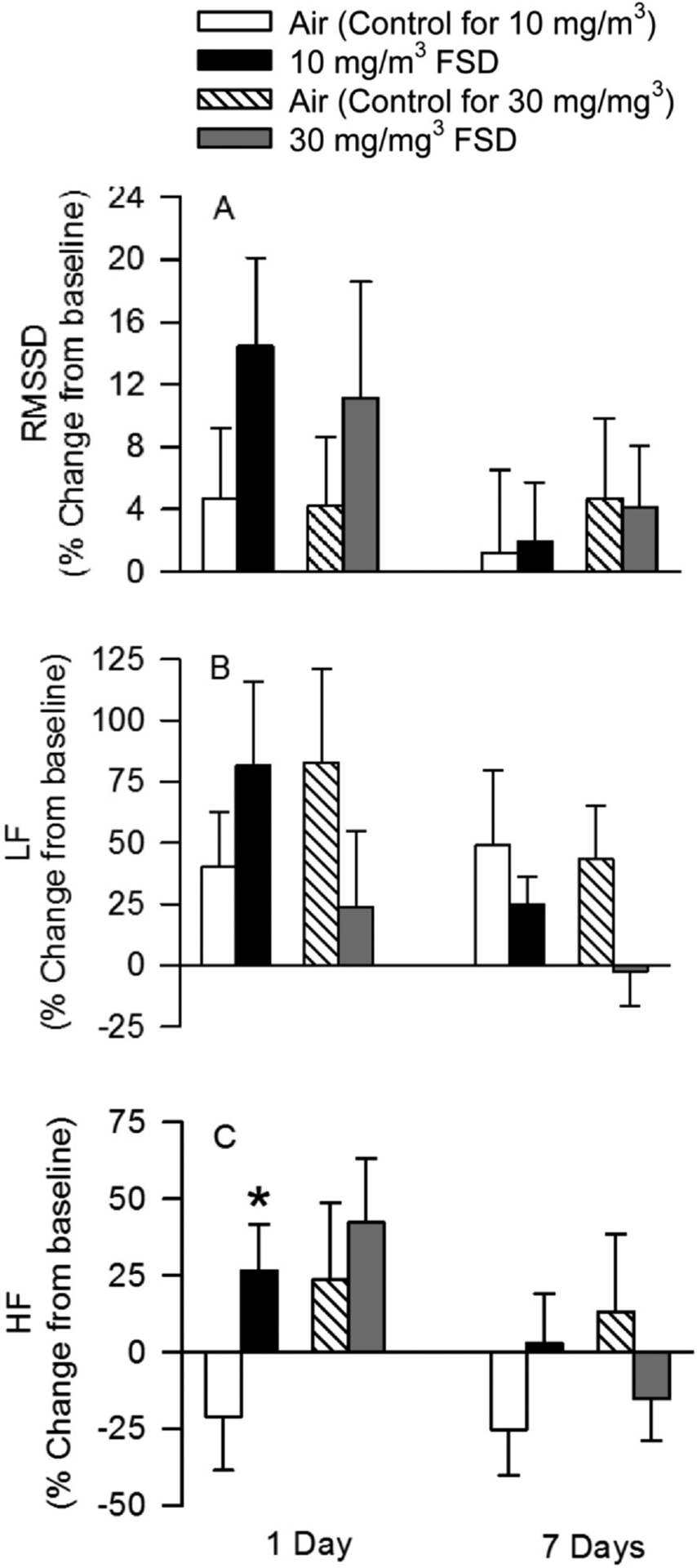
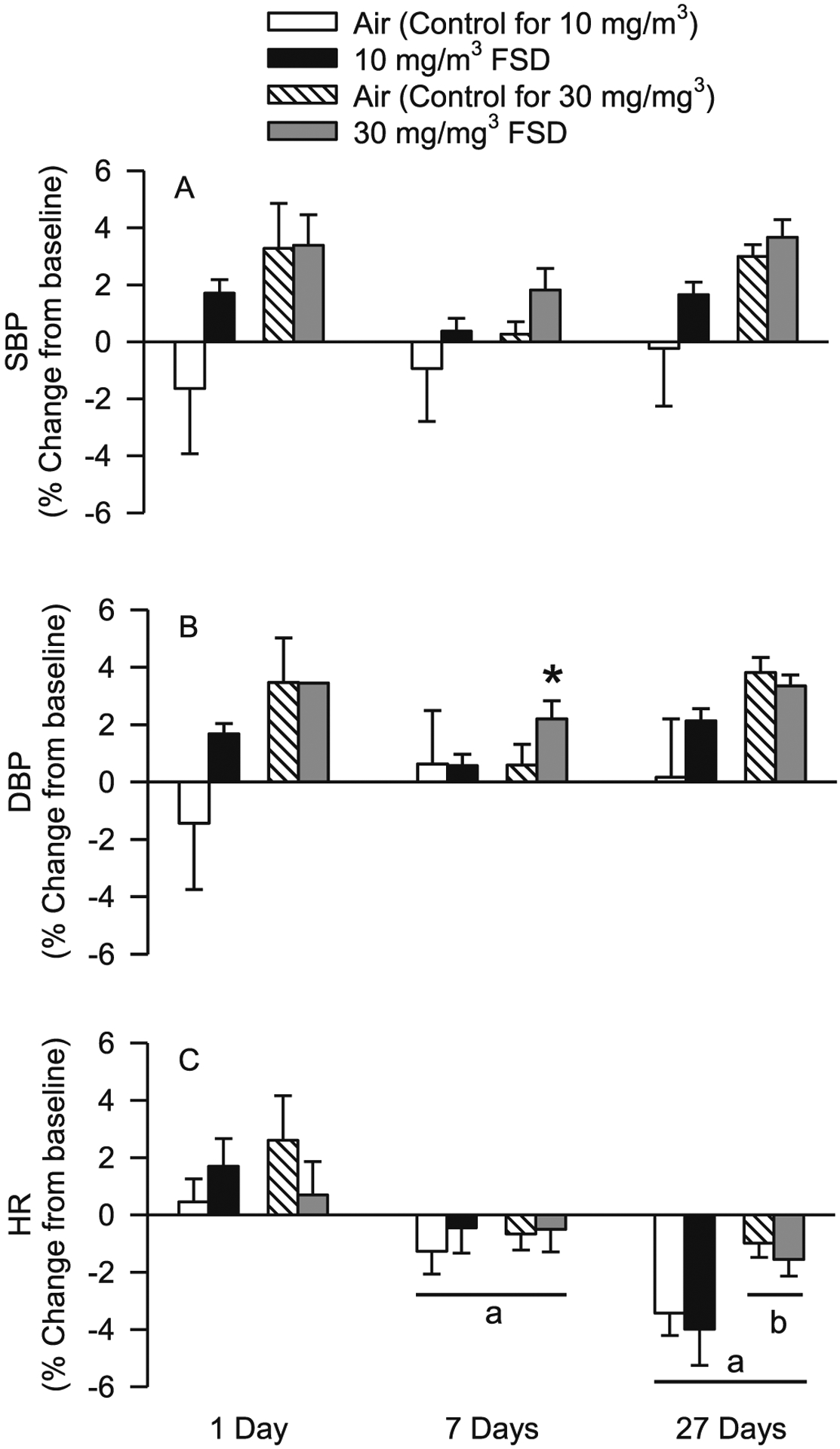
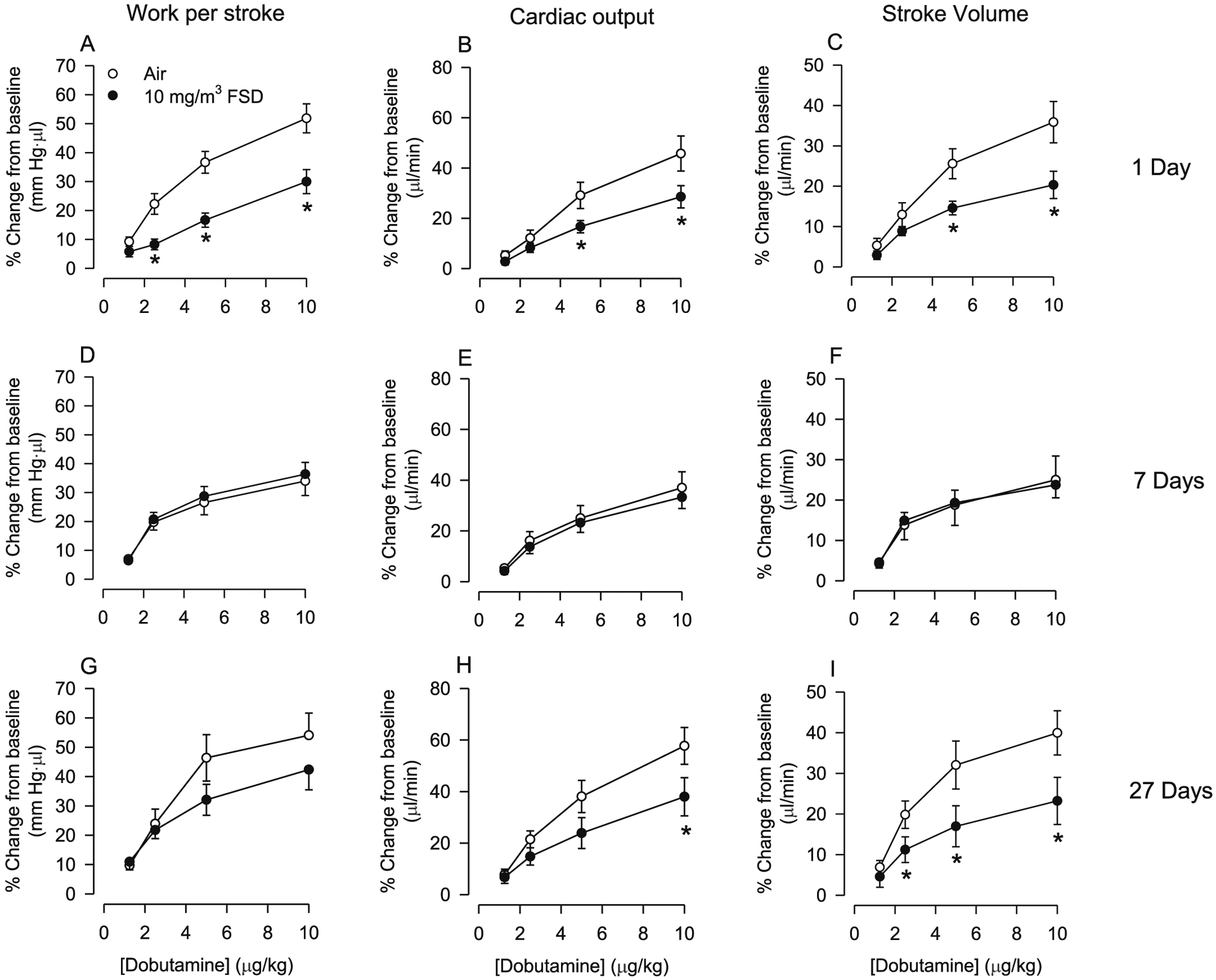
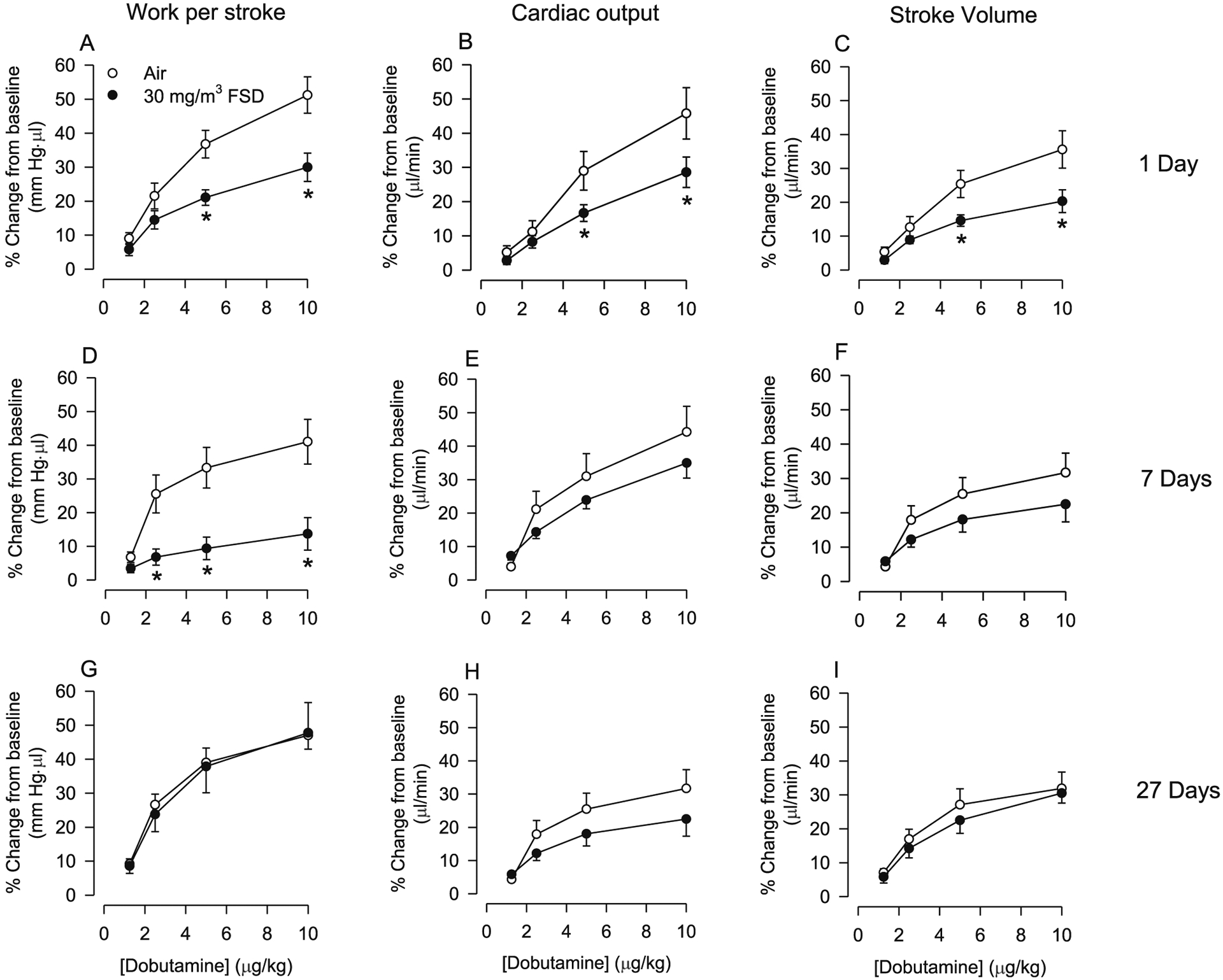
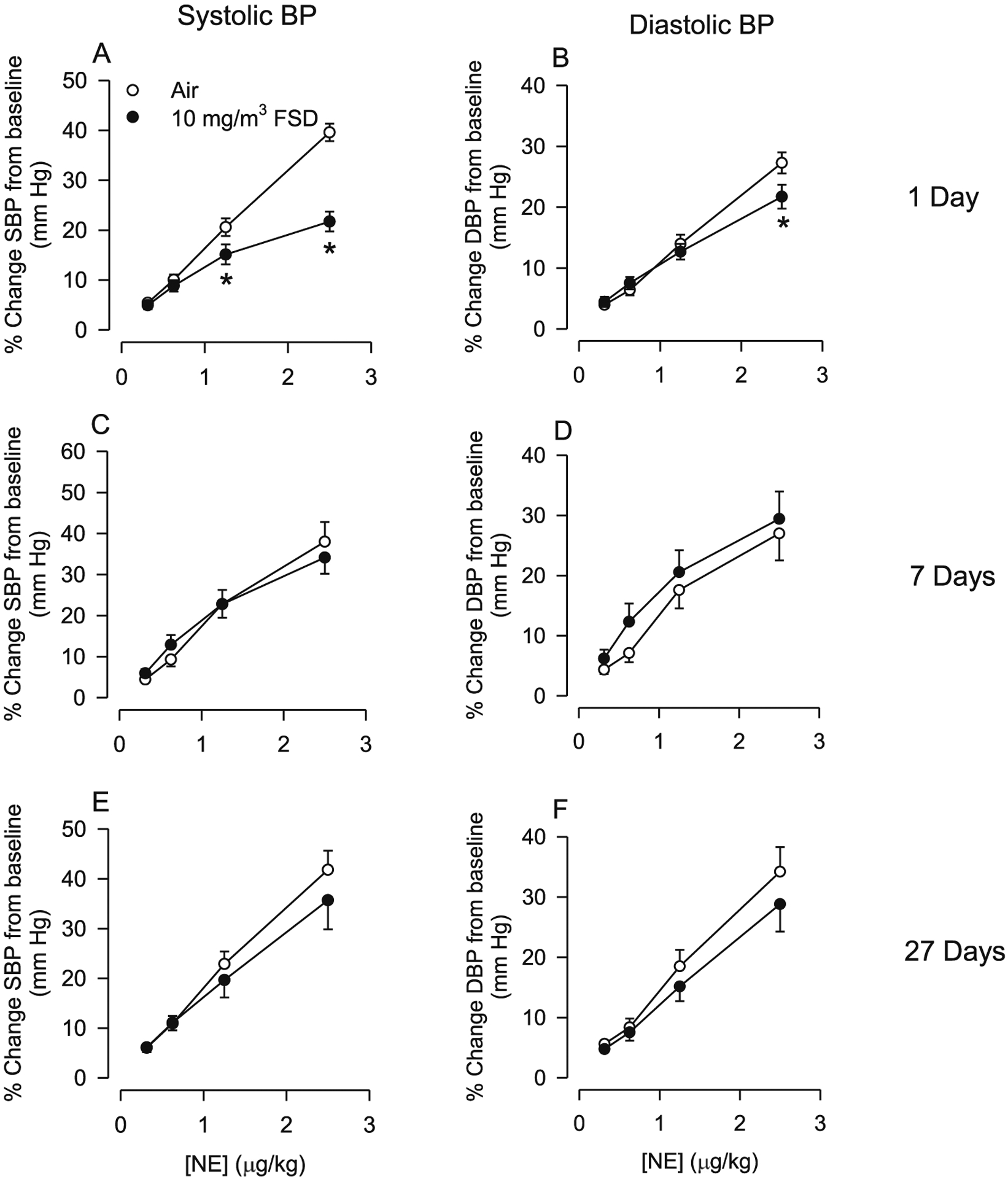
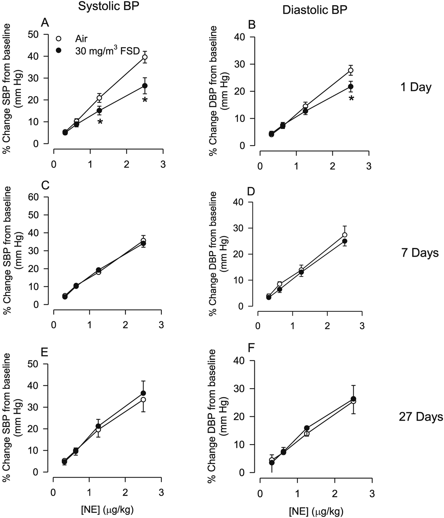
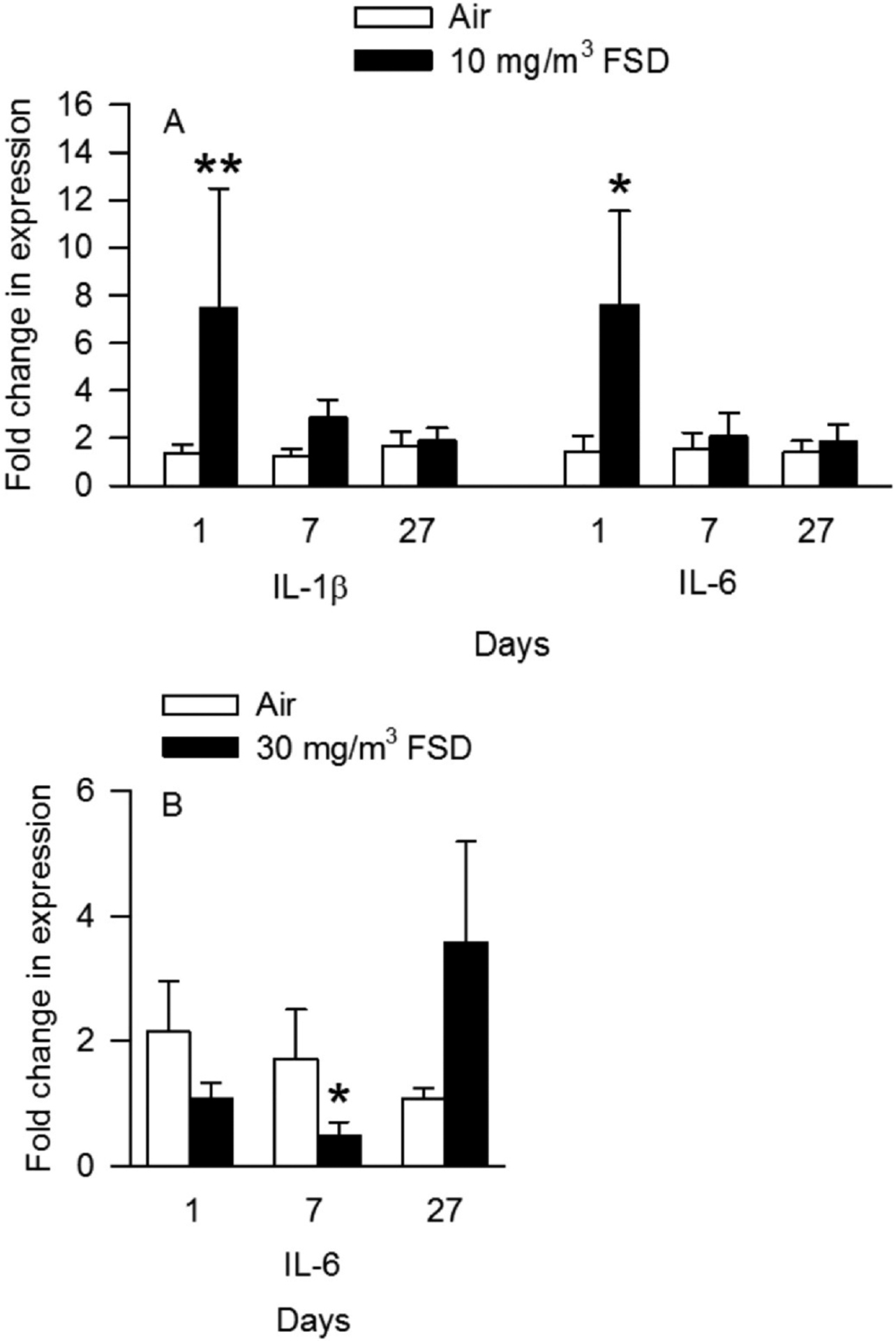
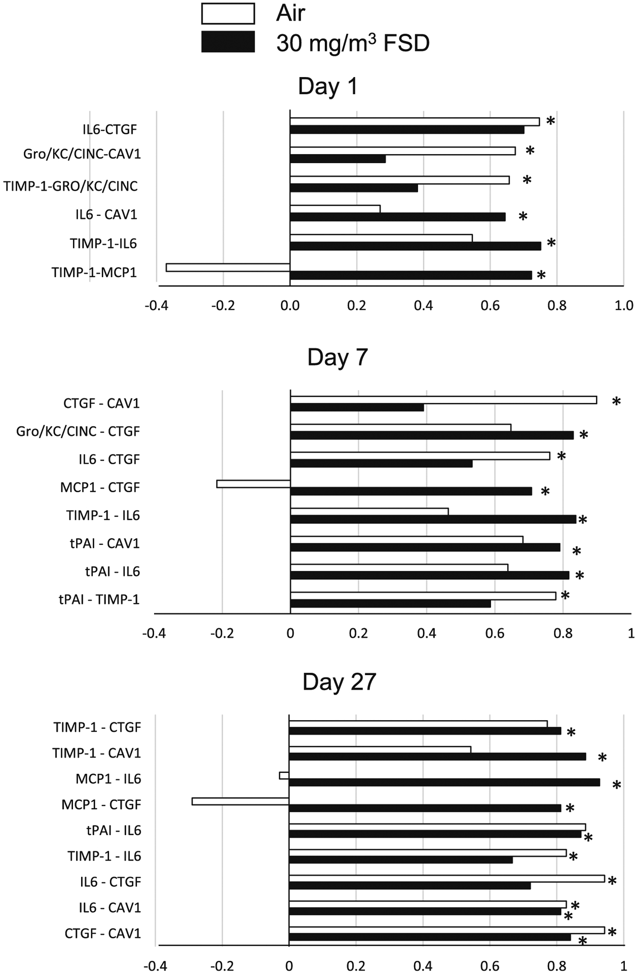
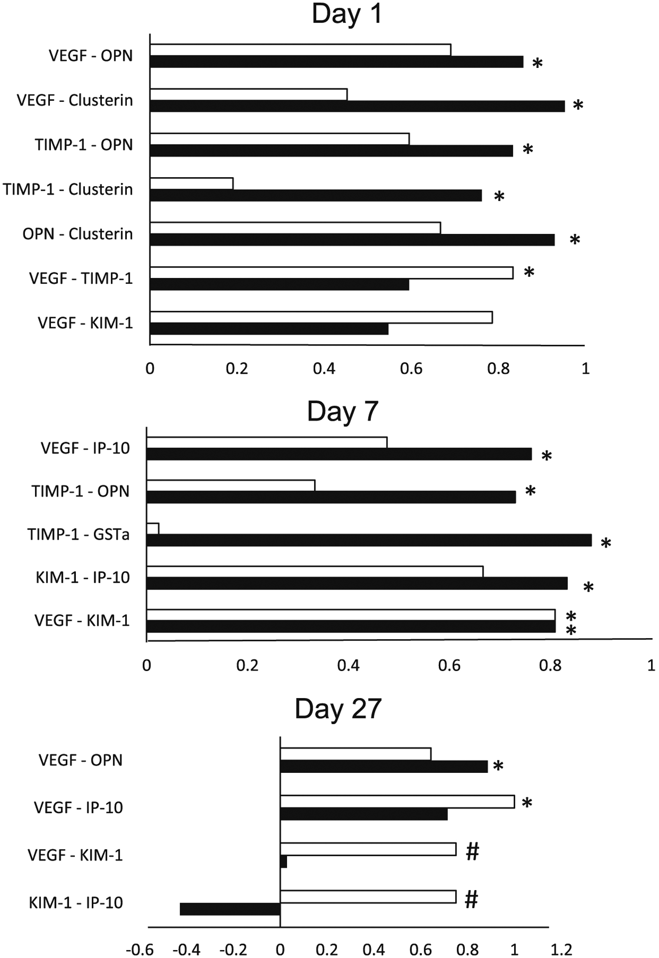
Hydraulic fracturing is used to access oil and natural gas reserves. This process involves the high-pressure injection of fluid to fracture shale. Fracking fluid contains approximately 95% water, chemicals and 4.5% fracking sand. Workers may be exposed to fracking sand dust (FSD) during the manipulation of the sand, and negative health consequences could occur if FSD is inhaled. In the absence of any information about its potential toxicity, a comprehensive rat animal model study (see Fedan et al., 2020) was designed to investigate the bioactivities of several FSDs in comparison to MIN-U-SIL® 5, a respirable α-quartz reference dust used in previous animal models of silicosis, in several organ systems. The goal of this study was to assess the effects of inhalation of one FSD, i.e., FSD 8, on factors and tissues that affect cardiovascular function. Male rats were exposed to 10 or 30 mg/m3 FSD (6 h/d for 4 d) by whole body inhalation, with measurements made 1, 7 or 27 d post-exposure. One day following exposure to 10 mg/m3 FSD the sensitivity to phenylephrine-induced vasoconstriction in tail arteries in vitro was increased. FSD exposure at both doses resulted in decreases in heart rate (HR), HR variability, and blood pressure in vivo. FSD induced changes in hydrogen peroxide concentrations and transcript levels for pro-inflammatory factors in heart tissues. In kidney, expression of proteins indicative of injury and remodeling was reduced after FSD exposure. When analyzed using regression analysis, changes in proteins involved in repair and remodeling were correlated. Thus, it appears that inhalation of FSD does have some prolonged effects on cardiovascular, and, possibly, renal function. The findings also provide information regarding potential mechanisms that may lead to these changes, and biomarkers that could be examined to monitor physiological changes that could be indicative of impending cardiovascular dysfunction.

Keywords: Blood pressure; Heart rate variability; Oxidative stress; Renal injury; Vasoconstriction; Vasodilation.


Conflict of interest statement

Declaration of Competing Interest The authors declare that they have no conflicts of interest in relation to this publication.

Figures

Fig. 1.
Fig. 1.
Vascular responses to phenylephrine (PE) and acetylcholine (ACh) in ventral tail arteries collected from rats exposed to filtered air or 10 mg/m3 frack sand dust (FSD) 8 (in all figures, FSD 8 is abbreviated as FSD; n = 7–8 animals/group and at each time point). On days 1 and 7, the constriction induced by PE (−6.5 dose, M) was greater in arteries from FSD 8-compared to air-exposed rats (A and B; *P < 0.05). On day 27 there was no difference in PE-induced vasoconstriction (C). ACh-induced re-dilation was not different between the air- and FSD 8-exposed groups on any post-exposure day (ACh: Figs. D–F represent days 1, 7 and 27 respectively).
Fig. 2.
Fig. 2.
Vascular responses to phenylephrine (PE) and acetylcholine (ACh) in ventral tail arteries collected from rats exposed to air or 30 mg/m3 frack sand dust (FSD) 8 (n = 7–8 animals/group and at each time point. Neither PE-induced vasoconstriction nor ACh-induced re-dilation were affected by FSD 8 1, 7 or 27 d after the exposure (PE: panels A–C, respectively; ACh: panels D–F).
Fig. 3.
Fig. 3.
Nitrate/nitrite (Nox) and hydrogen peroxide (H2O2) concentrations in the hearts of rats exposed to air or frack sand dust (FSD) 8 (n = 8 animals/group and at each time point. Exposure to 10 mg/m3 FSD 8 did not affect Nox concentrations in the heart (A). However, H2O2 concentrations were reduced 7 and 27 d after exposure (B; *P < 0.05). There were no treatment-related differences in H2O2 concentrations with exposure to 30 mg/m3 FSD 8. However, H2O2 concentrations were reduced on days 7 and 27, in heart tissue from both air and FSD 8-treated rats (#P < 0.05). N ox concentrations were below the level of detection after exposure to the 30 mg/m3 FSD 8 in all animals.
Fig. 4.
Fig. 4.
Nitrate/nitrite [Nox; A, 10 mg/m3 frack sand dust (FSD) 8, or C, 30 mg/m3 FSD 8] and hydrogen peroxide (H2O2;B, 10 mg/m3 FSD 8, or D, 30 mg/m3 FSD 8) levels in the kidneys of rats at 1, 7 or 27 d after exposure (n = 8 animals/group and at each time point). Exposure to 10 mg/m3 FSD 8 did not significantly affect Nox (A) or H2O2 (B) concentrations in the kidneys. However, Nox concentrations were decreased in FSD 8-exposed compared to air-exposed rats 27 d after exposure to the 30 mg/m3 FSD 8 (C). Nox concentrations also were lower on day 7 and 27 than on day 1 in air-exposed rats (C and D; #P < 0.05). FSD 8 exposure at 30 mg/m3 did not alter H2O2 concentrations in the kidney.
Fig. 5.
Fig. 5.
Estimates of heart rate (HR) variability recorded using telemetry in rats after exposure to air or frack sand dust (FSD) 8 (n = 6–8 rats/air or FSD8 exposure). Data are presented as the % change from baseline. Baseline data are measures that were collected prior to air or FSD 8 exposure. RMSSD (A; the R-R interval) was variable, but there were no significant effects of treatment or day after recovery on this measure. There were also no significant differences in the low frequency signal (B). However, a significant increase in the high frequency component of the EEG signal occurred one day after exposure (C) to 10 mg/m3 FSD 8.
Fig. 6.
Fig. 6.
Effects of frack sand dust (FSD) 8 exposure on blood pressure and heart rate (HR). Data are presented as the % change from baseline (n = 6–8 air or FSD 8 rats per time point). Exposure to FSD 8 did not affect systolic blood pressure (SBP) on day 1 (A). Diastolic blood pressure (DBP) was not affected by exposure to FSD 8 on days 1 and 27 but was increased in animals treated with 30 mg/m3 FSD-8 (B). HR was lower on days 7 and 27 after exposure compared to 1 d after exposure, and HR was lower 27 days after exposure than 7 days after exposure (C). Twenty-seven d after exposure, both air controls and animals treated with 30 mg/m3 FSD 8 had lower HR values than animals in the 10 mg/m3 group (aless than day 1 values; bless than 10 m/mg3; *P < 0.05).
Fig. 7.
Fig. 7.
Effects of frack sand dust (FSD) 8 on cardiac work (A, D and G), cardiac output (B, E and H) and stroke volume (C, F and I) on responses to dobutamine in rats exposed to air or 10 mg/m3 FSD 8 (n = 6–8 air or FSD 8 rats per time point). One d after exposure to FSD 8, cardiac work, cardiac output and stroke volume responses to dobutamine were reduced (A–C). However, 7 d after exposure to FSD8 responsiveness to dobutamine returned to baseline levels (D–F). Twenty-seven d following exposure to FSD 8, cardiac output and stroke volume were again reduced in response to dobutamine (H and I). *P < 0.05.
Fig. 8.
Fig. 8.
Effects of frack sand dust (FSD) 8 on cardiac work (A, D and G), cardiac output (B, E and H) and stroke volume (C, F and I) responses to dobutamine in rats exposed to air or 30 mg/m3 FSD 8 (n = 6–8 air or FSD 8 rats per time point). One d after exposure to FSD 8, responsiveness to dobutamine was reduced (A–C). Seven d after exposure to FSD 8, dobutamine-induced increases in cardiac work were lower in exposed than in air-treated rats. Twenty-seven d following exposure to FSD 8, cardiac output and stroke volume returned to control levels (G-I). *P < 0.05.
Fig. 9.
Fig. 9.
Changes in systolic blood pressure (SBP) and diastolic blood pressure (DBP) in response to norepinephrine (NE) in rats treated with 10 mg/m3 frack sand dust (FSD) 8 (n = 6–8 air or FSD 8 rats per time point). One day following FSD 8 exposure there was a reduction in NE-induced increases in SBP and DBP (A and B). By 7 and 27 d following FSD 8 exposure, SBP and DBP returned to control levels. *P < 0.05.
Fig. 10.
Fig. 10.
Changes in systolic blood pressure (SBP) and diastolic blood pressure (DBP) in response to norepinephrine (NE) in rats treated with 30 mg/m3 frack sand dust (FSD) 8 (n = 6–8 air or FSD 8 rats per time point). One day following FSD 8 exposure there was a reduction in NE-induced increases in SBP and DBP (A and B). By 7 and 27 d following FSD 8 exposure, SBP and DBP returned to control levels. *P < 0.05.
Fig. 11.
Fig. 11.
Effects of exposure to 10 and 30 mg/m3 frack sand dust (FSD) 8 on interleukin (IL) 1-β and IL-6 in the heart after 1, 7 or 27 d of exposure. (n = 8 animals/group and at each time point *P < 0.05).
Fig. 12.
Fig. 12.
Correlations between various proteins measured in the heart of rats exposed to frack sand dust (FSD) 8. The white bars represent correlations between proteins in air-exposed rats and the black bars represent correlations between proteins in the FSD 8-exposed rats (n = 8 animals/group and at each time point). Abbreviations: connective tissue growth factor (CTFG), Gro/KC/CINC (cytokine-induced neutrophil chemoattractant protein), caveolin-1 (CAV-1), tissue inhibitor of matrix-metalloproteinase (TIMP-1), monocyte chemoattractant protein (MCP-1), tissue plasminogen activating inhibitor (tPAI). *P < 0.05.
Fig. 13.
Fig. 13.
Correlations between various proteins measured in the kidneys of rats exposed to frack sand dust (FSD) 8. The white bars represent correlations between proteins in air exposed rats and the black bars represent correlations between proteins in the FSD 8-exposed rats. Abbreviations: vascular endothelial growth factor (VEGF), osteopontin gene (OPN), kidney injury molecule-1 (KIM-1), glutathione-S-transferase (GST)-α, IFN-γ-induced 10 (IP-10).

References

    1. Anderson SE, Shane H, Long C, Marrocco A, Lukomska E, Roberts JR, Marshall N, Fedan JS, 2020. Biological effects of inhaled hydraulic fracturing sand dust. VIII. Immunotoxicity. Toxicol. Appl. Pharmacol (Manuscript submitted to this journal as a tandem paper to accompany this manuscript.). - PMC - PubMed
    1. Cauley E, Wang X, Dyavanapalli J, Sun K, Garrott K, Kuzmiak-Glancy S, Kay MW, Mendelowitz D, 2015. Neurotransmission to parasympathetic cardiac vagal neurons in the brain stem is altered with left ventricular hypertrophy-induced heart failure. Am. J. Physiol. Heart Circ. Physiol 309, H1281–H1287. - PMC - PubMed
    1. Esswein E, Kiefer M, Snawder J, Breitenstein M, 2012. Worker exposure to crystalline silica during hydraulic fracturing. In: NIOSH/CDC. NIOSH Science Blog. http://blogs.cdc.gov/niosh-science-blog/2012/05/23/silica-fracking. - PubMed
    1. Esswein EJ, Breitenstein M, Snawder J, Kiefer M, Sieber WK, 2013. Occupational exposures to respirable crystalline silica during hydraulic fracturing. J. Occup. Environ. Hyg 10, 347–356. - PubMed
    1. Fedan JS, 2020. Biological effects of inhaled hydraulic fracturing sand dust. I. Scope of the investigation. Toxicol. Appl. Pharmacol (Manuscript submitted to this journal as a tandem paper to accompany this manuscript.). - PMC - PubMed

Publication types