

Pediatric chest x-ray in covid-19 infection
- PMID: 32932176
- PMCID: PMC7448740
- DOI: 10.1016/j.ejrad.2020.109236
Pediatric chest x-ray in covid-19 infection
Abstract
Background: The outbreak of COVID-19 has become pandemic. Pediatric population has been less studied than adult population and prompt diagnosis is challenging due to asymptomatic or mild episodes. Radiology is an important complement to clinical and epidemiological features.
Objective: To establish the most common CXR patterns in children with COVID-19, evaluate interobserver correlation and to discuss the role of imaging techniques in the management of children.
Materials and methods: Forty-four patients between 0 and 16 years of age with confirmed SARS-Cov-2 infection and CXR were selected. Two paediatric radiologists independently evaluated the images and assessed the type of abnormality, distribution and evolution when available.
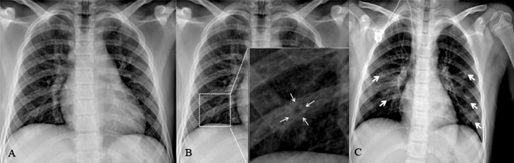
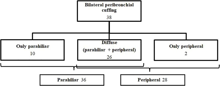
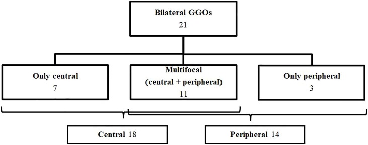
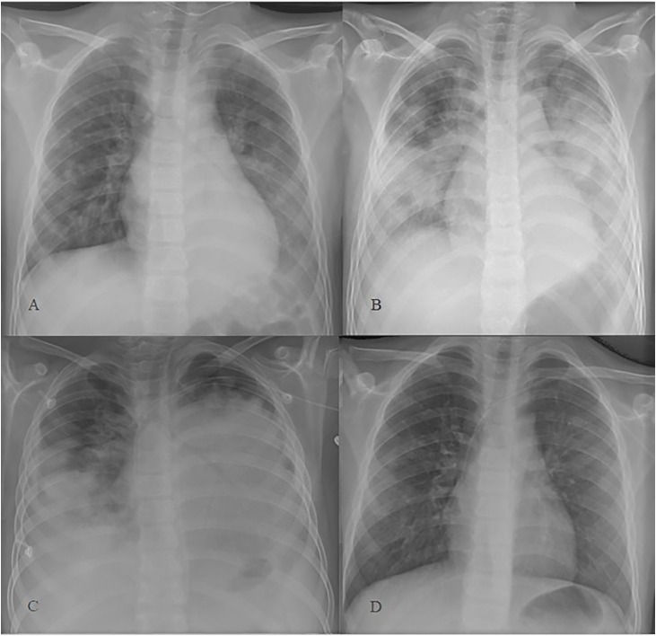
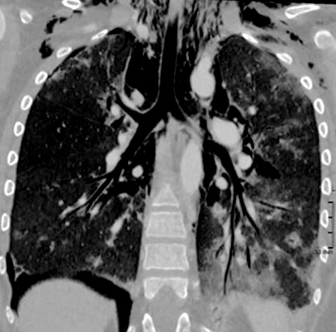
Results: Median age was 79.8 months (ranging from 2 weeks to 16 years of age). Fever was the most common symptom (43.5 %). 90 % of CXR showed abnormalities. Peribronchial cuffing was the most common finding (86.3 %) followed by GGOs (50 %). In both cases central distribution was more common than peripheral. Consolidations accounted for 18.1 %. Normal CXR, pleural effusion, and altered cardiomediastinal contour were the least common.
Conclusion: The vast majority of CXR showed abnormalities in children with COVID-19. However, findings are nonspecific. Interobserver correlation was good in describing consolidations, normal x-rays and GGOs. Imaging techniques have a role in the management of children with known or suspected COVID-19, especially in those with moderate or severe symptoms or with underlying risk factors.
Keywords: COVID 19; Outbreak; Paediatric; Paediatric imaging; Pneumonia; Radiology; SARS-CoV-2; Thoracic imaging.
Copyright © 2020 Elsevier B.V. All rights reserved.
Figures
References
-
- World Health Organization . 2020. Coronavirus Disease (COVID-19). Events As They Happen.https://www.who.int/emergencies/diseases/novel-coronavirus-2019/events-a...
-
- Na Zhu, Zhang Dingyu, Wang Wenling, Li Xingwang, Bo Yang, Song Jingdong, Zhao Xiang, Huang Baoying, Shi Weifeng, Roujian Lu, Niu Peihua, Zhan Faxian, et al. For the China novel coronavirus investigating and research team. A novel coronavirus from patients with pneumonia in China, 2019. N. Engl. J. Med. 2020;382(8):727–733. doi: 10.1056/NEJMoa2001017. - DOI - PMC - PubMed
-
- International Committee on Taxonomy Viruses . 2020. Naming the 2019 Coronavirus.https://talk.ictvonline.org/
-
- World Health Organization . 2020. Naming the Coronavirus Disease (COVID-19) and the Virus That Causes It.https://www.who.int/emergencies/diseases/novel-coronavirus-2019/technica...
-
- Kar SujitaKumar, Verma Nishant, Saxena ShailendraK. In: Coronavirus Disease 2019 (COVID-19). Epidemiology, Pathogenesis, Diagnosis and Therapeutics. 1st ed. Saxena ShailendraK., editor. Centre for Advanced Research King George’s Medical University; Lucknow, India: 2020.
MeSH terms
LinkOut - more resources
Full Text Sources
Miscellaneous
