Review
. 2021 Jan;22(1):86-96.
doi: 10.3348/kjr.2020.0144. Epub 2020 Sep 10.

Spectral Computed Tomography: Fundamental Principles and Recent Developments

Affiliations
Review

Spectral Computed Tomography: Fundamental Principles and Recent Developments

Aaron So et al. Korean J Radiol. 2021 Jan.

Abstract

CT is a diagnostic tool with many clinical applications. The CT voxel intensity is related to the magnitude of X-ray attenuation, which is not unique to a given material. Substances with different chemical compositions can be represented by similar voxel intensities, making the classification of different tissue types challenging. Compared to the conventional single-energy CT, spectral CT is an emerging technology offering superior material differentiation, which is achieved using the energy dependence of X-ray attenuation in any material. A specific form of spectral CT is dual-energy imaging, in which an additional X-ray attenuation measurement is obtained at a second X-ray energy. Dual-energy CT has been implemented in clinical settings with great success. This paper reviews the theoretical basis and practical implementation of spectral/dual-energy CT.

Keywords: Computed tomography; Dual energy; Spectral; X-rays.


Figures

Fig. 1
Fig. 1. CT scanner systems that are currently available for dual-energy/spectral imaging.
A. Dual-source. B. Single-source with ultrafast kV switching. C. Single-source without ultrafast kV switching. D. Single-source with dual-layer detector. E. Single-source with split-filter. F. Single-source with photon-counting detector. CT = computed tomography
Fig. 2
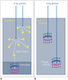
Fig. 2. (A) Scintillator-based energy-integrating detector versus (B) semiconductor-based photon counting detector.
Fig. 3
Fig. 3. Flow diagram for generation of material-density and virtual monochromatic images from a dual-energy scan.
Fig. 4
Fig. 4. Mass attenuation coefficients of iodine (solid blue curve) and water (solid orange curve) as a function of X-ray photon energy.
The mass attenuation coefficient of an unknown material (calcium in this example, represented by a dotted purple curve) over the X-ray energy range in diagnostic CT can be approximately represented as a linear combination of two basis materials, iodine and water, except at the K-edge energy of iodine. The amount of iodine and water required to represent the material of interest can be estimated at two different X-ray photon energies (marked by the yellow and green bars).
Fig. 5
Fig. 5. Comparison of the mass attenuation coefficients of iodine and gold and their respective K-edge energies.
The graph illustrates that multi-energy CT acquisition can be used to separate the attenuation signals between conventional iodine-based contrast agent and novel gold-based lipoprotein nanoparticle contrast agent. Preliminary results from preclinical studies suggest that the goldbased contrast agent may be useful for assessing the contents of an atherosclerotic plaque to determine if it is vulnerable to rupture (44).
Fig. 6
Fig. 6. Illustration of three-material decomposition with twodimensional mapping of the CT numbers (in HU) measured from a dual-energy scan.
In this example, any unknown material that falls within the triangle defined by the HU-pairs of the three selected basis materials can be represented as a linear combination of these basis materials. The volume fraction of each basis material can be determined from the corresponding vertices in the map.

References

    1. Flohr TG, McCollough CH, Bruder H, Petersilka M, Gruber K, Süss C, et al. First performance evaluation of a dual-source CT (DSCT) system. Eur Radiol. 2006;16:256–268. - PubMed
    1. Flohr TG, Bruder H, Stierstorfer K, Petersilka M, Schmidt B, McCollough CH. Image reconstruction and image quality evaluation for a dual source CT scanner. Med Phys. 2008;35:5882–5897. - PMC - PubMed
    1. Flohr TG. CT systems. Curr Radiol Rep. 2013;1:52–63.
    1. Kyriakou Y, Kalender WA. Intensity distribution and impact of scatter for dual-source CT. Phys Med Biol. 2007;52:6969–6989. - PubMed
    1. Petersilka M, Stierstorfer K, Bruder H, Flohr T. Strategies for scatter correction in dual source CT. Med Phys. 2010;37:5971–5992. - PubMed