. 2020 Sep 12;25(18):4191.
doi: 10.3390/molecules25184191.

Novel Acetylcholinesterase Inhibitors Based on Uracil Moiety for Possible Treatment of Alzheimer Disease

Affiliations

Novel Acetylcholinesterase Inhibitors Based on Uracil Moiety for Possible Treatment of Alzheimer Disease

Vyacheslav E Semenov et al. Molecules. .

Abstract

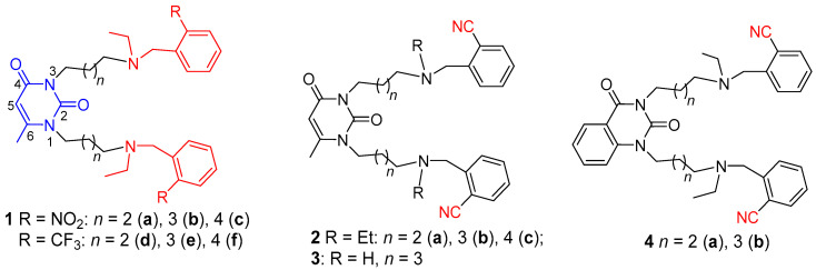
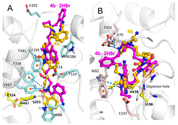
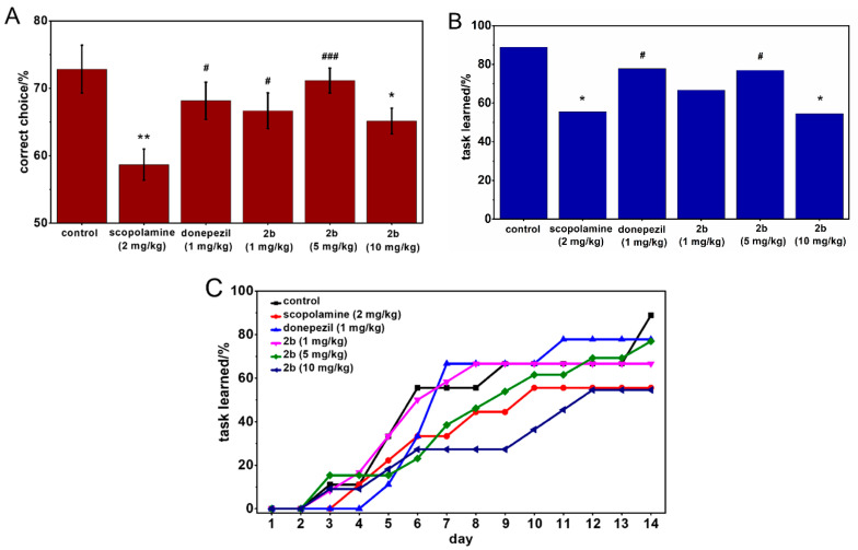
In this study, novel derivatives based on 6-methyluracil and condensed uracil were synthesized, namely, 2,4-quinazoline-2,4-dione with ω-(ortho-nitrilebenzylethylamino) alkyl chains at the N atoms of the pyrimidine ring. In this series of synthesized compounds, the polymethylene chains were varied from having tetra- to hexamethylene chains, and secondary NH, tertiary ethylamino, and quaternary ammonium groups were introduced into the chains. The molecular modeling of the compounds indicated that they could function as dual binding site acetylcholinesterase inhibitors, binding to both the peripheral anionic site and active site. The data from in vitro experiments show that the most active compounds exhibit affinity toward acetylcholinesterase within a nanomolar range, with selectivity for acetylcholinesterase over butyrylcholinesterase reaching four orders of magnitude. In vivo biological assays demonstrated the potency of these compounds in the treatment of memory impairment using an animal model of Alzheimer disease.

Keywords: 6-methyluracil; Alzheimer disease; acetylcholinesterase; inhibitors; peripheral anionic site.


Conflict of interest statement

The authors declare no conflict of interest regarding the publication of this paper.

Figures

Figure 1
Figure 1
Structures of previous reported acetylcholinesterase (AChE) inhibitors 1af with 6-methyluracil, nitro-, and trifluoromethyl moieties [25] and herein reported AChE inhibitors 2ac, 3, 4a,b with 6-methyluracil, quinazoline-2,4-dione and nitrile moieties. Numbering of pyrimidine ring atoms is shown.
Scheme 1
Scheme 1
Synthesis of title compounds with 6-methyluracil moietes and tertiary amino groups. Reagents and conditions: (a) Ethyl amine, K2CO3, 2-propanol, room temperature (RT); (b) o-nitrilebenzyl bromide, K2CO3, CH3CN.
Scheme 2
Scheme 2
Synthesis of title compounds with quinazoline-2,4-dione moieties and tertiary amino groups. Reagents and conditions: (a) Ethylamine, K2CO3, 2-propanol, RT; (b) o-nitrilebenzyl bromide, K2CO3, CH3CN.
Scheme 3
Scheme 3
Synthesis of title compounds with secondary amino groups. Reagents and conditions: (a) NaN3, dimethylformamide (DMF), 50–60 °C; (b) 1. triphenylphosphine, tetrahydrofuran; 2. HCl (H2O); (c) 1. CH3ONa, CH3OH, 2. 2-nitrilebenzaldehyde, CHCl3; (d) NaBH4, CH3OH.
Figure 2
Figure 2
Binding poses of 6-methyluracil 2b (carbon atoms are shown in green) and its charged counterpart 2b·2HBr (carbon atoms are shown in violet) inside active site gorges of (A) hAChE and (B) hBChE. Yellow, dashed lines represent hydrogen bonds, orange represents π–π stacking interactions, and blue lines indicate ionic and π–cation interactions. The catalytic triads of the enzymes are indicated using yellow carbon atoms.
Figure 3
Figure 3
Binding poses of quinazoline-2,4-dione 4b (carbon atoms are shown gold) and its charged counterpart 4b·2HBr (carbon atoms are shown in magenta) inside active site gorges of (A) hAChE and (B) hBChE. Yellow, dashed lines represent hydrogen bonds, orange represents π–π stacking interactions, and blue lines are ionic and π–cation interactions. The catalytic triads of the enzymes are shown with yellow carbon atoms.
Figure 4
Figure 4
Binding poses of quinazoline-2,4-dione 4b (carbon atoms are shown in gold) and its analog with shorter linker 4a (carbon atoms are shown in orange) inside the hAChE active site gorge. Yellow, dashed lines show hydrogen bonds, orange represents π–π stacking interactions, and blue lines indicate ionic and π–cation interactions.
Figure 5
Figure 5
Effect of compound 2b with 6-methyluracil moiety, pentamethylene chains, and tertiary amino groups on spatial memory performance in an Alzheimer disease (AD) scopolamine mouse model. Graphs represent percentage of correct choices in the T-maze test. Data are expressed as mean ± standard error. Statistical analysis was performed using the Mann–Whitney test (A), percentage of mice reaching the criterion of learning the task by day 14 in the T-maze test. Statistical analysis was performed using Fisher’s exact test (B) and dynamic of reaching the criterion of learning the task (C); * p < 0.05, ** p < 0.01 compared to control group; # p < 0.05, ### p < 0.001 compared to scopolamine-treated group.

References

    1. van Cauwenberghe C., van Broeckhoven C., Sleegers K. The genetic landscape of Alzheimer disease: Clinical implications and perspectives. Genet. Med. 2015;18:421–430. doi: 10.1038/gim.2015.117. - DOI - PMC - PubMed
    1. Moreira P., Santos M., Oliveira C., Shenk J., Nunomura A., Smith M., Zhu X., Perry G. Alzheimer disease and the role of free radicals in the pathogenesis of the disease. CNS Neurol. Disord. Drug Targets. 2008;7:3–10. - PubMed
    1. Finder V.H. Alzheimer’s Disease: A general introduction and pathomechanism. J. Alzheimer’s Dis. 2010;22:S5–S19. doi: 10.3233/JAD-2010-100975. - DOI - PubMed
    1. Goedert M., Spillantini M.G. A century of Alzheimer’s disease. Science. 2006;314:777–781. doi: 10.1126/science.1132814. - DOI - PubMed
    1. Davies P., Maloney A.J. Selective loss of central cholinergic neurons in Alzheimer’s disease. Lancet. 1976;2:1403. doi: 10.1016/S0140-6736(76)91936-X. - DOI - PubMed

MeSH terms