

Effects of the Delta Opioid Receptor Agonist DADLE in a Novel Hypoxia-Reoxygenation Model on Human and Rat-Engineered Heart Tissue: A Pilot Study
- PMID: 32932811
- PMCID: PMC7565486
- DOI: 10.3390/biom10091309
Effects of the Delta Opioid Receptor Agonist DADLE in a Novel Hypoxia-Reoxygenation Model on Human and Rat-Engineered Heart Tissue: A Pilot Study
Abstract
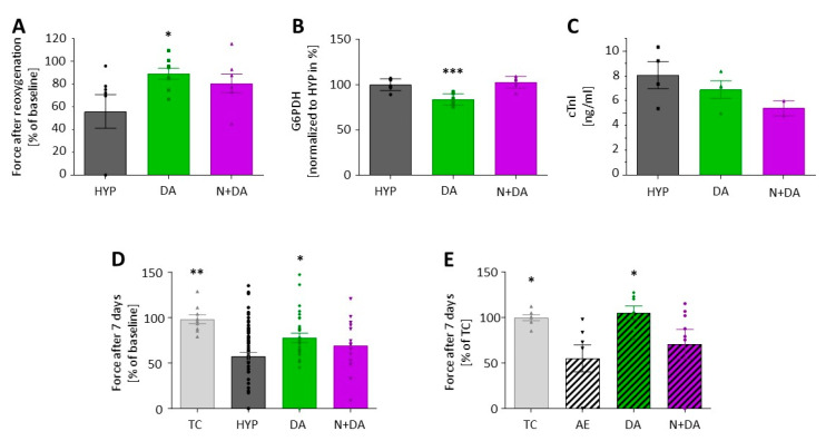
Intermittent hypoxia and various pharmacological compounds protect the heart from ischemia reperfusion injury in experimental approaches, but the translation into clinical trials has largely failed. One reason may lie in species differences and the lack of suitable human in vitro models to test for ischemia/reperfusion. We aimed to develop a novel hypoxia-reoxygenation model based on three-dimensional, spontaneously beating and work performing engineered heart tissue (EHT) from rat and human cardiomyocytes. Contractile force, the most important cardiac performance parameter, served as an integrated outcome measure. EHTs from neonatal rat cardiomyocytes were subjected to 90 min of hypoxia which led to cardiomyocyte apoptosis as revealed by caspase 3-staining, increased troponin I release (time control vs. 24 h after hypoxia: cTnI 2.7 vs. 6.3 ng/mL, ** p = 0.002) and decreased contractile force (64 ± 6% of baseline) in the long-term follow-up. The detrimental effects were attenuated by preceding the long-term hypoxia with three cycles of 10 min hypoxia (i.e., hypoxic preconditioning). Similarly, [d-Ala2, d-Leu5]-enkephalin (DADLE) reduced the effect of hypoxia on force (recovery to 78 ± 5% of baseline with DADLE preconditioning vs. 57 ± 5% without, p = 0.012), apoptosis and cardiomyocyte stress. Human EHTs presented a comparable hypoxia-induced reduction in force (55 ± 5% of baseline), but DADLE failed to precondition them, likely due to the absence of δ-opioid receptors. In summary, this hypoxia-reoxygenation in vitro model displays cellular damage and the decline of contractile function after hypoxia allows the investigation of preconditioning strategies and will therefore help us to understand the discrepancy between successful conditioning in vitro experiments and its failure in clinical trials.
Keywords: 3D tissue model; cardiac hypertrophy; cardioprotection; human induced pluripotent stem cells; opioids; preconditioning; reperfusion injury; tissue engineering; translational medicine.
Conflict of interest statement
T.E., A.H., and M.N.H. are co-founders of EHT Technologies GmbH.
Figures


Similar articles
-
Myocardial protection by ischemic preconditioning and delta-opioid receptor activation in the isolated working rat heart.J Thorac Cardiovasc Surg. 2001 Nov;122(5):986-92. doi: 10.1067/mtc.2001.116950. J Thorac Cardiovasc Surg. 2001. PMID: 11689805
-
HL-1 myocytes exhibit PKC and K(ATP) channel-dependent delta opioid preconditioning.J Surg Res. 2003 Oct;114(2):187-94. doi: 10.1016/s0022-4804(03)00248-8. J Surg Res. 2003. PMID: 14559445
-
Genomic analysis of [d-Ala2, d-Leu5] enkephalin preconditioning in cortical neuron and glial cell injury after oxygen deprivation.Brain Res. 2012 Apr 4;1447:91-105. doi: 10.1016/j.brainres.2012.01.049. Epub 2012 Jan 28. Brain Res. 2012. PMID: 22356887
-
[ROLE OF OPIOID RECEPTORS IN THE REGULATION OF RESISTANCE OF HEART TO IMPACT OF ISCHEMIA-REPERFUSION].Ross Fiziol Zh Im I M Sechenova. 2017 Mar;103(3):230-49. Ross Fiziol Zh Im I M Sechenova. 2017. PMID: 30199204 Review. Russian.
-
Multifaceted Effects of Delta Opioid Receptors and DADLE in Diseases of the Nervous System.Curr Drug Discov Technol. 2018;15(2):94-108. doi: 10.2174/1570163814666171010114403. Curr Drug Discov Technol. 2018. PMID: 29032758 Review.
Cited by
-
Myocardial infarction from a tissue engineering and regenerative medicine point of view: A comprehensive review on models and treatments.Biophys Rev (Melville). 2022 Sep;3(3):031305. doi: 10.1063/5.0093399. Epub 2022 Aug 30. Biophys Rev (Melville). 2022. PMID: 36091931 Free PMC article. Review.
-
[D-Ala2, D-Leu5]-enkephalin (DADLE) provides protection against myocardial ischemia reperfusion injury by inhibiting Wnt/β-Catenin pathway.BMC Cardiovasc Disord. 2024 Feb 19;24(1):115. doi: 10.1186/s12872-024-03790-6. BMC Cardiovasc Disord. 2024. PMID: 38373914 Free PMC article.
-
Effects of delta-opioid receptor agonist pretreatment on the cardiotoxicity of bupivacaine in rats.BMC Anesthesiol. 2022 Jan 12;22(1):19. doi: 10.1186/s12871-022-01568-x. BMC Anesthesiol. 2022. PMID: 35021986 Free PMC article.
-
Mitophagy is induced in human engineered heart tissue after simulated ischemia and reperfusion.J Cell Sci. 2025 May 1;138(9):jcs263408. doi: 10.1242/jcs.263408. Epub 2025 Mar 19. J Cell Sci. 2025. PMID: 39912384 Free PMC article.
-
Cardioprotection by poloxamer 188 is mediated through increased endothelial nitric oxide production.Sci Rep. 2025 Apr 30;15(1):15170. doi: 10.1038/s41598-025-97079-z. Sci Rep. 2025. PMID: 40307302 Free PMC article.
References
-
- Zhao Z.Q., Corvera J.S., Halkos M.E., Kerendi F., Wang N.P., Guyton R.A., Vinten-Johansen J. Inhibition of myocardial injury by ischemic postconditioning during reperfusion: Comparison with ischemic preconditioning. Am. J. Physiol Heart Circ. Physiol. 2003;285:H579–H588. doi: 10.1152/ajpheart.01064.2002. - DOI - PubMed
Publication types
MeSH terms
Substances
Grants and funding
LinkOut - more resources
Full Text Sources
Research Materials
