. 2020 Sep 11;9(9):2076.
doi: 10.3390/cells9092076.

Retinoic Acid Improves the Recovery of Replication-Competent Virus from Latent SIV Infected Cells

Affiliations

Retinoic Acid Improves the Recovery of Replication-Competent Virus from Latent SIV Infected Cells

Omalla A Olwenyi et al. Cells. .

Abstract

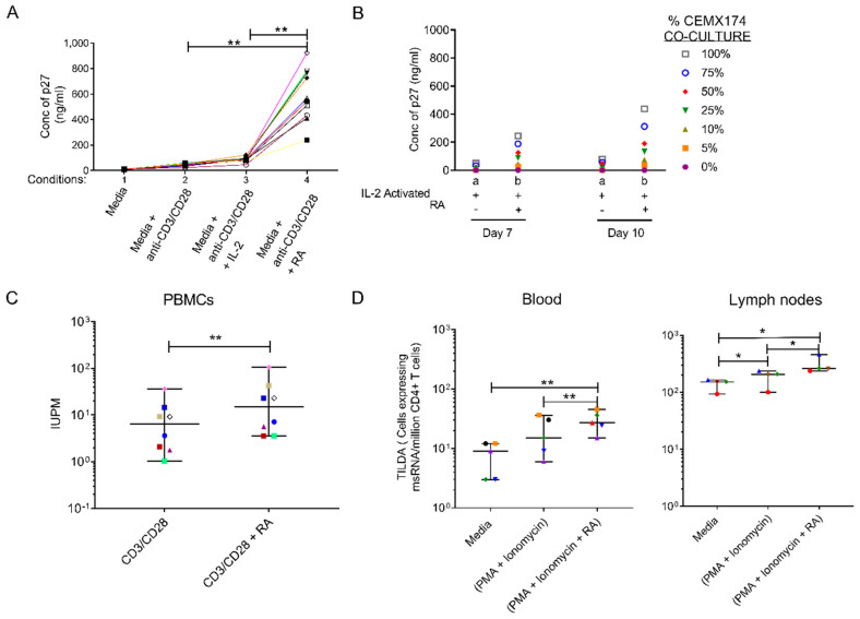
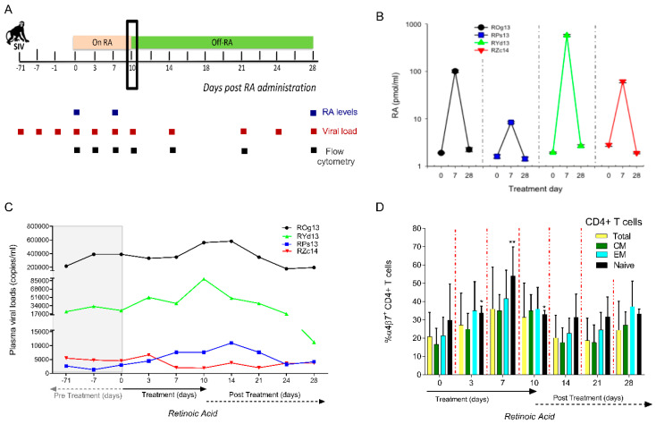
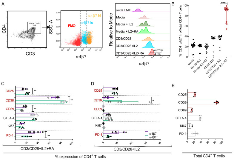
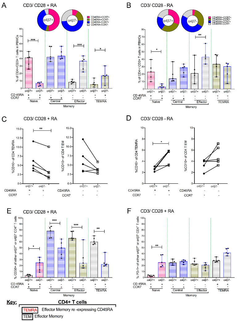
The accurate estimation and eradication of Human Immunodeficiency Virus (HIV) viral reservoirs is limited by the incomplete reactivation of cells harboring the latent replication-competent virus. We investigated whether the in vitro and in vivo addition of retinoic acid (RA) enhances virus replication and improves the detection of latent virus. Peripheral blood mononuclear cells (PBMCs) from naive and anti-retroviral therapy (ART)-treated SIV-infected rhesus macaques (RMs) were cultured in vitro with anti-CD3/CD28 + IL-2 in the presence/absence of RA. Viral RNA and p27 levels were quantified using RT-qPCR and ELISA, respectively. Viral reservoirs were estimated using the Tat/Rev-Induced Limited Dilution Assay (TILDA) and Quantitative Viral Outgrowth Assay (QVOA). In vitro and in vivo measures revealed that there was also an increase in viral replication in RA-treated versus without RA conditions. In parallel, the addition of RA to either CD3/CD28 or phorbol myristate acetate (PMA)/ionomycin during QVOA and TILDA, respectively, was shown to augment reactivation of the replication-competent viral reservoir in anti-retroviral therapy (ART)-suppressed RMs as shown by a greater than 2.3-fold increase for QVOA and 1 to 2-fold increments for multi-spliced RNA per million CD4+ T cells. The use of RA can be a useful approach to enhance the efficiency of current protocols used for in vitro and potentially in vivo estimates of CD4+ T cell latent reservoirs. In addition, flow cytometry analysis revealed that RA improved estimates of various viral reservoir assays by eliciting broad CD4 T-cell activation as demonstrated by elevated CD25 and CD38 but reduced CD69 and PD-1 expressing cells.

Keywords: CD3/CD28 treatment; CD4 T cells; PBMCs; SIV; immune activation; retinoic acid; viral load and provirus.


Conflict of interest statement

The authors declare no conflict of interest.

Figures

Figure 1
Figure 1
Retinoic Acid (RA) enhances viral replication in vitro and improves detection of the viral reservoir. (A) P27 levels were measured in supernatant of 10 randomly selected naive Peripheral blood mononuclear cells (PBMCs) infected with SIVmac239 after in vitro culturing with (1) media, unstimulated (2) anti-CD3/CD28 beads to activate the T-cell receptor (3) anti-CD3/CD28 + IL-2 and 4) anti-CD3/CD28 beads + IL-2 together with RA added under separate conditions on day 10. (B) Levels of p27 detected in supernatant fluids from pooled (n = 10 RMs) enriched CD4 T cells that were treated with either (a) anti-CD3/CD28 and IL-2 only or (b) anti-CD3/CD28 + IL-2 and RA. Thereafter, viral expansion was carried out by the co-culture with CEMX174 cells across different concentrations (0 to 100%) on days 7 and 10 respectively. (C) Quantitative Viral Outgrowth Assay (QVOA) of CD4+ T cells purified from PBMCs of anti-retroviral therapy (ART)-suppressed macaques indicating levels of Infectious Units per Million (IUPM) in CD3/CD28 versus CD3/CD28 + RA conditions (n = 8). (D) levels of msRNA transcripts obtained from enriched CD4 T cells collected from PBMC (n = 5 RMs) or auxiliary lymph node CD4+ T cells (n = 4 RMs) that were cultured in media only, media plus phorbol myristate acetate (PMA) + ionomycin and media plus PMA + ionomycin supplemented with retinoic acid. * shows p < 0.05 and ** represents p < 0.001 significant difference across studied groups obtained using Wilcoxon matched pairs signed rank tests.
Figure 2
Figure 2
Administration of RA affects SIV replication kinetics by causing modest increases in peripheral SIV replication and the expansion of naive CD4+ T cells (n = 4 RMs). (A) Pictorial schema indicating that RMs were provided with a daily regimen of 10 mg/kg of all trans RA orally for 10 days and then followed for 28 days. (B) Levels of RA in plasma were measured at baseline, on day 7 of RA treatment and day 28 (18 days post RA treatment). (C) Plasma viral loads measurements were carried out at prospective timepoints prior to and following the administration and later withdrawal of RA. (D) Prospective box and whisker plots showing differences in total CD4+ T cells and CD4+ T cell subsets. CD4+ T cell phenotypes were grouped based on their CD28 and CD95 expression. Naive CD4+ T cells were categorized as CD28+/CD95, central memory CD4+ T cells as CD28+/CD95+ and effector memory CD4+ T cells as CD28/CD95+.
Figure 3
Figure 3
The addition of RA upregulates α4β7 hi expression on CD4+ T cells cultured under different conditions resulting in distinct immune activation profiles between α4β7+ and α4β7 CD4+ T cells. Flow cytometry gating strategy showing: (A) CD4+ T cells derived from CD3+/lymphocyte/live and single cells and later profiled for the extent of α4β7 expression. Following this is an overlay dot plot showing the spread of α4β7 (Fluorescence minus one (FMO)—red, α4β7 lo—blue and α4β7 hi—orange) within the CD4+ population. Then, adjacent to this is a representative overlay histogram generated after flow cytometry analysis indicating levels of α4β7hi expression on CD4 T cells normalized to mode following the culturing of PBMCs under the different conditions: media only, media + IL-2, media + IL-2 + RA, anti-CD3/CD28, anti-CD3/CD28 + IL-2, anti-CD3/CD28 + IL-2 + RA and α4β7 FMO as a control. (B) Aligned dot plot showing the frequency of CD4 + α4β7hi T cells in total CD4+ T cells obtained from 16 naive RMs and cultured under different conditions named in (A). (C) Percent expression of differences in IA markers CD38, CD69, CTLA 4, Ki67 and PD-1 expressed on α4β7+ vs. α4β7 CD4+ T cells after cell culture in media comprising of anti CD3/CD28 beads, IL-2 and RA (n = 5 rhesus macaque PBMCs) and (D) α4β7+ vs. α4β7 CD4+ T cells following cell culture media containing anti CD3/CD28 beads and IL-2 (n = 5 rhesus macaque PBMCs). (E) Levels of percent expression of several immune activation (IA) markers (Ki67, CD25, CD38, CD69 and PD-1) on total CD4+ T cells. * shows p < 0.05, ** indicates p < 0.001 while *** denotes p < 0.0001 resulting from Wilcoxon matched pairs signed rank tests between paired groups of the different comparisons.
Figure 4
Figure 4
Addition of 1uM RA to PBMCs that were previously activated with anti-CD3/CD28 beads and cultured in IL-2 enriched media leads to different memory/differentiation profiles in α4β7+ vs. α4β7 CD4+ T cells (n = 5). Combined bar graph and pie chart showing the comparative distribution of naive, central memory, effector memory (EM) and terminal effector memory cells re-expressing CD45RA (TEMRA) subsets within α4β7+ vs. α4β7 CD4+ T cells obtained from PBMC treated with (A) CD3/CD28 beads + RA or (B) CD3/CD28 beads-RA. (C) Dot plots indicating paired comparisons for the quantification of CD10+ levels in α4β7+ vs. α4β7 partitions of CD4+ TEMRA cells and CD4+ TEM cells in CD3/CD28 + RA or (D) CD3/CD28 − RA treatment conditions. (E) The distribution of CD38 and (F) PD-1 markers of immune activation amongst CD4+ T cell subsets obtained from PBMC treated with CD3/CD28 beads + RA conditions. * shows p < 0.05, ** indicates p < 0.001 while *** denotes p < 0.0001 obtained using Wilcoxon matched pairs signed rank tests.

Similar articles

Cited by

References

    1. Cohen C. Second ‘Cured’ HIV Patient Goes Public. [(accessed on 25 June 2020)]; Available online: https://www.sciencemag.org/news/2020/03/second-cured-hiv-patient-goes-pu....
    1. Gupta R.K., Abdul-Jawad S., McCoy L.E., Mok H.P., Peppa D., Salgado M., Martinez-Picado J., Nijhuis M., Wensing A.M.J., Lee H., et al. HIV-1 remission following CCR5Δ32/Δ32 haematopoietic stem-cell transplantation. Nature. 2019;568:244–248. doi: 10.1038/s41586-019-1027-4. - DOI - PMC - PubMed
    1. Henrich T.J., Hanhauser E., Marty F.M., Sirignano M.N., Keating S., Lee T.H., Robles Y.P., Davis B.T., Li J.Z., Heisey A., et al. Antiretroviral-free HIV-1 remission and viral rebound after allogeneic stem cell transplantation: Report of 2 cases. Ann. Intern. Med. 2014;161:319–327. doi: 10.7326/M14-1027. - DOI - PMC - PubMed
    1. Mavigner M., Watkins B., Lawson B., Lee S.T., Chahroudi A., Kean L., Silvestri G. Persistence of virus reservoirs in ART-treated SHIV-infected rhesus macaques after autologous hematopoietic stem cell transplant. PLoS Pathog. 2014;10:e1004406. doi: 10.1371/journal.ppat.1004406. - DOI - PMC - PubMed
    1. Bruner K.M., Hosmane N.N., Siliciano R.F. Towards an HIV-1 cure: Measuring the latent reservoir. Trends Microbiol. 2015;23:192–203. doi: 10.1016/j.tim.2015.01.013. - DOI - PMC - PubMed

Publication types