. 2020 Sep 11;20(18):5183.
doi: 10.3390/s20185183.

Fabrication of Parylene-Coated Microneedle Array Electrode for Wearable ECG Device

Affiliations

Fabrication of Parylene-Coated Microneedle Array Electrode for Wearable ECG Device

Afraiz Tariq Satti et al. Sensors (Basel). .

Abstract

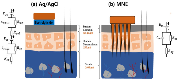
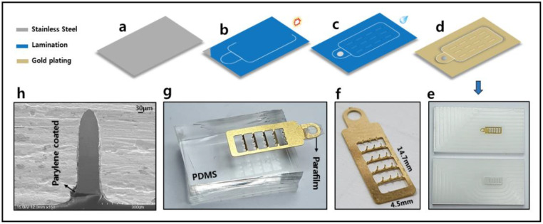
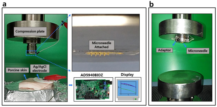
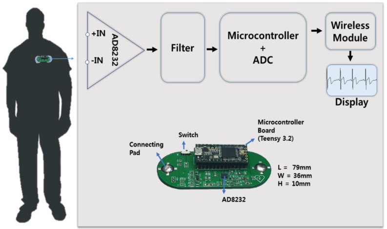
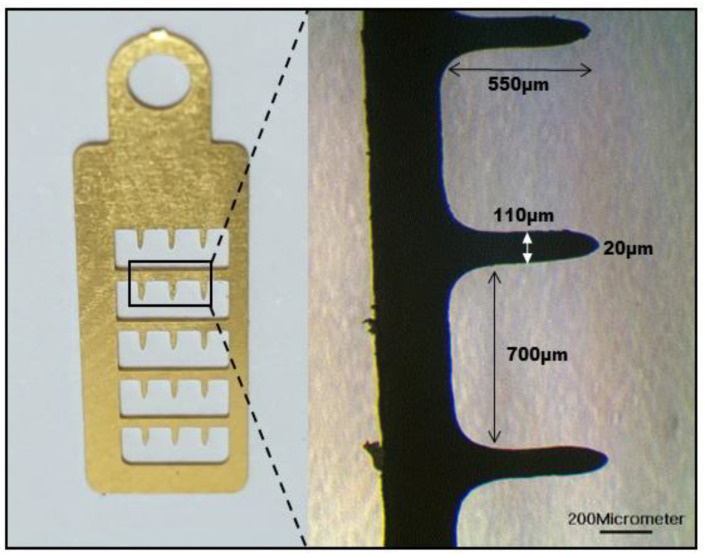
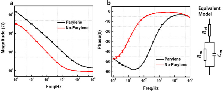
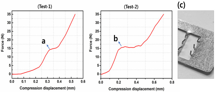
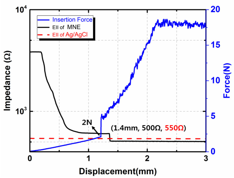
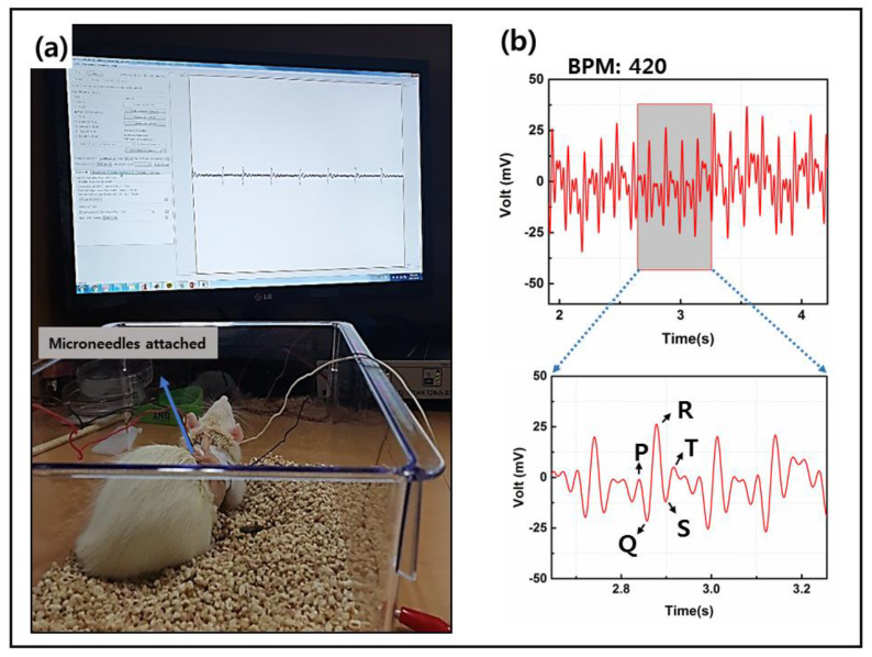
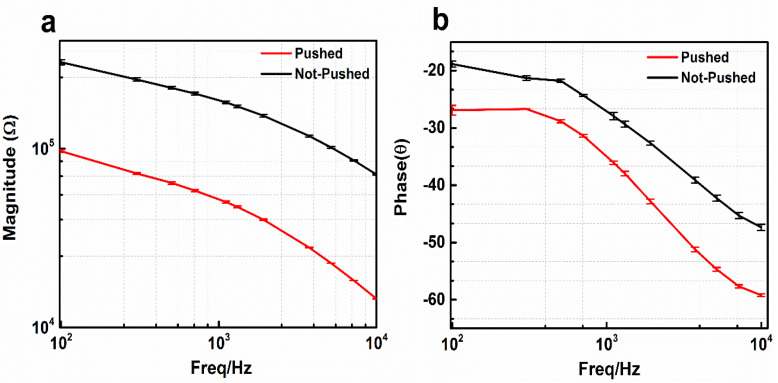
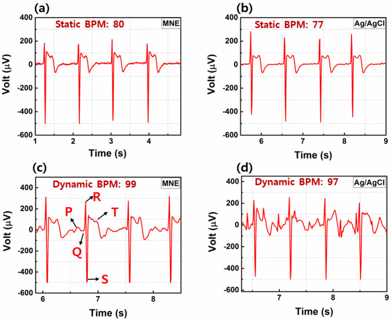
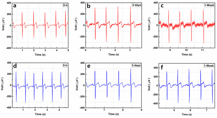
Microneedle array electrodes (MNE) showed immense potential for the sensitive monitoring of the bioelectric signals by penetrating the stratum corneum with high electrical impedance. In this paper, we introduce a rigid parylene coated microneedle electrode array and portable electrocardiography (ECG) circuit for monitoring of ECG reducing the motion artifacts. The developed MNE showed stability and durability for dynamic and long-term ECG monitoring in comparison to the typical silver-silver chloride (Ag/AgCl) wet electrodes. The microneedles showed no mechanical failure under the compression force up-to 16 N, but successful penetration of skin tissue with a low insertion force of 5 N. The electrical characteristics of the fabricated MNE were characterized by impedance spectroscopy with equivalent circuit model. The designed wearable wireless ECG monitoring device with MNE proved feasibility of the ECG recording which reduces the noise of movement artifacts during dynamic behaviors.

Keywords: ECG; cardiovascular diseases; impedance; microneedle electrode; parylene.


Conflict of interest statement

The authors declare no conflict of interest.

Figures

Figure 1
Figure 1
Schematic of the interface between (a) the skin and typical Ag/AgCl electrode, (b) the skin and microneedle array electrode (MNE).
Figure 2
Figure 2
Fabrication process of MNE; (a) medical-grade 316L stainless steel; (b) lamination of light sensitive photo resist and exposure to UV light; (c) developing for the positive image print; (d) gold electroplated; (e) jig to move microneedles at 90 degrees; (f) MNE with standing microneedles; (g) microneedles inserted into polydimethylsiloxane (PDMS) and connecting pad covered with Parafilm M® for parylene coating the surface area of MNE; (h) SEM image of single needle showing the parylene coating.
Figure 3
Figure 3
(a) Electrode–skin interface impedance (EII) recording setup during the insertion test. (b) Fracture test setup.
Figure 4
Figure 4
Developed wireless electrocardiography (ECG) monitoring system.
Figure 5
Figure 5
MNE dimensions taken by microscope at 200-micrometer scale.
Figure 6
Figure 6
Parylene-coating test with 0.9% NaCl. (a) Magnitude. (b) Phase. The data represents the average of three independent measurements with error shown in SD (n = 3).
Figure 7
Figure 7
Fracture test of the MNE. (a,b) Resistance force before mechanical failure of the microneedles. (c) Microneedles after the fracture test.
Figure 8
Figure 8
Insertion force and EII recorded during MNE insertion into the porcine cadaver skin.
Figure 9
Figure 9
(a) Rat with electrodes fixed on the chest. (b) ECG of the rat.
Figure 10
Figure 10
Electrical impedance spectra. (a) Impedance magnitude and (b) phase value before and after penetration of MNE inside skin. The data represents the average of three independent measurements with error shown in SD (n = 3).
Figure 11
Figure 11
ECG using MNE compared with Ag/AgCl electrode for (a,b) static or (c,d) dynamic behaviors.
Figure 12
Figure 12
Long-term use of electrode for ECG recording with (a) Ag/AgCl at 0 h, (b) Ag/AgCl after 3 days, (c) Ag/AgCl after 1 week, (d) MNE at 0 h, (e) MNE after 3 days, (f) MNE after 1 week.

References

    1. Virani S.S., Alonso A., Benjamin E.J., Bittencourt M.S., Callaway C.W., Carson A.P., Chamberlain A.M., Chang A.R., Cheng S., Delling F.N., et al. Heart disease and stroke statistics—2020 update: A report from the American Heart Association. Circulation. 2020;141:E139–E596. doi: 10.1161/CIR.0000000000000757. - DOI - PubMed
    1. Paramasivam A., Priyadharsini J.V., Raghunandhakumar S., Elumalai P. A novel COVID-19 and its effects on cardiovascular disease. Hypertens. Res. 2020;43:729–730. doi: 10.1038/s41440-020-0461-x. - DOI - PMC - PubMed
    1. Ganatra S., Hammond S.P., Nohria A. The Novel Coronavirus Disease (COVID-19) threat for patients with cardiovascular disease and cancer. JACC CardioOncol. 2020;2:350–355. doi: 10.1016/j.jaccao.2020.03.001. - DOI - PMC - PubMed
    1. Searle A., Kirkup L. A direct comparison of wet, dry and insulating bioelectric recording electrodes. Physiol. Meas. 2000;21:271–283. doi: 10.1088/0967-3334/21/2/307. - DOI - PubMed
    1. O’Mahony C., Pini F., Blake A., Webster C., O’Brien J., McCarthy K.G. Microneedle-based electrodes with integrated through-silicon via for biopotential recording. Sens. Actuators A Phys. 2012;186:130–136. doi: 10.1016/j.sna.2012.04.037. - DOI

LinkOut - more resources