. 2020 Sep 11;12(9):2777.
doi: 10.3390/nu12092777.

Attenuating Effects of Pyrogallol-Phloroglucinol-6,6-Bieckol on Vascular Smooth Muscle Cell Phenotype Changes to Osteoblastic Cells and Vascular Calcification Induced by High Fat Diet

Affiliations

Attenuating Effects of Pyrogallol-Phloroglucinol-6,6-Bieckol on Vascular Smooth Muscle Cell Phenotype Changes to Osteoblastic Cells and Vascular Calcification Induced by High Fat Diet

Myeongjoo Son et al. Nutrients. .

Abstract

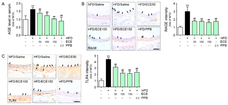
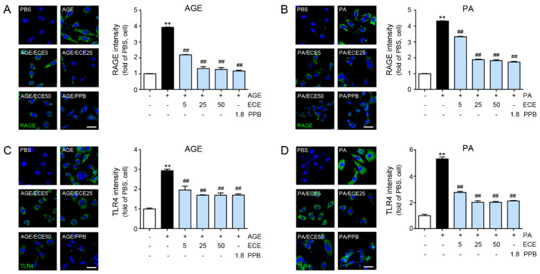
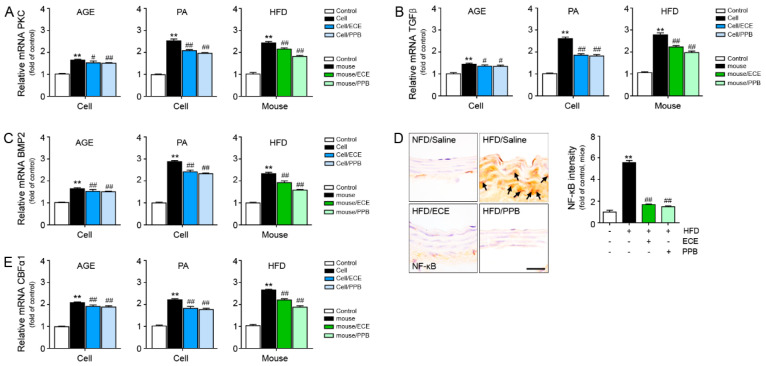
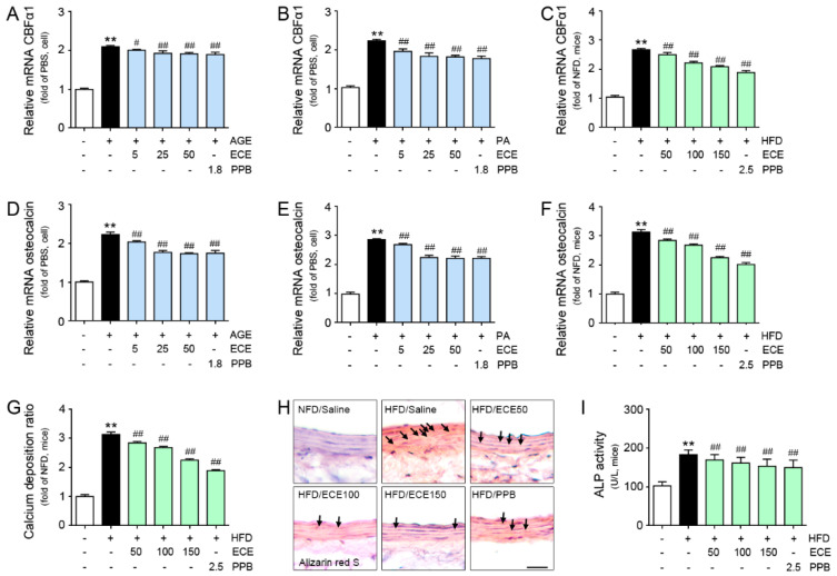
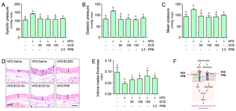
Advanced glycation end products/receptor for AGEs (AGEs/RAGEs) or Toll like receptor 4 (TLR4) induce vascular smooth muscle cell (VSMC) phenotype changes in osteoblast-like cells and vascular calcification. We analyzed the effect of Ecklonia cava extract (ECE) or pyrogallol-phloroglucinol-6,6-bieckol (PPB) on VSMC phenotype changes and vascular calcification prompted by a high-fat diet (HFD). HFD unregulated RAGE, TLR4, transforming growth factor beta (TGFβ), bone morphogenetic protein 2 (BMP2), protein kinase C (PKC), and nuclear factor kappa-light-chain-enhancer of activated B cells (NF-κB) signals in the aorta of mice. ECE and PPB restored the increase of those signal pathways. AGE- or palmitate-treated VSMC indicated similar changes with the animal. HFD increased osteoblast-like VSMC, which was evaluated by measuring core-binding factor alpha-1 (CBFα-1) and osteocalcin expression and alkaline phosphatase (ALP) activity in the aorta. ECE and PPB reduced vascular calcification, which was analyzed by the calcium deposition ratio, and Alizarin red S stain was increased by HFD. PPB and ECE reduced systolic, diastolic, and mean blood pressure, which increased by HFD. PPB and ECE reduced the phenotype changes of VSMC to osteoblast-like cells and vascular calcification and therefore lowered the blood pressure.

Keywords: Ecklonia cava extract; Toll-like receptor 4; pyrogallol-phloroglucinol-6,6-bieckol; receptor of AGEs; vascular calcification.


Conflict of interest statement

The authors declare no conflict of interest.

Figures

Figure 1
Figure 1
Comparative analysis of concentration-dependent ECE and PPB administration on the reduction of AGE and its receptor expression level in HFD-fed mice. (A) AGE levels were calculated in HFD-fed mice with or without ECE/PPB administration. (B) Light microscopic images showing RAGE expression (brown, arrow) in the aorta of HFD-fed mice. Quantitative graphs showing RAGE intensity from representative images. Scale bar = 50 μm. (C) Light microscopic images showing TLR4 expression (brown, arrow) in the aorta of HFD-fed mice. Quantitative graphs showing TLR4 intensity from representative images. Scale bar = 50 μm. **, p < 0.01 vs. NFD/Saline group; ##, p < 0.01 vs. HFD/Saline group. AGE, advanced glycation end products; ECE, Ecklonia cava extract; HFD, high-fat diet; NFD, normal fat diet; PPB, pyrogallol-phloroglucinol-6,6-bieckol; RAGE, receptor for advanced glycation end products; TLR4, toll-like receptor 4.
Figure 2
Figure 2
Comparative analysis of concentration-dependent ECE and PPB treatment on the reduction of RAGE and TLR4 receptor expression level in AGE or PA-treated VSMC. (A,B) Confocal fluorescence microscopic images demonstrating RAGE expression (green) and nuclei (DAPI, blue) in AGE or PA-treated VSMC. Quantitative graphs demonstrating RAGE intensity from representative images. Scale bar = 50 μm. (C,D) Confocal fluorescence microscopic images demonstrating TLR4 expression (green) and nuclei (DAPI, blue) in AGE or PA-treated VSMC. Quantitative graphs demonstrating TLR4 intensity from representative images. Scale bar = 50 μm; **, p < 0.01 vs. PBS group; ##, p < 0.01 vs. AGE or PA group. AGE, advanced glycation end products; DAPI, 4′,6-diamidino-2-phenylindole; ECE, Ecklonia cava extract; PA, palmitate; PBS, phosphate-buffered saline; PPB, pyrogallol-phloroglucinol-6,6-bieckol; RAGE, receptor for advanced glycation end products; TLR4, toll-like receptor 4; VSMC, vascular smooth muscle cell.
Figure 3
Figure 3
Comparative analysis of ECE and PPB treatment on the reduction of vascular smooth muscle cell phenotypic switching to osteoblast-like cell in AGE or palmitate-treated VSMC and HFD-fed mice. mRNA levels of the phenotypic switching to osteoblast-like cell-related molecules (A) PKC, (B) TGFβ, (C) BMP2, and (E) CBFα1 were determined using qRT-PCR. (D) Arrows indicate NF-κB protein expression of HFD-fed mice and quantified graph showing intensity. We incubated 25 μg/mL ECE or 1.8 μg/mL PPB with AGE or PA-treated VSMC (cell) and 100 mg/kg ECE or 2.5 mg/kg PPB administered with HFD-fed mice. scale bar = 25 μm. **, p < 0.01 vs. PBS or NFD/Saline group; #, p < 0.05, ##, p < 0.01 vs. AGE, PA or HFD/Saline group. AGE, advanced glycation end products; BMP2, Bone morphogenetic protein 2; CBFα1, core-binding factor alpha 1; ECE, Ecklonia cava extract; HFD, high-fat diet; NFD, normal fat diet; NF-κB, nuclear factor kappa-light-chain-enhancer of activated B cells; PA, palmitate; PBS, phosphate-buffered saline; PKC, protein kinase C; PPB, pyrogallol-phloroglucinol-6,6-bieckol; qRT-PCR, real-time quantitative reverse transcription polymerase chain reaction; RAGE, receptor for advanced glycation end products; TGFβ, transforming growth factor beta; VSMC, vascular smooth muscle cell.
Figure 4
Figure 4
Comparative analysis of ECE and PPB treatment on the reduction of osteoblast-like cell marker expression in AGE or palmitate-treated VSMC and HFD-fed mice. (AC) mRNA levels of the osteoblast-like markers CBFα1 in AGE- or PA-treated VSMC (cell) and HFD-fed mice were determined by qRT-PCR. (DF) mRNA levels of the osteoblast-like markers osteocalcin in AGE- or PA-treated VSMC (cell) and HFD-fed mice were determined by qRT-PCR. (G) Calcium deposition was calculated using the calcium deposition assay and (H) Alizarin red S staining in HFD-fed mice. Positive signal (red dot, arrow) was marked with an arrow in images. Scale bar = 50 μm. (I) ALP activity was measured with the use of the aorta of HFD-fed mice. **, p < 0.01 vs. PBS or NFD/Saline group; #, p < 0.05, ##, p < 0.01 vs. AGE, PA or HFD/Saline group. AGE, advanced glycation end products; ALP, alkaline phosphatase; CBFα1, core-binding factor alpha 1; ECE, Ecklonia cava extract; HFD, high-fat diet; NFD, normal fat diet; PA, palmitate; PBS, phosphate-buffered saline; PPB, pyrogallol-phloroglucinol-6,6-bieckol; qRT-PCR, real-time quantitative reverse transcription polymerase chain reaction; VSMC, vascular smooth muscle cell.
Figure 5
Figure 5
Comparative analysis of ECE and PPB administration on the regulation of blood pressure in HFD-fed mice. (AC) Systolic, diastolic, and mean artery pressures were measured prior to sacrifice. (D) Light microscopic images showing H&E stained blood vessels and (E) Intima-media thickness acquired using representative H&E images. Arrows indicate media thickness. Scale bar = 50 μm. (F) Summary illustration image showing inhibitory effects of ECE and PPB on vascular smooth muscle cell calcification in HFD condition. Means denoted by a different letter indicate significant differences between groups. AGE, advanced glycation end products; BMP2, bone morphogenetic protein 2; CBFα1, core-binding factor alpha 1; ECE, Ecklonia cava extract; HFD, high-fat diet; H&E, hematoxylin and eosin stain; NFD, normal fat diet; PPB, pyrogallol-phloroglucinol-6,6-bieckol; NF-κB, nuclear factor kappa-light-chain-enhancer of activated B cells; RAGE, receptor for advanced glycation end products; TGFβ, transforming growth factor beta; TLR4, toll like receptor 4.

Similar articles

Cited by

References

    1. Bostrom K.I., Rajamannan N.M., Towler D.A. The regulation of valvular and vascular sclerosis by osteogenic morphogens. Cir. Res. 2011;109:564–577. doi: 10.1161/CIRCRESAHA.110.234278. - DOI - PMC - PubMed
    1. Wong N.D., Vo A., Abrahamson D., Tobis J.M., Eisenberg H., Detrano R.C. Detection of coronary artery calcium by ultrafast computed tomography and its relation to clinical evidence of coronary artery disease. Am. J. Cardiol. 1994;73:223–227. doi: 10.1016/0002-9149(94)90223-2. - DOI - PubMed
    1. Goodman W.G., Goldin J., Kuizon B.D., Yoon C., Gales B., Sider D., Wang Y., Chung J., Emerick A., Greaser L., et al. Coronaryartery calcification in young adults with end-stage renal disease who are undergoing dialysis. N. Eng. J. Med. 2000;342:1478–1483. doi: 10.1056/NEJM200005183422003. - DOI - PubMed
    1. Fadini G.P., Albiero M., Menegazzo L., Boscaro E., de Kreutzenberg S.V., Agostini C., Cabrelle A., Binotto G., Rattazzi M., Bertacco E., et al. Widespread increase in myeloid calcifying cells contributes to ectopic vascular calcification in type 2 diabetes. Cir. Res. 2011;108:1112–1121. doi: 10.1161/CIRCRESAHA.110.234088. - DOI - PubMed
    1. Chellan B., Sutton N.R., Bowman M.A.H. S100/RAGE-Mediated Inflammation and Modified Cholesterol Lipoproteins as Mediators of Osteoblastic Differentiation of Vascular Smooth Muscle Cells. Front. Cardiovasc. Med. 2018;5:163. doi: 10.3389/fcvm.2018.00163. - DOI - PMC - PubMed

LinkOut - more resources