. 2020 Sep 11;9(9):2082.
doi: 10.3390/cells9092082.

Intravitreal Co-Administration of GDNF and CNTF Confers Synergistic and Long-Lasting Protection against Injury-Induced Cell Death of Retinal Ganglion Cells in Mice

Affiliations

Intravitreal Co-Administration of GDNF and CNTF Confers Synergistic and Long-Lasting Protection against Injury-Induced Cell Death of Retinal Ganglion Cells in Mice

Simon Dulz et al. Cells. .

Abstract

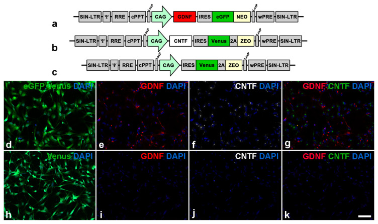
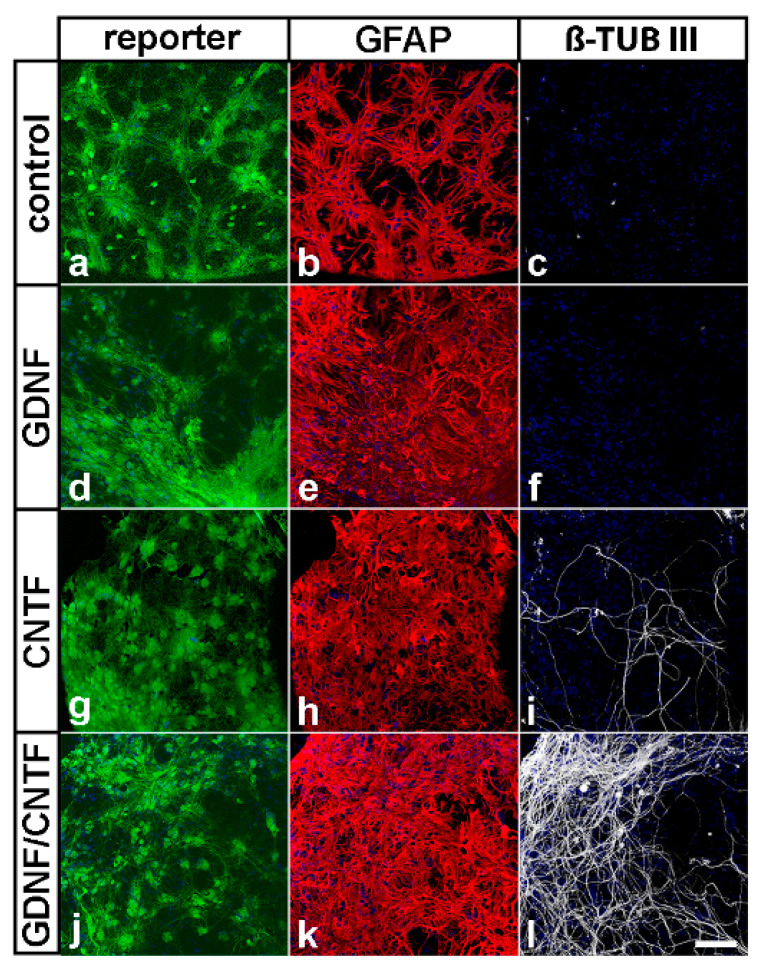
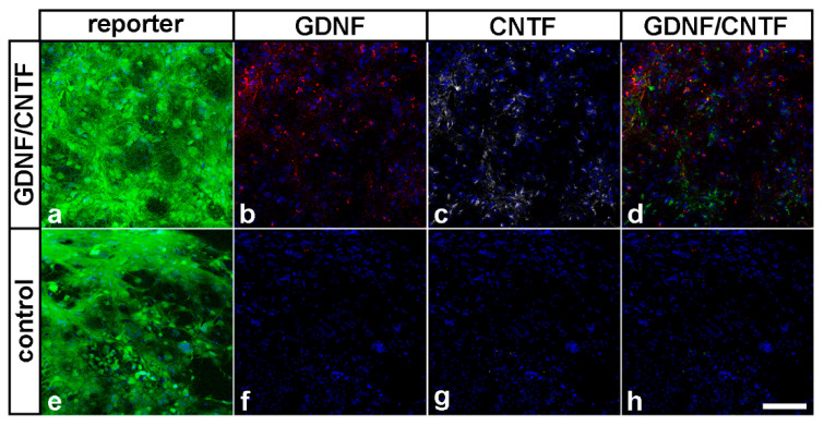
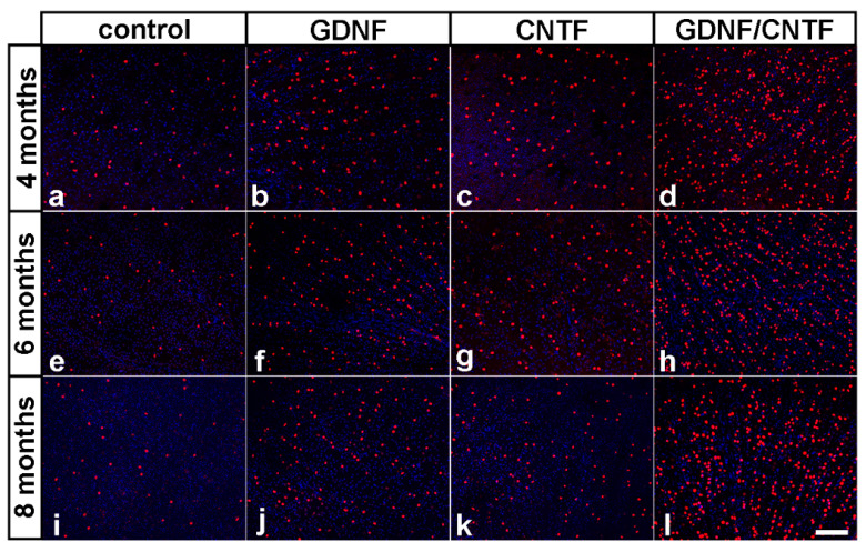
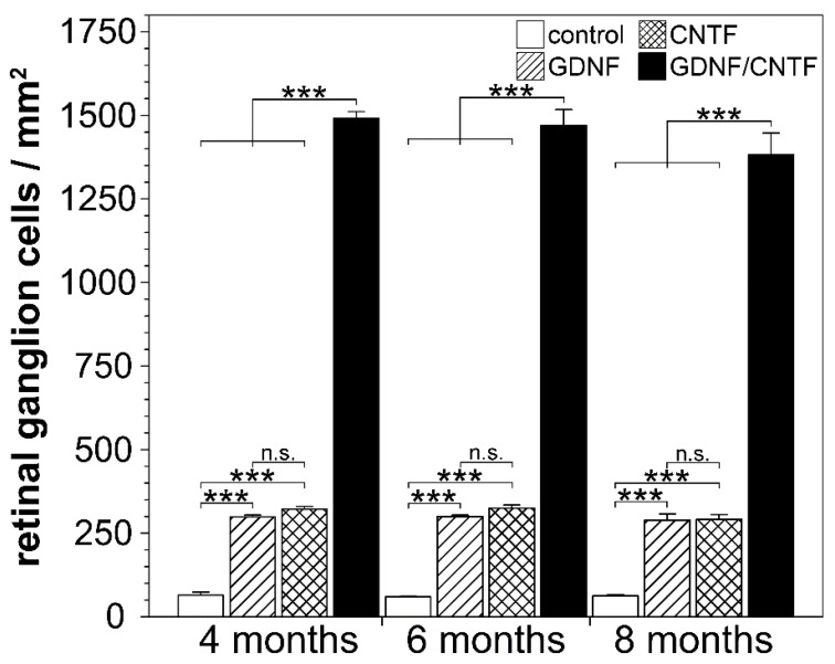
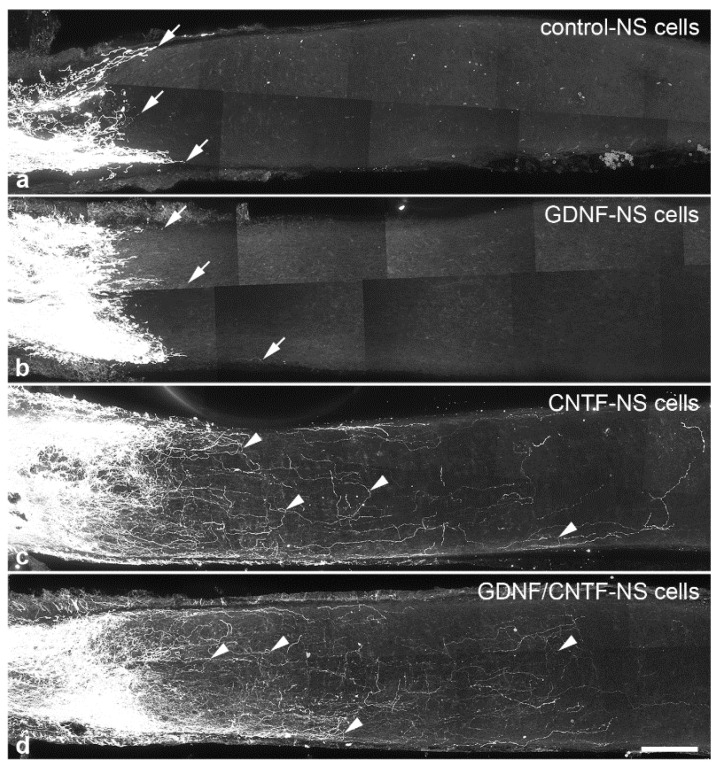
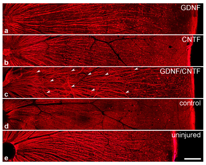
We have recently demonstrated that neural stem cell-based intravitreal co-administration of glial cell line-derived neurotrophic factor (GDNF) and ciliary neurotrophic factor (CNTF) confers profound protection to injured retinal ganglion cells (RGCs) in a mouse optic nerve crush model, resulting in the survival of ~38% RGCs two months after the nerve lesion. Here, we analyzed whether this neuroprotective effect is long-lasting and studied the impact of the pronounced RGC rescue on axonal regeneration. To this aim, we co-injected a GDNF- and a CNTF-overexpressing neural stem cell line into the vitreous cavity of adult mice one day after an optic nerve crush and determined the number of surviving RGCs 4, 6 and 8 months after the lesion. Remarkably, we found no significant decrease in the number of surviving RGCs between the successive analysis time points, indicating that the combined administration of GDNF and CNTF conferred lifelong protection to injured RGCs. While the simultaneous administration of GDNF and CNTF stimulated pronounced intraretinal axon growth when compared to retinas treated with either factor alone, numbers of regenerating axons in the distal optic nerve stumps were similar in animals co-treated with both factors and animals treated with CNTF only.

Keywords: CNTF; GDNF; axotomy; lentiviral vectors; neural stem cells; neuroprotection; optic nerve; regeneration; retinal ganglion cells.


Conflict of interest statement

The authors declare no conflict of interest. The sponsors had no role in the design, execution, interpretation or writing of the study.

Figures

Figure 1
Figure 1
Lentiviral vectors and modified clonal neural stem cell lines. Neural stem cells were transduced with a lentiviral vector encoding glial cell line-derived neurotrophic factor (GDNF) and enhanced green fluorescent protein (eGFP) (a) or ciliary neurotrophic factor (CNTF) and Venus (b). Cells for control experiments were transduced with a vector encoding Venus only (c). Immunocytochemical analysis of a culture composed of a 1:1 mixture of a GDNF- and a CNTF-overexpressing neural stem cell line revealed that cells co-expressed either eGFP and GDNF (d,e) or Venus and CNTF (d,f). Cells for control experiments expressed Venus (h) but neither GDNF (i) nor CNTF (j). The CNTF signal in (g,k) (overlays of (e,f) and (i,j), respectively) is shown in green. Scale bar: 50 µm.
Figure 2
Figure 2
Survival and differentiation of intravitreally grafted neural stem cells. Grafted eGFP- or Venus-positive cells (a,d,g,j) were attached to the posterior surface of the lenses, where they survived for 8 months after transplantation. All NS cell lines were preferentially differentiated into GFAP-positive astrocytes (b,e,h,k). A few CNTF- (i) and a significant fraction of GDNF/CNTF-NS cells (l) were additionally differentiated into β-tubulin III-positive nerve cells. Neuronal differentiation of control-NS (c) or GDNF-NS cells (f) was not observed. Note the numerous β-tubulin III-positive neurites in animals with co-grafted GDNF- and CNTF-NS cells. β-TUB III: β-tubulin III; GFAP: glial fibrillary acidic protein. Scale bar: 100 µm.
Figure 3
Figure 3
Transgene expression in grafted NS cells eight months after transplantation. Eight months after transplantation, GDNF/CNTF-NS cells expressed eGFP or Venus (a) together with GDNF (b) or CNTF (c). Control-NS cells expressed Venus (e) but not GDNF (f) or CNTF (g). The CNTF signal in (d) and (h) (overlays of (b,c) and (f,g), respectively) is shown in green. Scale bar: 100 µm.
Figure 4
Figure 4
Long-term survival of axotomized ganglion cells in GDNF-, CNTF- and GDNF/CNTF-treated retinas. BRN-3A-positive ganglion cells in flat-mounted retinas 4 (ad), 6 (eh) and 8 (il) months after an optic nerve lesion and intravitreal transplantations of control-NS (a,e,i), GDNF-NS (b,f,j), CNTF-NS (c,g,k) or GDNF/CNTF-NS cells (d,h,l). GDNF- or CNTF-treated retinas contained significantly more ganglion cells than control retinas at all post-lesion time points. Note the markedly increased density of surviving ganglion cells in retinas co-treated with GDNF and CNTF when compared with retinas treated with either factor alone. All images were taken close to the optic disc. Scale bar: 50 µm.
Figure 5
Figure 5
Quantitative analysis of retinal ganglion cell survival. The density of BRN-3A-positive ganglion cells was determined in flat-mounted retinas 4, 6 and 8 months after an optic nerve crush and intravitreal transplantations of control-NS (open bars), GDNF-NS (hatched bars), CNTF-NS (cross-hatched bars) or GDNF/CNTF-NS cells (filled bars). The density of retinal ganglion cells in GDNF- or CNTF-treated retinas was significantly higher than in control retinas at all post-lesion time points. Note that the number of surviving ganglion cells was markedly increased in retinas simultaneously treated with GDNF and CNTF when compared to retinas treated with either GDNF or CNTF. Note also the similar density of ganglion cells in each experimental group at the different post-lesion time points. Each bar represents the mean number (±SEM) of retinal ganglion cells per mm2 from six retinas. n.s.: not significant; ***: p < 0.001 according to two-way ANOVA followed by Bonferroni post-hoc test.
Figure 6
Figure 6
Regeneration of intraorbitally lesioned ganglion cell axons into the distal optic nerve stump. RGC axons were anterogradely labeled 1 month after an optic nerve crush in animals with grafted control- (a), GDNF- (b), CNTF- (c) or GDNF/CNTF- (d) NS cells. In mice with grafted control- or GDNF-NS cells, only a few axons regrew for only a short distance into the distal nerve stump (white arrows in (a) and (b) label the tips of some regrown axons). In animals with grafted CNTF- and GDNF/CNTF-NS cells, in comparison, significantly more RGC axons were regrown across the lesion site and extended for significantly longer distances into the distal nerve stumps. Axons in CNTF- and GDNF/CNTF-treated mice followed irregular trajectories and some made U-turns (arrowheads in (c,d)) and grew back towards the retina. Scale bar: 100 µm.
Figure 7
Figure 7
Quantitative analyses of axon regeneration. The length of the longest regrown axon was determined in the distal optic nerve stumps of mice with grafted control- (circles), GDNF- (diamonds), CNTF- (squares) and GDNF/CNTF- (pentagons) NS cells (a). While axons in control and GDNF-treated animals extended for only around 500 µm into the distal nerve stumps, axons in CNTF- and GDNF/CNTF-treated animals extended for up to 3400 and 2600 µm, respectively, distal to the lesion site. Arrowheads indicate mean values (n = 6 for each experimental group). The number of regrown axons in optic nerve sections from CNTF- (open bars) and GDNF/CNTF- (filled bars) treated animals at different positions distal to the lesion site (b). Note the similar number of regenerating axons in both experimental groups at all positions analyzed. Each bar represents the mean number (±SEM) of axons per 100 µm nerve width in 25 µm thick nerve sections from 6 animals. n.s.: not significant; ***: p < 0.001 according to one-way ANOVA followed by Bonferroni post-hoc test.
Figure 8
Figure 8
Intraretinal growth of retinal ganglion cell axons. β-tubulin III-positive ganglion cell axons in flat-mounted retinas 8 months after an intraorbital crush and intravitreal transplantations of GDNF- (a), CNTF- (b), GDNF/CNTF- (c) or control- (d) NS cells. A retina from an adult animal with an uninjured optic nerve is shown for comparison (e). Axon fascicles in GDNF-, CNTF- and GDNF/CNTF-treated retinas (ac) were significantly thicker than in control retinas (d). Note the aberrant trajectories of some axon fascicles (some labeled with white arrowheads in (c)) in animals with grafted GDNF/CNTF-NS cells. Scale bar: 200 µm.

References

    1. Sanes J.R., Masland R.H. The types of retinal ganglion cells: Current status and implications for neuronal classification. Annu. Rev. Neurosci. 2015;38:221–246. doi: 10.1146/annurev-neuro-071714-034120. - DOI - PubMed
    1. Baden T., Berens P., Franke K., Roman Roson M., Bethge M., Euler T. The functional diversity of retinal ganglion cells in the mouse. Nature. 2016;529:345–350. doi: 10.1038/nature16468. - DOI - PMC - PubMed
    1. Rheaume B.A., Jereen A., Bolisetty M., Sajid M.S., Yang Y., Renna K., Sun L., Robson P., Trakhtenberg E.F. Single cell transcriptome profiling of retinal ganglion cells identifies cellular subtypes. Nat. Commun. 2018;9:2759. doi: 10.1038/s41467-018-05134-3. - DOI - PMC - PubMed
    1. Morin L.P., Studholme K.M. Retinofugal projections in the mouse. J. Comp. Neurol. 2014;522:3733–3753. doi: 10.1002/cne.23635. - DOI - PMC - PubMed
    1. Berry M., Ahmed Z., Logan A. Return of function after CNS axon regeneration: Lessons from injury-responsive intrinsically photosensitive and alpha retinal ganglion cells. Prog. Retin. Eye Res. 2019;71:57–67. doi: 10.1016/j.preteyeres.2018.11.006. - DOI - PubMed

Publication types

MeSH terms

Substances

LinkOut - more resources