Review
. 2020 Sep 11;10(9):1816.
doi: 10.3390/nano10091816.

Tailoring Iron Oxide Nanoparticles for Efficient Cellular Internalization and Endosomal Escape

Affiliations
Review

Tailoring Iron Oxide Nanoparticles for Efficient Cellular Internalization and Endosomal Escape

Laura Rueda-Gensini et al. Nanomaterials (Basel). .

Abstract

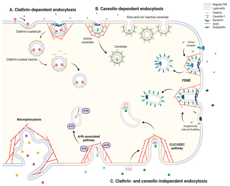
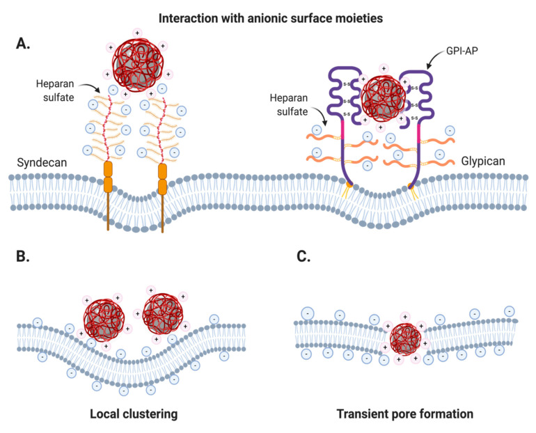
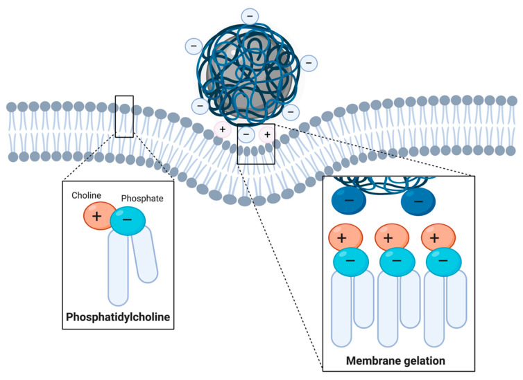
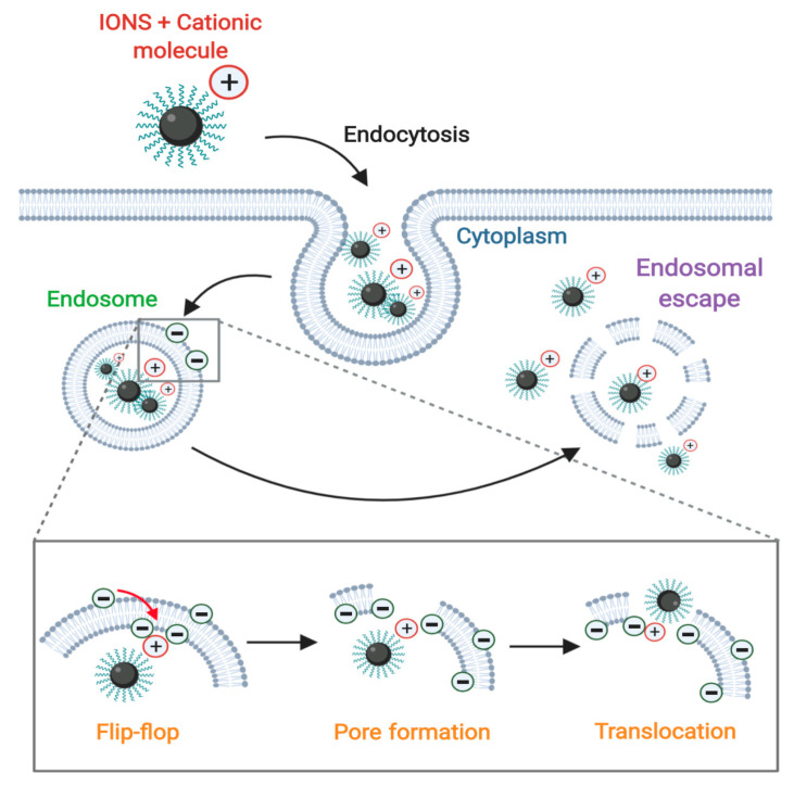
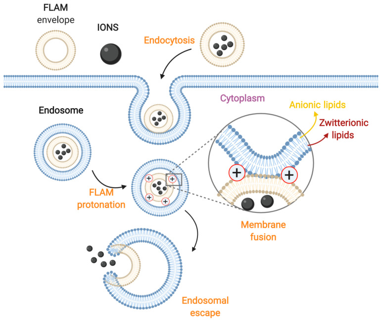
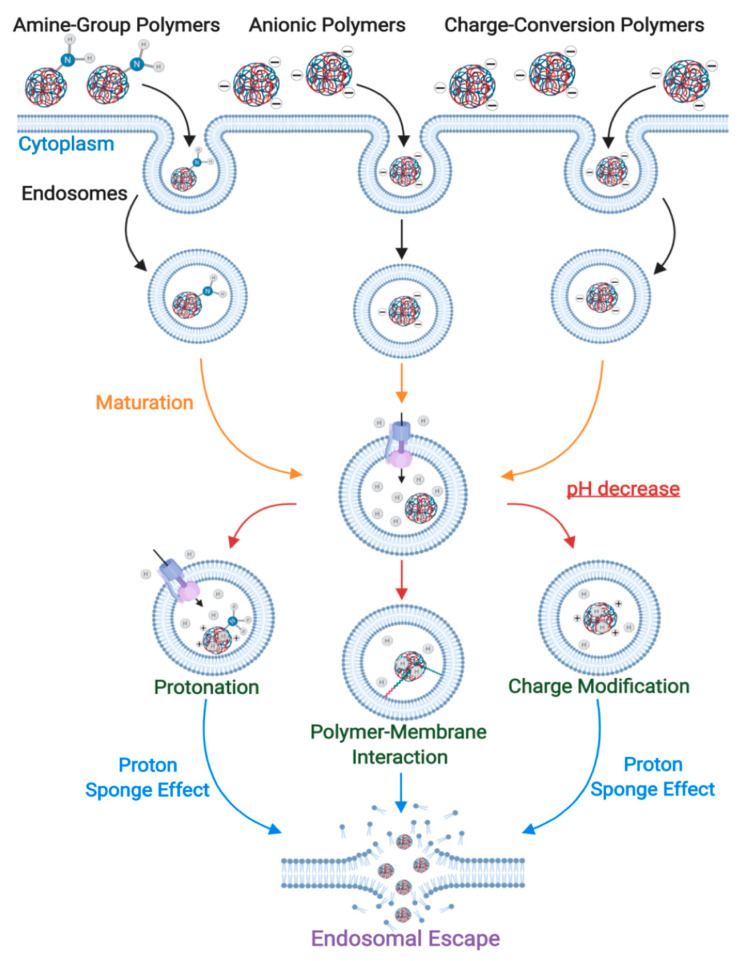
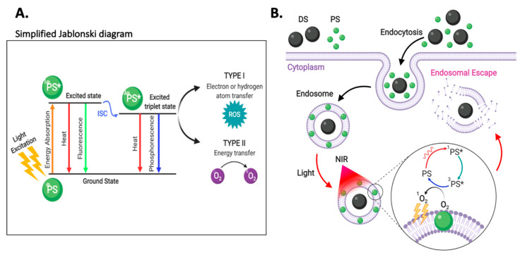
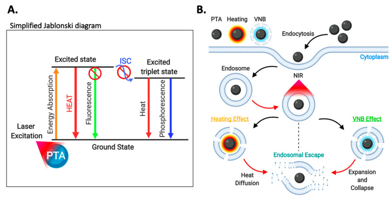
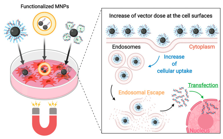
Iron oxide nanoparticles (IONs) have been widely explored for biomedical applications due to their high biocompatibility, surface-coating versatility, and superparamagnetic properties. Upon exposure to an external magnetic field, IONs can be precisely directed to a region of interest and serve as exceptional delivery vehicles and cellular markers. However, the design of nanocarriers that achieve an efficient endocytic uptake, escape lysosomal degradation, and perform precise intracellular functions is still a challenge for their application in translational medicine. This review highlights several aspects that mediate the activation of the endosomal pathways, as well as the different properties that govern endosomal escape and nuclear transfection of magnetic IONs. In particular, we review a variety of ION surface modification alternatives that have emerged for facilitating their endocytic uptake and their timely escape from endosomes, with special emphasis on how these can be manipulated for the rational design of cell-penetrating vehicles. Moreover, additional modifications for enhancing nuclear transfection are also included in the design of therapeutic vehicles that must overcome this barrier. Understanding these mechanisms opens new perspectives in the strategic development of vehicles for cell tracking, cell imaging and the targeted intracellular delivery of drugs and gene therapy sequences and vectors.

Keywords: drug delivery; endocytosis; endosomal escape; iron oxide nanoparticles.


Conflict of interest statement

The authors declare no conflict of interest.

Figures

Figure 1
Figure 1
General schematic of the different endocytic mechanisms by pinocytosis, classified as (A) clathrin-dependent, (B) caveolin-dependent and (C) clathrin- and caveolin-independent (FEME, CLIC/GEEC pathway, Arf6 and macropinocytosis). (Created with BioRender.com).
Figure 2
Figure 2
Adsorptive interactions between charged nanoparticles (CNPs) and the plasma membrane. (A) Electrostatic interactions with anionic syndecans and glypicans rich in heparan sulfate. (B) Cooperative membrane wrapping phenomena by cumulative CNP interactions with anionic phospholipids. (C) Transient pore formation by small CNPs (≤20 nm) due to strong attraction to the inner membrane layer in phosphatidylserine-rich regions. (Created with BioRender.com).
Figure 3
Figure 3
Local membrane gelation induced by ANPs in phosphatidylcholine-rich membrane microdomains. (Created with BioRender.com).
Figure 4
Figure 4
Endosomal escape mediated by the Proton-Sponge Effect. Surface coatings of IONs sequester protons from the endosomal lumen and create an osmotic gradient. The increase in osmotic pressure, coupled with destabilizing interactions between cationic surfaces of IONs and the endosomal membrane ultimately leads to the lysis of endosomal vesicles. (Created with BioRender.com).
Figure 5
Figure 5
Schematic illustration of the translocation mechanism. Nanoparticles (IONS coated with cationic molecules) are endocytosed by the cell and internalized inside endosomes. The positive charge of the coated nanoparticles generates a flip-flop of the endosome’s cytosolic anionic lipids, which induces the generation of pores through which the nanoparticles can cross the endosomal membrane to reach the cytosol. (Created with BioRender.com).
Figure 6
Figure 6
Schematic illustration of the membrane fusion mechanism of fusogenic lipids or amphiphilic molecules (FLAM)-ION complexes. The nanoparticles are encapsulated within a FLAM envelope for subsequent internalization by endocytosis. Within the endosome, the FLAM phospholipids protonate, thereby inducing the fusion of this envelope with the Zwitterionic luminal lipids of the endosomal vesicles. This process ultimately leads to endosomal escape. (Created with BioRender.com).
Figure 7
Figure 7
pH-triggered endosomal escape strategies via polymers susceptible to protonation. This includes polymers with pendant uncharged amino-groups at physiological pH, anionic polymers and charge-conversion polymers (Created with BioRender.com).
Figure 8
Figure 8
(A) Simplified Jablonski diagram showing the different energy transfer events involved in PCI. PS: photosensitizer, ICS: intersystem crossing. (B) Schematic illustration of the PCI process. Delivery systems (DS) and photosensitizers (PS) are endocytosed by the cell and colocalized into the endosomal vesicles. PS are mainly localized in the endosomal membranes due to their amphiphilic properties. After NIR irradiation, PS absorb the light energy and transfer it to molecular oxygen, thereby generating highly toxic singlet oxygen. These molecules cause important oxidative damage in the endocytic membranes, which ultimately leads to endosomal escape by their disruption. (Created with BioRender.com).
Figure 9
Figure 9
Schematic illustration of the PCI process by using upconverted nanoparticles (UCNPs). UCNPs and photosensitizers (PS) are endocytosed by the cell and colocalized with endosomal vesicles. PS intercalate within endosomal membranes due to their amphiphilic properties. After NIR irradiation, sensitizer ions (S) absorb the energy and transfer it to activator ions (A) capable of emitting radiation (UV or Vis). PS then absorb the energy and transfer it to molecular oxygen, thereby generating highly toxic singlet oxygen. These molecules cause important oxidative damage in the endocytic membranes, which ultimately leads to endosomal escape by their disruption. ET: energy transfer, H: host matrix (Created with BioRender.com)
Figure 10
Figure 10
(A) Simplified Jablonski diagram describing the different energy transfer mechanisms involved in PTT. PTA: photothermal transduction agent, ICS: intersystem crossing. (B) Schematic illustration of the PTT process. Photothermal transduction agents (PTAs) are taken up by endocytosis and trapped into the endosomes. After NIR irradiation, PTAs absorb the light energy and transform it into heat, which could lead to endosomal escape by two major mechanisms. In the first one, also known as the heating effect leads to the destabilization of endosomal membranes by a localized increase in temperature. In the second one, the released heat is high enough to generate a vapor layer surrounding the PTAs such that it expands as a vapor nanobubble (VNB) that eventually collapses to induce endocytic membrane disruption. (Created with BioRender.com).
Figure 11
Figure 11
Schematic illustration of the transfection mechanism. After the endosomal escape of the vehicles, the cargo (usually DNA) is released into the cytoplasm. The cargo interacts with the nuclear pore complex (NPC), where importin proteins activate to mediate nuclear internalization (1). Inside the nucleus, internalized molecules interact with nuclear structures (2). Subsequently, the remaining molecules bind to RanG proteins (3) for their recycling (4) and release into the cytoplasm (5). (Created with BioRender.com).
Figure 12
Figure 12
Schematic of the magnetofection principle. Under the effect of a magnetic field, functionalized IONs are guided directly to target cells. This generates an increase in the vector availability at the cell surfaces that leads to an increase in cellular uptake. Endosomal escape occurs by the action of the specific molecules used to functionalize IONs. High transfection rates can be achieved: More nucleic acids loaded IONs into the cytoplasm leads to an increase of free nucleic acids generating more efficient transfection rates. (Created with BioRender.com).

References

    1. Tong S., Zhu H., Bao G. Magnetic Iron Oxide Nanoparticles for Disease Detection and Therapy. Mater. Today. 2019;31:86–99. doi: 10.1016/j.mattod.2019.06.003. - DOI - PMC - PubMed
    1. Price P.M., Mahmoud W.E., Al-Ghamdi A.A., Bronstein L.M. Magnetic Drug Delivery: Where the Field Is Going. Front. Chem. 2018;6 doi: 10.3389/fchem.2018.00619. - DOI - PMC - PubMed
    1. Zoppellaro G. Magnetic Nanoheterostructures. Springer International Publishing; Cham, Switzerland: 2020. Iron Oxide Magnetic Nanoparticles (NPs) Tailored for Biomedical Applications; pp. 57–102. - DOI
    1. Bohara R.A., Singh P. Magnetic Nanoheterostructures. Springer International Publishing; Cham, Switzerland: 2020. Multiple Myeloma: Role of Magnetic Nanoparticles; pp. 479–494. - DOI
    1. Gupta A.K., Gupta M. Synthesis and Surface Engineering of Iron Oxide Nanoparticles for Biomedical Applications. Biomaterials. 2005;26:3995–4021. doi: 10.1016/j.biomaterials.2004.10.012. - DOI - PubMed

LinkOut - more resources