. 2020 Sep 13;9(9):2090.
doi: 10.3390/cells9092090.

Ionocytes and CFTR Chloride Channel Expression in Normal and Cystic Fibrosis Nasal and Bronchial Epithelial Cells

Affiliations

Ionocytes and CFTR Chloride Channel Expression in Normal and Cystic Fibrosis Nasal and Bronchial Epithelial Cells

Paolo Scudieri et al. Cells. .

Abstract

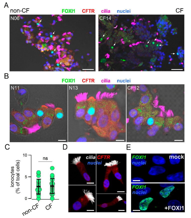
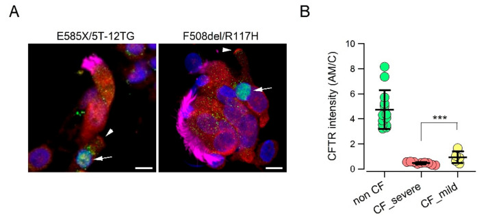
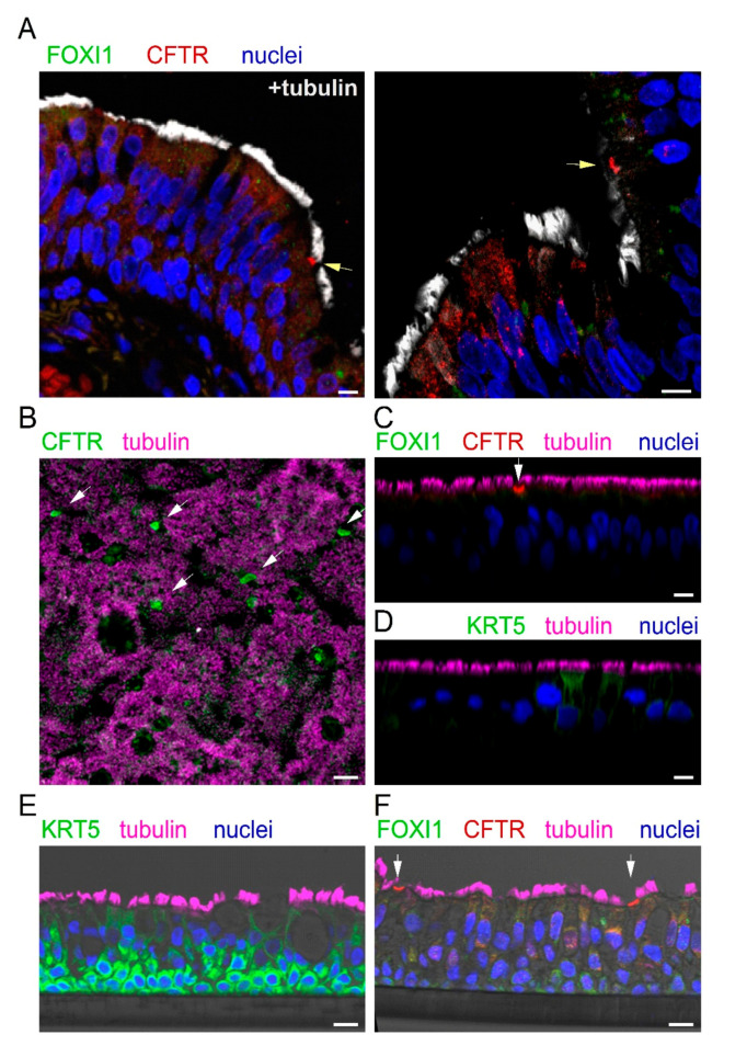
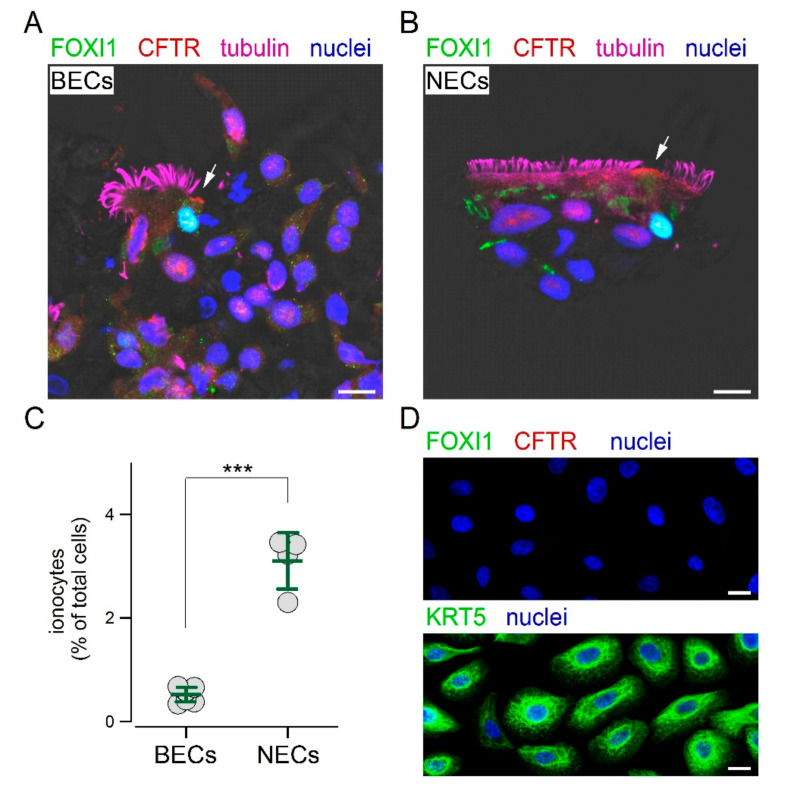
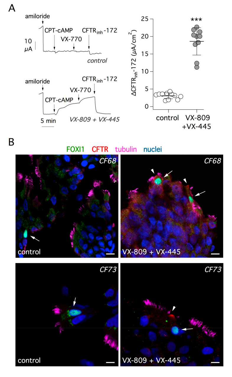
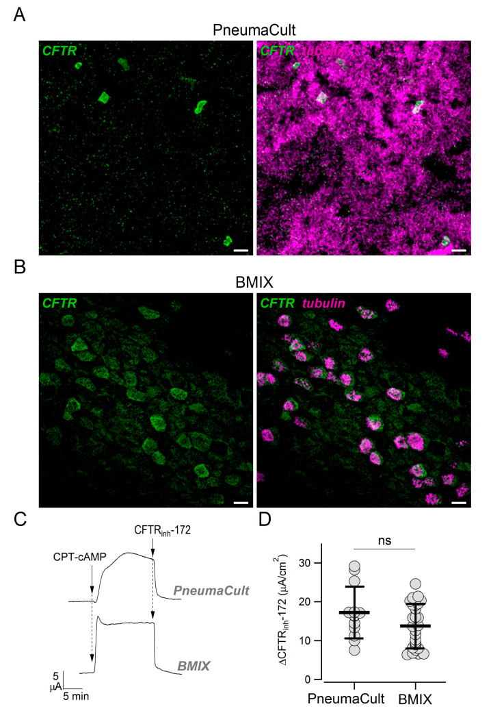
The airway epithelium contains ionocytes, a rare cell type with high expression of Forkhead Box I1 (FOXI1) transcription factor and Cystic Fibrosis Transmembrane conductance Regulator (CFTR), a chloride channel that is defective in cystic fibrosis (CF). Our aim was to verify if ionocyte development is altered in CF and to investigate the relationship between ionocytes and CFTR-dependent chloride secretion. We collected nasal cells by brushing to determine ionocyte abundance. Nasal and bronchial cells were also expanded in vitro and reprogrammed to differentiated epithelia for morphological and functional studies. We found a relatively high (~3%) ionocyte abundance in ex vivo nasal samples, with no difference between CF and control individuals. In bronchi, ionocytes instead appeared very rarely as previously reported, thus suggesting a possible proximal-distal gradient in human airways. The difference between nasal and bronchial epithelial cells was maintained in culture, which suggests an epigenetic control of ionocyte development. In the differentiation phase of the culture procedure, we used two media that resulted in a different pattern of CFTR expression: confined to ionocytes or more broadly expressed. CFTR function was similar in both conditions, thus indicating that chloride secretion equally occurs irrespective of CFTR expression pattern.

Keywords: CFTR; airway epithelium; chloride secretion; cystic fibrosis; ionocytes.


Conflict of interest statement

The authors declare no conflict of interest.

Figures

Figure 1
Figure 1
Detection of ionocytes in the human nasal epithelium. (A) Representative low magnification images showing staining for CFTR, cilia, FOXI1, and nuclei. The two images are from a control individual and a cystic fibrosis (CF) patient. Arrows indicate nuclei positive for FOXI1. Scale bar: 25 µm. (B) High magnification images for two control individuals and one CF patient. Ionocytes were clearly identified in non-CF samples as non-ciliated cells with apical positivity for CFTR and nuclear positivity for FOXI1. In most CF samples, CFTR could not be detected due to mutations impairing protein synthesis or trafficking. Scale bar: 5 µm. (C) Abundance of ionocytes in non-CF vs. non-CF samples. Each dot represents the mean percentage of ionocytes over total cells in a single individual (n = 18 and 22 for non-CF and CF samples, respectively). (D) Representative images from a nasal sample devoid of ionocytes and with CFTR expressed in ciliated cells. Scale bar: 5 µm. (E) Validation of the FOXI1 antibody. Images show nuclear signal only in CFBE41o- cells transfected with the FOXI1 plasmid. Scale bar: 5 µm.
Figure 2
Figure 2
CFTR protein expression in the apical membrane of nasal ionocytes from CF patients with “mild” mutations. (A) Representative images of nasal epithelial cells collected by brushing from CF patients with indicated genotypes. Cells were stained for FOXI1 (green), CFTR (red), cilia (magenta), and nuclei (blue). Arrows indicate nuclei positive for FOXI1. Arrowheads indicate CFTR signal in the apical membrane. (B) Scatter dot plot reporting the intensity of CFTR signal in the apical membrane (relative to signal in the cytosol, AM/C) of FOXI1-positive cells. This parameter was significantly higher (***, p < 0.01) in cells with “mild” mutations compared to cells with severe mutations.
Figure 3
Figure 3
CFTR expression in bronchial epithelium in vivo and in vitro. (A) Representative immunofluorescence images of bronchial sections showing staining for CFTR (red), cilia (white), FOXI1 (green), and nuclei (blue). Arrows indicate the presence of putative ionocytes, i.e., non-ciliated cells with strong CFTR positivity in the apical membrane. Nuclear FOXI1 staining was never detected in these histological preparations. (BD) Immunofluorescence analysis on cultured bronchial epithelia, after fixation and permeabilization. CFTR was expressed in isolated non-ciliated cells ((B), xy scan; (C), xz scan). FOXI1 was instead undetectable (C). KRT5, a marker of basal cells, was also absent ((D), xz scan). (E,F) Immunofluorescence analysis on cultured bronchial epithelia, after inclusion in paraffin and sectioning. In contrast to intact epithelium, sectioning allowed the detection of KRT5 in basal cells (E). In contrast, FOXI1 signal was still absent in CFTR-expressing cells (F). Scale bar: 10 µm in (A) and (CF) and 25 µm in (B).
Figure 4
Figure 4
Detection of ionocytes in cultured epithelial cells. (A,B) Immunofluorescence analysis of cultured bronchial (BECs) and nasal (NECs) epithelial cells after brushing. Under this condition, FOXI1 and CFTR were detected within the same non-ciliated cells. Arrows indicate CFTR signal in the apical membrane. Scale bar: 10 µm. (C) Abundance of ionocytes in cultured BECs and NECs. The percentage of ionocytes was significantly (***, p < 0.001) higher in NECs. (D) Immunofluorescence analysis of airway epithelial cells in the expansion phase of the culture procedure. These cells expressed the basal cell marker KRT5, but not CFTR or FOXI1. Scale bar: 5 µm.
Figure 5
Figure 5
Rescue of F508del-CFTR function and trafficking by a combination of correctors. (A) Representative short-circuit current recordings (left) and summary of results (right) from experiments on CF bronchial epithelial cells (F508del/F508del genotype). Cells were treated for 24 h with vehicle alone or with a combination of correctors: VX-809 (1 µM) plus VX-445 (5 µM). During experiments, the epithelial Na+ channel (ENaC) was blocked with amiloride (10 µM). CFTR-dependent Cl secretion was then stimulated with 8-(4-chlorophenylthio)adenosine 3′,5′-cyclic monophosphate (CPT-cAMP, 100 µM) and the potentiator VX-770 (1 µM). Finally, CFTR activity was inhibited with CFTRinh-172 (10 µM). The scatter dot plot on the right reports the amplitude of CFTRinh-172 effect, which reflects the extent of CFTR function. The results demonstrate significant (***, p < 0.001) rescue of F508del-CFTR by the combination of correctors. (B) Analysis of F508del-CFTR protein rescue by immunofluorescence. The figure shows representative immunofluorescence images of cultured bronchial epithelial cells from two CF patients (CF68 and CF73). Cells were treated with vehicle (control, left) or with VX-809 plus VX-445 (right) and then stained for FOXI1 (arrows), CFTR, tubulin (cilia), and nuclei. The treatment with correctors led to the appearance of CFTR signal in the apical membrane (arrowheads). Scale bar: 10 µm.
Figure 6
Figure 6
Effect of culture medium on CFTR expression pattern. (A,B) Detection of CFTR and cilia in epithelia differentiated in PneumaCult ALI or BMIX medium. Scale bar: 10 µm. (C) Representative short-circuit current recordings showing activation (CPT-cAMP, 100 µM) and inhibition (CFTRinh-172, 10 µM) of CFTR in bronchial epithelia generated with PneumaCult ALI or BMIX. (D) CFTR activity measured in PneumaCult ALI and BMIX epithelia. The plot reports the size of the effect of CFTRinh-172 after maximal activation of CFTR with CPT-cAMP. The two groups of data were not significantly different.

References

    1. Crystal R.G., Randell S.H., Engelhardt J.F., Voynow J., Sunday M.E. Airway epithelial cells. Proc. Am. Thorac. Soc. 2008;5:772–777. doi: 10.1513/pats.200805-041HR. - DOI - PMC - PubMed
    1. Curran D.R., Cohn L. Advances in mucous cell metaplasia: A plug for mucus as a therapeutic focus in chronic airway disease. Am. J. Respir. Cell. Mol. Biol. 2010;42:268–275. doi: 10.1165/rcmb.2009-0151TR. - DOI - PMC - PubMed
    1. Park K.-S., Korfhagen T.R., Bruno M.D., Kitzmiller J.A., Wan H., Wert S.E., Hershey G.K.K., Chen G., Whitsett J.A. SPDEF regulates goblet cell hyperplasia in the airway epithelium. J. Clin. Investig. 2007;117:978–988. doi: 10.1172/JCI29176. - DOI - PMC - PubMed
    1. Scudieri P., Caci E., Bruno S., Ferrera L., Schiavon M., Sondo E., Tomati V., Gianotti A., Zegarra-Moran O., Pedemonte N., et al. Association of TMEM16A chloride channel overexpression with airway goblet cell metaplasia. J. Physiol. 2012;590:6141–6155. doi: 10.1113/jphysiol.2012.240838. - DOI - PMC - PubMed
    1. Boers J.E., Ambergen A.W., Thunnissen E. Number and Proliferation of Clara Cells in Normal Human Airway Epithelium. Am. J. Respir. Crit. Care Med. 1999;159:1585–1591. doi: 10.1164/ajrccm.159.5.9806044. - DOI - PubMed

Publication types

MeSH terms

Substances