Review
. 2020 Sep 13;21(18):6702.
doi: 10.3390/ijms21186702.

Host Synthesized Carbohydrate Antigens on Viral Glycoproteins as "Achilles' Heel" of Viruses Contributing to Anti-Viral Immune Protection

Affiliations
Review

Host Synthesized Carbohydrate Antigens on Viral Glycoproteins as "Achilles' Heel" of Viruses Contributing to Anti-Viral Immune Protection

Uri Galili. Int J Mol Sci. .

Abstract

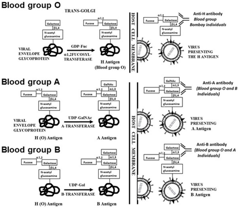
The glycans on enveloped viruses are synthesized by host-cell machinery. Some of these glycans on zoonotic viruses of mammalian reservoirs are recognized by human natural antibodies that may protect against such viruses. These antibodies are produced mostly against carbohydrate antigens on gastrointestinal bacteria and fortuitously, they bind to carbohydrate antigens synthesized in other mammals, neutralize and destroy viruses presenting these antigens. Two such antibodies are: anti-Gal binding to α-gal epitopes synthesized in non-primate mammals, lemurs, and New World monkeys, and anti-N-glycolyl neuraminic acid (anti-Neu5Gc) binding to N-glycolyl-neuraminic acid (Neu5Gc) synthesized in apes, Old World monkeys, and many non-primate mammals. Anti-Gal appeared in Old World primates following accidental inactivation of the α1,3galactosyltransferase gene 20-30 million years ago. Anti-Neu5Gc appeared in hominins following the inactivation of the cytidine-monophosphate-N-acetyl-neuraminic acid hydroxylase gene, which led to the loss of Neu5Gc <6 million-years-ago. It is suggested that an epidemic of a lethal virus eliminated ancestral Old World-primates synthesizing α-gal epitopes, whereas few mutated offspring lacking α-gal epitopes and producing anti-Gal survived because anti-Gal destroyed viruses presenting α-gal epitopes, following replication in parental populations. Similarly, anti-Neu5Gc protected few mutated hominins lacking Neu5Gc in lethal virus epidemics that eliminated parental hominins synthesizing Neu5Gc. Since α-gal epitopes are presented on many zoonotic viruses it is suggested that vaccines elevating anti-Gal titers may be of protective significance in areas endemic for such zoonotic viruses. This protection would be during the non-primate mammal to human virus transmission, but not in subsequent human to human transmission where the virus presents human glycans. In addition, production of viral vaccines presenting multiple α-gal epitopes increases their immunogenicity because of effective anti-Gal-mediated targeting of vaccines to antigen presenting cells for extensive uptake of the vaccine by these cells.

Keywords: Covid-19; Neu5Gc; anti-Gal; anti-Neu5Gc; blood groups ABO and Bombay; glycan shield; viral epidemics; viral vaccines; zoonotic virus; α-gal epitopes.


Conflict of interest statement

The author is the inventor in US patents 9662383 and 10201601 (Assignee, University of Massachusetts), which include some of the methods described in this review.

Figures

Figure 1
Figure 1
Synthesis of α-gal epitopes (A) and N-glycolyl-neuraminic acid (Neu5Gc) (B) on glycans of enveloped virus glycoproteins. The “capping” of the glycans occurs in the trans-Golgi compartment of host-cells and is mediated by α1,3galactosyltransferase and α2,3 and α2,6 sialyltransferases, using UDP-Gal and CMP-Neu5Gc as high-energy sugar donors, respectively. The hydroxyl of Neu5Gc differentiating it from Neu5Ac is indicated. Viral glycoproteins are assembled on cell membranes to form the virus envelope, followed by budding of the virus. Antibodies binding to these glycans and the species producing them are indicated. Modified from ref. [16].
Figure 2
Figure 2
Amplifying the specific anti-virus immune response by formation of immune complexes between natural anti-carbohydrate antibodies and lysed virus, as demonstrated by the natural anti-Gal antibody. Anti-Gal binding to α-gal epitopes on viral glycans activates the complement cascade causing virolysis and neutralization of the virus and recruitment of antigen presenting cells (APC) such as dendritic cells and macrophages by complement cleavage peptides. Subsequent binding of the anti-Gal/virus immune complex to the Fcγ receptor (FcγR) on dendritic cells and macrophages induces extensive uptake of the opsonized virions by these APC and transport of the internalized virus to the regional lymph nodes. The APC further process and present the viral immunogenic peptides on major histocompatibility complex (MHC) molecules for the activation of CD8+ cytolytic T cells (CTL) and CD4+ helper T cells. The latter cells provide help to B cells producing virus specific neutralizing antibodies. The CTL detect various host cells infected with the virus and kill them. Modified from “The natural anti-Gal antibody as foe turned friend in medicine” by U. Galili, 2018, Elsevier/Academic Press, p. 153.
Figure 3
Figure 3
Suggested evolutionary periods (indicated by arrows) in which the α-gal epitope and Neu5Gc carbohydrate antigens were eliminated and the anti-Gal and anti-N-glycolyl neuraminic acid (anti-Neu5Gc) antibodies appeared as a result of viral epidemics. The presence of α-gal epitopes only in mammals and in no other vertebrates implies that synthesis of α-gal epitopes initiated in mammals at an early evolutionary period before marsupials and placental mammals diverged from a common ancestor. The absence of α-gal epitopes only in humans, apes and Old World monkeys implies that the elimination of primates synthesizing this epitope and the corresponding production of the natural anti-Gal antibody occurred in ancestral Old World primates (catarrhini) after they diverged from New World monkeys (platirrhini). N-5-glycolyl-neuraminic acid (Neu5Gc) is synthesized in most mammals (including apes and Old World monkeys) and other vertebrates, but it is absent in humans. In contrast, humans produce the natural anti-Neu5Gc antibody. This strongly suggests that elimination of Neu5Gc occurred after hominins and ancestors of chimpanzee diverged from a common ancestor. Modified from “The natural anti-Gal antibody as foe turned friend in medicine” by U. Galili, 2018, Elsevier/Academic Press, p. 25.
Figure 4
Figure 4
Hypothesis on the association between the evolutionary elimination of α-gal epitopes in ancestral Old World primates and an epidemic caused by a virus that was lethal to these ancestral primates. Early ancestral Old World primates synthesized α-gal epitopes, as New World monkeys, lemurs and non-primate mammals. Accidental base deletion mutation in one early Old World primate resulted in the inactivation of the α1,3GT gene (GGTA1). After few generations, a small offspring population homozygous for this inactivating mutation produced the natural anti-Gal antibody. An epidemic by a lethal virus caused the extinction of the parental populations synthesizing α-gal epitopes. The virus produced in parental populations presented multiple α-gal epitopes on the glycan shield (“α-gal” in the rectangles). Anti-Gal in individuals of the mutated population protected against the virus by binding to the α-gal epitopes on its glycan shield. This ultimately resulted in the replacement of the parental populations synthesizing α-gal epitopes with Old World primate progeny lacking this epitope and producing the natural anti-Gal antibody. Modified from ref. [89].
Figure 5
Figure 5
Synthesis of H (O), A and B antigens on the glycan shield of enveloped virus glycoproteins as a result of replication in human host-cells of various blood groups. The corresponding antibodies are according to the blood group. Blood group Bombay individuals have mutations that inactivate the two α1,2FT genes FUT1 and FUT2, therefore they cannot synthesize the H antigen (blood group O) and they produce natural anti-H (anti-O) antibody in addition to the production of anti-A and anti-B antibodies. Blood group A and B antigens are synthesized on the H antigen by the corresponding A and B transferases. The synthesized carbohydrate antigens are presented on the glycan shield of the budding virus and bind natural antibodies, as indicated. Modified from ref. [16].

Similar articles

Cited by

References

    1. Vigerust D.J., Shepherd V.L. Virus glycosylation: Role in virulence and immune interactions. Trends Microbiol. 2007;15:211–218. doi: 10.1016/j.tim.2007.03.003. - DOI - PMC - PubMed
    1. Walls A.C., Tortorici M.A., Frenz B., Snijder J., Li W., Rey F.A., DiMaio F., Bosch B.-J., Veesler D. Glycan shield and epitope masking of a coronavirus spike protein observed by cryo-electron microscopy. Nat. Struct. Mol. Biol. 2016;23:899–905. doi: 10.1038/nsmb.3293. - DOI - PMC - PubMed
    1. Xiong X., Tortorici M.A., Snijder J., Yoshioka C., Walls A.C., Li W., McGuire A.T., Rey F.A., Bosch B.J., Veesler D. Glycan Shield and Fusion Activation of a Deltacoronavirus Spike Glycoprotein Fine-Tuned for Enteric Infections. J. Virol. 2018;92:e01628-17. doi: 10.1128/JVI.01628-17. - DOI - PMC - PubMed
    1. Wei X., Decker J.M., Wang S., Hui H., Kappes J.C., Wu X., Salazar-Gonzalez M.G., Salazar J.F., Kilby J.M., Saag M.S., et al. Antibody neutralization and escape by HIV-1. Nature. 2003;422:307–312. doi: 10.1038/nature01470. - DOI - PubMed
    1. Crooks E., Tong T., Chakrabarti B., Narayan K., Georgiev I., Menis S. Vaccine-elicited tier 2 HIV-1 neutralizing antibodies bind to quaternary epitopes involving glycan-deficient patches Proximal to the CD4 Binding Site. PLoS Pathog. 2015;11:e1004932. doi: 10.1371/journal.ppat.1004932. - DOI - PMC - PubMed