. 2020 Sep 13;12(9):2808.
doi: 10.3390/nu12092808.

Effects of Human Milk Oligosaccharides on the Adult Gut Microbiota and Barrier Function

Affiliations

Effects of Human Milk Oligosaccharides on the Adult Gut Microbiota and Barrier Function

Tanja Šuligoj et al. Nutrients. .

Abstract

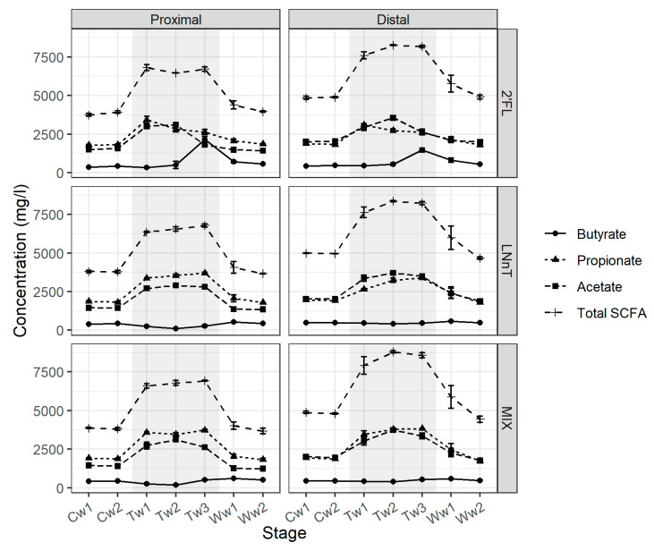
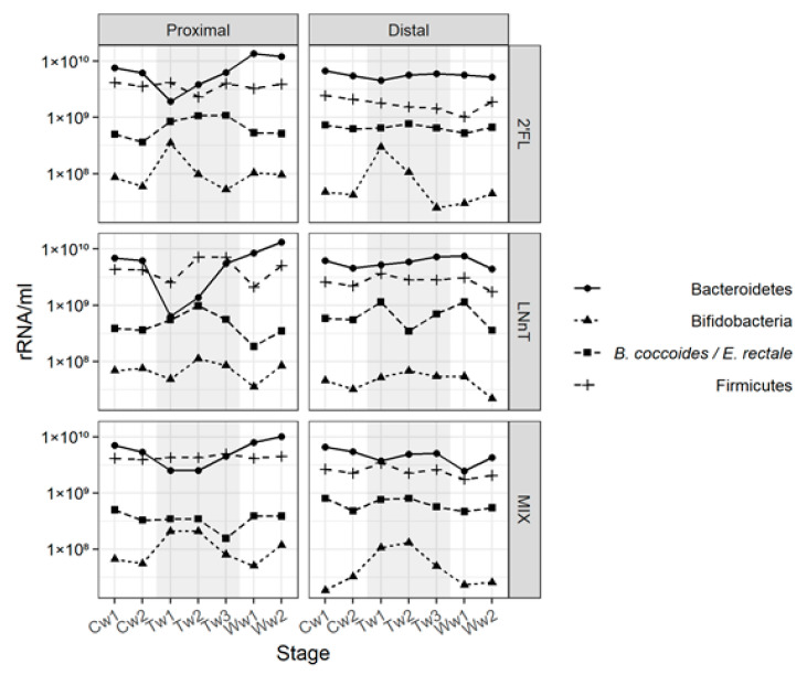
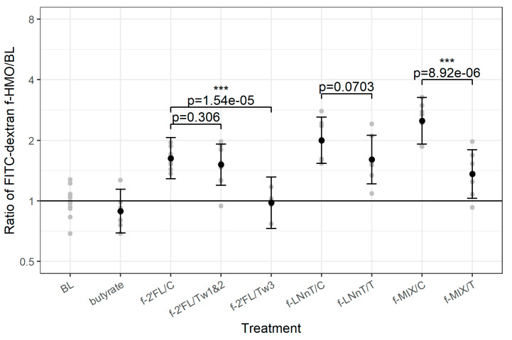
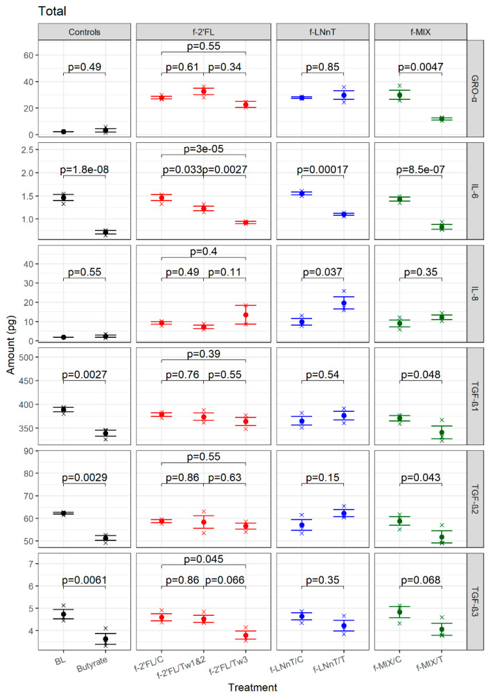
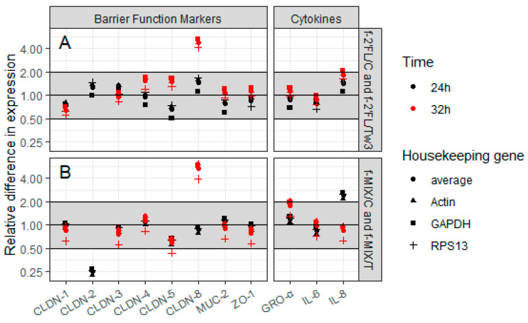
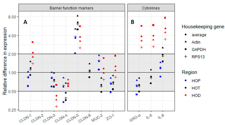
Human milk oligosaccharides (HMOs) shape the gut microbiota in infants by selectively stimulating the growth of bifidobacteria. Here, we investigated the impact of HMOs on adult gut microbiota and gut barrier function using the Simulator of the Human Intestinal Microbial Ecosystem (SHIME®), Caco2 cell lines, and human intestinal gut organoid-on-chips. We showed that fermentation of 2'-O-fucosyllactose (2'FL), lacto-N-neotetraose (LNnT), and combinations thereof (MIX) led to an increase of bifidobacteria, accompanied by an increase of short chain fatty acid (SCFA), in particular butyrate with 2'FL. A significant reduction in paracellular permeability of FITC-dextran probe was observed using Caco2 cell monolayers with fermented 2'FL and MIX, which was accompanied by an increase in claudin-8 gene expression as shown by qPCR, and a reduction in IL-6 as determined by multiplex ELISA. Using gut-on-chips generated from human organoids derived from proximal, transverse, and distal colon biopsies (Colon Intestine Chips), we showed that claudin-5 was significantly upregulated across all three gut-on-chips following treatment with fermented 2'FL under microfluidic conditions. Taken together, these data show that, in addition to their bifidogenic activity, HMOs have the capacity to modulate immune function and the gut barrier, supporting the potential of HMOs to provide health benefits in adults.

Keywords: SHIME®; adult gut microbiota; gut barrier function; gut-on-chips; human milk oligosaccharides.


Conflict of interest statement

The authors declare no conflict of interest. L.K.V. and B.M.C. participated in the design of the study, the interpretation of the data, and the writing of the manuscript but did not participate in the collection and analyses of data and encouraged publication of the study.

Figures

Figure 1
Figure 1
Impact of human milk oligosaccharides (HMOs) on short chain fatty acid (SCFA) production using the Simulator of the Human Intestinal Microbial Ecosystem (SHIME®) model. Absolute values of acetic acid, propionic acid, butyric acid, and total SCFA associated with 2’FL (top panel), LNnT (middle panel), and MIX (bottom panel) in the proximal and distal colon reactors are presented. Samples for SCFA analysis were collected during two weeks of control period (Cw1 and Cw2), three weeks of treatment period (Tw1, Tw2, and Tw3), and two weeks of washout period (Ww1 and Ww2). Three samples were collected each week. Error bars correspond to standard errors calculated from the 3 measurements per relevant week.
Figure 2
Figure 2
Impact of HMOs, 2’FL (top panel), LNnT (middle panel), and MIX (bottom panel), on microbial populations in the SHIME® model. The relative proportion of Bifidobacterium, B. coccoides/E. rectale, Firmicutes and Bacteroidetes in the lumen of the proximal and distal colon vessel was determined by qPCR. Samples for qPCR analysis were collected once a week during two weeks of control period (Cw1 and Cw2), three weeks of treatment period (Tw1, Tw2 and Tw3), and two weeks of washout period (Ww1 and Ww2).
Figure 3
Figure 3
Effect of f-HMO treatment on FD4 permeability on Caco2 cells. The data show results for FD4 concentration in basolateral medium (µg/mL) of f-2’FL/C, f-2’FL/Tw1&2, f-2’FL/Tw3, f-LNnT/C, f-LNnT/T, f-MIX/C, and f-MIX/T, medium-only treated Caco2 cells (BL) and butyrate as the control. Comparison of FD4 permeability for each f-HMO to BL is shown. Grey points represent individual replicates. Black points and error bars (95% confidence levels) represent estimates of treatment effect as compared with BL, with outcomes scaled such that the horizontal line at y = 1 represents the average BL level. p-values correspond to pairwise comparison of FD4 permeability for each of the following f-HMO sample pairs: f-2’FL/C versus f-2’FL/Tw1&2, f-2’FL/C versus f-2’FL/Tw3, f-LNnT/C versus f-LNnT/T, and f-MIX/C versus f-MIX/T. The p-values shown for each treatment vs. control comparison are FDR corrected (see text for details). *** shown for p < 0.001.
Figure 4
Figure 4
Impact of f-HMO treatment on cytokine secretion of Caco2 cells. Caco2 cells grown on transwells were treated with f-2’FL, f-LNnT, and f-MIX samples, for 24 h. Total amount of secreted cytokines GRO-α, IL-6, IL-8, and TGF-β1–3 are shown (pg). Medium only treated cells (BL) and 5 mM butyrate-treated cells were included as controls. The p-values shown alongside each comparison are FDR-corrected (see Section 2 for details).
Figure 5
Figure 5
Effect of f-HMO treatment on gene expression of barrier function proteins and gene expression of cytokines of Caco2. The 2^(−∆∆Ct) method data are shown for Caco2 cells grown on transwells that were treated with f-2’FL (A) or f-MIX (B) for 24 h (black) and 32 h (red). Actin, GAPDH, and RPS13 were used as reference genes to normalize the data. CLDN-2 was not expressed in the f-MIX samples at 32 h time point.
Figure 6
Figure 6
Effect of the f-HMO treatment on gene expression of barrier function proteins of the gut-on-chip. (A) Effect of 32 h treatment with f-2’FL on gene expression of barrier function proteins of proximal (HOP), transverse (HOT), and distal gut-on-chips (HOD); (B) Effect of 32 h treatment with f-2’FL on gene expression of cytokines and chemokines. Data are shown using 2^(−∆∆Ct) method. Actin, GAPDH, and RPS13 were used as reference genes to normalize the data. CLDN-2 was not expressed in any of the three gut-on-chips. CLDN-8 expression could not be determined in HOP and HOD, and IL-6 in HOP.

References

    1. Bode L. Human milk oligosaccharides: Every baby needs a sugar mama. Glycobiology. 2012;22:1147–1162. doi: 10.1093/glycob/cws074. - DOI - PMC - PubMed
    1. Cabrera-Rubio R., Kunz C., Rudloff S., García-Mantrana I., Crehuá-Gaudiza E., Martínez-Costa C., Collado M.C. Association of Maternal Secretor Status and Human Milk Oligosaccharides with Milk Microbiota: An Observational Pilot Study. J. Pediatr. Gastroenterol. Nutr. 2019;68:256–263. doi: 10.1097/MPG.0000000000002216. - DOI - PubMed
    1. Bode L. Human Milk Oligosaccharides: Next-Generation Functions and Questions. Nestle Nutr. Inst. Workshop Ser. 2019;90:191–201. - PubMed
    1. Elwakiel M., Hageman J.A., Wang W., Szeto I.M., van Goudoever J.B., Hettinga K.A., Schols H.A. Human Milk Oligosaccharides in Colostrum and Mature Milk of Chinese Mothers: Lewis Positive Secretor Subgroups. J. Agric. Food Chem. 2018;66:7036–7043. doi: 10.1021/acs.jafc.8b02021. - DOI - PMC - PubMed
    1. Egge H., Dell A., Von Nicolai H. Fucose containing oligosaccharides from human milk. I. Separation and identification of new constituents. Arch. Biochem. Biophys. 1983;224:235–253. doi: 10.1016/0003-9861(83)90207-2. - DOI - PubMed

MeSH terms

LinkOut - more resources