. 2020 Sep 15;13(1):35.
doi: 10.1186/s13072-020-00349-5.

Systematic genetic and proteomic screens during gametogenesis identify H2BK34 methylation as an evolutionary conserved meiotic mark

Affiliations

Systematic genetic and proteomic screens during gametogenesis identify H2BK34 methylation as an evolutionary conserved meiotic mark

Marion Crespo et al. Epigenetics Chromatin. .

Abstract

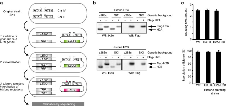
Background: Gametes are highly differentiated cells specialized to carry and protect the parental genetic information. During male germ cell maturation, histone proteins undergo distinct changes that result in a highly compacted chromatin organization. Technical difficulties exclude comprehensive analysis of precise histone mutations during mammalian spermatogenesis. The model organism Saccharomyces cerevisiae possesses a differentiation pathway termed sporulation which exhibits striking similarities to mammalian spermatogenesis. This study took advantage of this yeast pathway to first perform systematic mutational and proteomics screens on histones, revealing amino acid residues which are essential for the formation of spores.

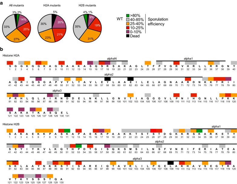
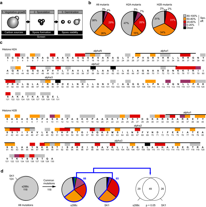
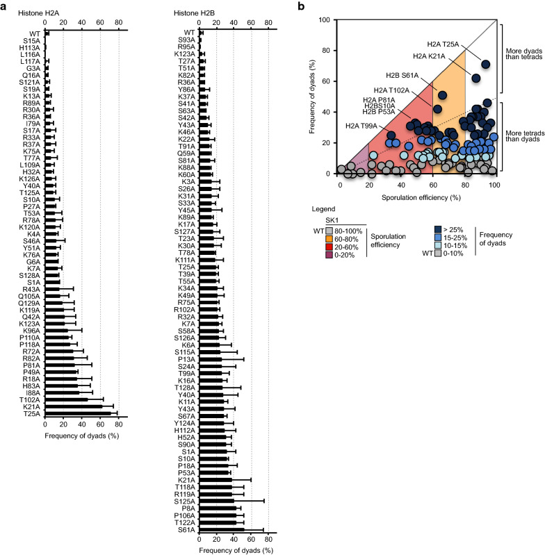
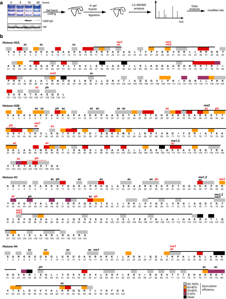
Methods: A systematic mutational screen has been performed on the histones H2A and H2B, generating ~ 250 mutants using two genetic backgrounds and assessing their ability to form spores. In addition, histones were purified at key stages of sporulation and post-translational modifications analyzed by mass spectrometry.

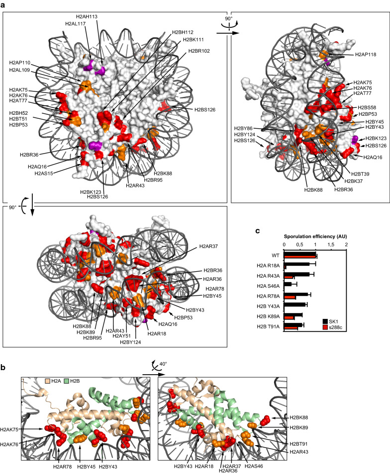
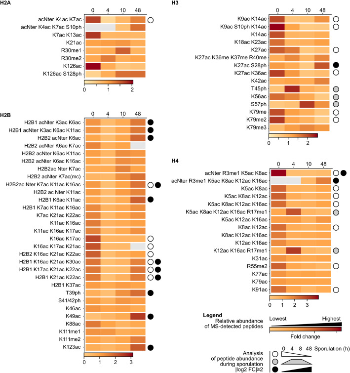
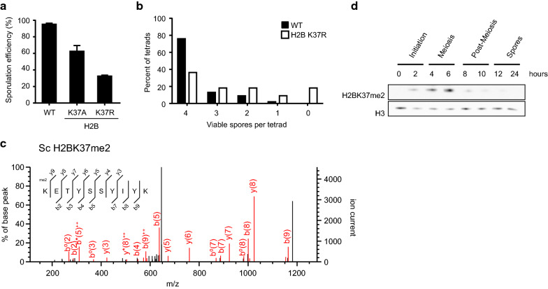
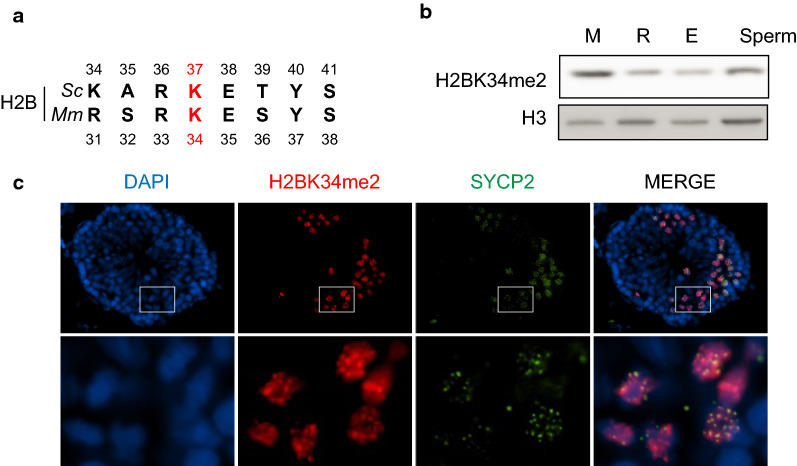
Results: The mutation of 75 H2A H2B residues affected sporulation, many of which were localized to the nucleosome lateral surface. The use of different genetic backgrounds confirmed the importance of many of the residues, as 48% of yeast histone mutants exhibited impaired formation of spores in both genetic backgrounds. Extensive proteomic analysis identified 67 unique post-translational modifications during sporulation, 27 of which were previously unreported in yeast. Furthermore, 33 modifications are located on residues that were found to be essential for efficient sporulation in our genetic mutation screens. The quantitative analysis of these modifications revealed a massive deacetylation of all core histones during the pre-meiotic phase and a close interplay between H4 acetylation and methylation during yeast sporulation. Methylation of H2BK37 was also identified as a new histone marker of meiosis and the mouse paralog, H2BK34, was also enriched for methylation during meiosis in the testes, establishing conservation during mammalian spermatogenesis.

Conclusion: Our results demonstrate that a combination of genetic and proteomic approaches applied to yeast sporulation can reveal new aspects of chromatin signaling pathways during mammalian spermatogenesis.


Conflict of interest statement

The authors declare that they have no competing interests.

Figures

Fig. 1
Fig. 1
A systematic screen on the histones H2A H2B identifies residues essential for sporulation in the s228c genetic background. a Statistical analysis of the sporulation efficiency of the H2A H2B mutants. The severity of the sporulation defects is described using the following color code: a sporulation efficiency included within 0–10%, 10–25%, 25–40%, 40–80% and 80–100% is represented in purple, red, orange, grey and green, respectively. Lethal mutations are represented in black. This same color-coding is used throughout the figures of the manuscript for the s288c background. b Localization of the substitution mutants with affected sporulation efficiency. Histone fold is represented on the top of each histone based on the yeast nucleosome structure [81]
Fig. 2
Fig. 2
Creation of the histone H2A and H2B shuffle strain in the SK1 genetic background. a Creation strategy. Genomic copies encoding H2A (HTA1 and HTA2) and H2B (HTB1 and HBT2) have been deleted and the HTA1 and HTB1 genes are expressed from an autonomous plasmid. The original URA3 plasmid can be replaced by a new HIS3 plasmid with any desired mutation. b Validation of the H2A and H2B shuffle strain. Plasmids encoding Flag-tagged H2A or H2B have been introduced in the strain and these histones have been detected by western blot. They confirm that all genomic copies of H2A/H2B have been deleted and the only H2A/H2B proteins are expressed from the autonomous plasmid in both SK1 and s288c genetic backgrounds. c No growth or sporulation defects are detected in the H2A and H2B shuffle strain in the SK1 background. The genetic manipulation did not affect doubling times during vegetative growth (top) or sporulation efficiency (bottom, see Additional file 1: Figure S1 for more details) when comparing the H2A H2B shuffling strain to a WT or a previously constructed H3 H4 shuffle strain [33]
Fig. 3
Fig. 3
A systematic genetic screen identifies H2A and H2B residues essential for the formation of spores in the SK1 genetic background. a Schematic of the strategy used for the genetic screen which systematically mutated H2A and H2B residues to alanine. > 250 mutant strains with at least three independent isolates per mutation have been created and validated by sequencing. The growth of strains with non-lethal mutations was first assessed on acetate, a non-fermentable carbon source. Then, their ability to form spores and the viability and stress resistance of these spores has been characterized. b Statistical analysis of the sporulation efficiency of the H2A H2B mutants. The severity of the sporulation defects is described using the following color code: a sporulation efficiency included within 0–20%, 20–60%, 60–80%, and 80–100% is represented in purple, red, orange, and grey, respectively. Lethal mutations are represented in black. This same color-coding is used throughout the figures of the manuscript for the SK1 background in accordance with Ref. [33]. c Localization of the substitution mutants with affected sporulation efficiency. Histone fold is represented on the top of each histone based on the yeast nucleosome structure [81]. d Venn diagram representing the repartition of the residues important for sporulation in SK1 and s288c genetic backgrounds. 49 mutations are associated with sporulation defects in both s288c and SK1 background, out of a total of 88 and 85, respectively. This enrichment is statistically significant (hypergeometric test, p value of 0.05)
Fig. 4
Fig. 4
Increased formation of dyads in H2A or H2B mutant strains in the SK1 genetic background. a Frequencies of dyads observed for each mutant. Value for the WT strain is presented as the first entry. b Representation of the frequency of dyad and spores (dyads and tetrads). Residues with the most defects are highlighted
Fig. 5
Fig. 5
Representation of the nucleosome highlighting the residues essential for the completion of sporulation. a Residues which are important for sporulation are highlighted on the nucleosomal structure. Color scheme of the legend identical to Fig. 3. Structural information downloaded from entry 1ID3 [81] of the PDB database [105]. b Visualization of H2A H2B residues important for sporulation localized at the nucleosome–DNA interface. c Sporulation efficiency of a selection of residues presented in b. Sporulation efficiencies are expressed in arbitrary units, with WT normalized to 1 in SK1 and s288c backgrounds
Fig. 6
Fig. 6
Mass spectrometry identification of post-translational modifications on core histones during sporulation. a Histones were purified during sporulation and analyzed by mass spectrometry. Technical details are presented in the Method section. b Modifications identified for each histone during sporulation. Ac, acetylation; me1/me2/me3, mono-, di- and trimethylation; ph, phosphorylation. Sites which had not been identified in yeast are highlighted in red, based on Table 3. Their MS/MS spectra are presented in Additional file 3. Phosphorylated H2B residues with ambiguous localization are indicated with a red bar. All histones were identified as N-terminally acetylated. Color coding of the residues is from SK1 background (Fig. 3)
Fig. 7
Fig. 7
Quantification of the histone modifications identified by proteomics. The abundance of modified peptides was quantified and normalized to the signals of reference non-modified peptides for each histone, namely HLQLAIR and AGLTFPVGR for H2A, KETYSSYIYK and ETYSSYIYK for H2B, STELLIR for H3 and ISGLIYEETR and DNIQGITKPAIR for H4. The time after induction of sporulation is indicated in hours (4 h, meiosis; 10 h, post meiosis; 48 h, mature spores). The intensity of the brown color indicates the relative abundance during the progression of sporulation. Significant changes of abundance over the course of sporulation are highlighted with circles (| log2 of fold change | ≥ 2). Data are available in Additional file 5. acNter, N-terminal acetylation
Fig. 8
Fig. 8
H2BK37 is methylated during yeast meiosis. a Sporulation efficiency in H2BK37A and H2BK37R mutants. Both of them significantly affect the formation of spores. b Spores viability upon H2BK37R mutation. c MS/MS fragmentation spectrum identifying H2BK37me2 in yeast histone purified during meiosis. Sc, Saccharomyces cerevisiae. d Analysis of the abundance of H2BK37me2 during sporulation
Fig. 9
Fig. 9
Mouse H2BK34me2 is detected in meiosis during spermatogenesis. a Alignment of H2B sequences in yeast and human around the yeast H2BK37 residue. Sc, Saccharomyces cerevisiae. Mm, Mus musculus. b H2BK34me2 levels during spermatogenesis. Cells were purified at different stages and analyzed by western blot. M, meiosis (spermatocytes); R, round spermatids; E, elongating spermatids. C. H2BK34me2 detection in mouse seminiferous tubules analyzed by immunofluorescence. H2BK34me2 were co-detected with Sycp2, a classic marker of meiosis. Insets are magnified in the bottom images

Similar articles

Cited by

References

    1. Esteves SC. Novel concepts in male factor infertility: clinical and laboratory perspectives. J Assist Reprod Genet. 2016;33:1319–1335. - PMC - PubMed
    1. Carrell DT, Hammoud SS. The human sperm epigenome and its potential role in embryonic development. Mol Hum Reprod. 2010;16:37–47. - PubMed
    1. Carrell DT. Epigenetics of the male gamete. Fertil Steril. 2012;97:267–274. - PubMed
    1. Gannon JR, Emery BR, Jenkins TG, Carrell DT. The sperm epigenome: implications for the embryo. Adv Exp Med Biol. 2014;791:53–66. - PubMed
    1. Böhmdorfer G, Wierzbicki AT. Control of chromatin structure by long noncoding RNA. Trends Cell Biol. 2015;25:623–632. - PMC - PubMed

Publication types