. 2020 Dec 4;295(49):16545-16561.
doi: 10.1074/jbc.RA120.013998. Epub 2020 Sep 15.

Biochemical and biophysical analyses of hypoxia sensing prolyl hydroxylases from Dictyostelium discoideum and Toxoplasma gondii

Affiliations

Biochemical and biophysical analyses of hypoxia sensing prolyl hydroxylases from Dictyostelium discoideum and Toxoplasma gondii

Tongri Liu et al. J Biol Chem. .

Abstract

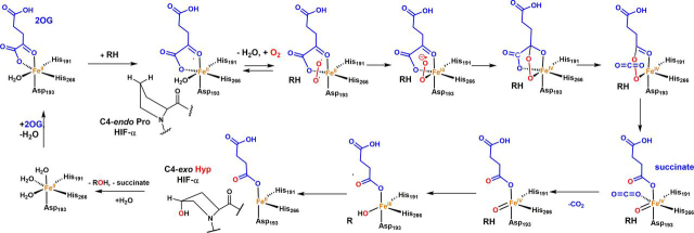
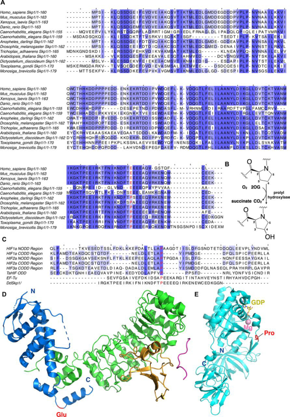
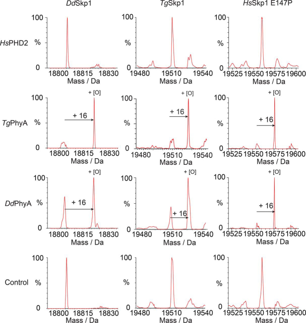
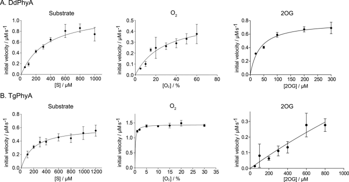
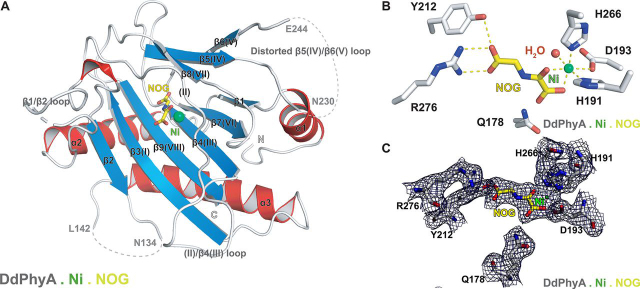
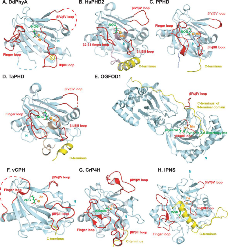
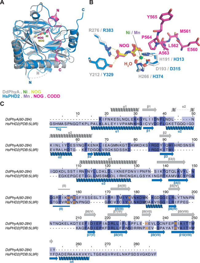
In animals, the response to chronic hypoxia is mediated by prolyl hydroxylases (PHDs) that regulate the levels of hypoxia-inducible transcription factor α (HIFα). PHD homologues exist in other types of eukaryotes and prokaryotes where they act on non HIF substrates. To gain insight into the factors underlying different PHD substrates and properties, we carried out biochemical and biophysical studies on PHD homologues from the cellular slime mold, Dictyostelium discoideum, and the protozoan parasite, Toxoplasma gondii, both lacking HIF. The respective prolyl-hydroxylases (DdPhyA and TgPhyA) catalyze prolyl-hydroxylation of S-phase kinase-associated protein 1 (Skp1), a reaction enabling adaptation to different dioxygen availability. Assays with full-length Skp1 substrates reveal substantial differences in the kinetic properties of DdPhyA and TgPhyA, both with respect to each other and compared with human PHD2; consistent with cellular studies, TgPhyA is more active at low dioxygen concentrations than DdPhyA. TgSkp1 is a DdPhyA substrate and DdSkp1 is a TgPhyA substrate. No cross-reactivity was detected between DdPhyA/TgPhyA substrates and human PHD2. The human Skp1 E147P variant is a DdPhyA and TgPhyA substrate, suggesting some retention of ancestral interactions. Crystallographic analysis of DdPhyA enables comparisons with homologues from humans, Trichoplax adhaerens, and prokaryotes, informing on differences in mobile elements involved in substrate binding and catalysis. In DdPhyA, two mobile loops that enclose substrates in the PHDs are conserved, but the C-terminal helix of the PHDs is strikingly absent. The combined results support the proposal that PHD homologues have evolved kinetic and structural features suited to their specific sensing roles.

Keywords: 2-oxoglutarate/α-ketoglutarate oxygenase; Dictyostelium discoideum; S-phase kinase-associated protein 1 (Skp1); Toxoplasma gondii; dioxygenase; hypoxia; hypoxia-inducible factor (HIF); hypoxia/oxygen sensor; prolyl-hydroxylase; protein evolution.


Conflict of interest statement

Conflict of interest—The authors declare no competing financial interests.

Figures

Figure 1
Figure 1
Outline of the consensus 2OG oxygenase mechanism. In the resting state, the active site iron is typically coordinated by a triad of residues (HX(D/E)…H motif) and 2-3 water molecules giving octahedral coordination. Sequential binding of 2OG, then substrate, then O2 to the active site occurs, causing displacement of the resting state metal-bound waters. Oxidative decarboxylation of 2OG (not all the proposed intermediates have been characterized) gives a ferryl intermediate that is directly responsible for substrate oxidation. In the case of the PHDs the substrate prolyl residue binds in a C4-endo conformation but changes to a C4-exo conformation on hydroxylation (61, 102).
Figure 2
Figure 2
Sequence alignment of eukaryotic Skp1 proteins.A, Skp1 sequences are from the following organisms: human (Homo sapiens, Gene ID NP_008861.2), mouse (Mus musculus, Gene ID NP_035673.3), frog (Xenopus laevis, Gene ID AAF65619.1), zebra fish (Danio rerio, Gene ID NP_957037.1), roundworm (Caenorhabditis elegans, Gene ID NP_510193.4), roundworm (Caenorhabditis elegans, Gene ID NP_507857.1), mosquito (Anopheles darlingi, Gene ID ETN58923.1), fruit fly (Drosophila melanogaster, Gene ID NP_477390.1), T. adhaerens (Gene ID EDV18896.1), Arabidopsis thaliana (Gene ID AEE35780.1), D. discoideum (Gene ID XP_644826.1), T. gondii (Gene ID CAJ20499.1), and Monosiga brevicollis (Gene ID EDQ90844.1, predicted protein). The (potentially) hydroxylated prolyl residue, as occurs for PhyA in D. discoideum and T. gondii, is in red. Residue 147 (HsSkp1, for example) is a glutamyl residue in many complex animals. B, prolyl hydroxylases catalyze prolyl-4-hydroxylation. C, sequence alignment of prolyl hydroxylase substrates. Human HIF-1α (Gene ID AAC68568.1), human HIF-2α (Gene ID Q99814.3), human HIF-3α (Gene ID Q9Y2N7.2), T. adhaerens HIF (Gene ID AFM37575.1), and P. putida EF-Tu (Gene ID B1JDW6.1). The (potentially) hydroxylated prolyl residue is in red. D, view from a structure of human Skp1 in the Skp1-Skp2-Cks1-p27 peptide complex (PDB code 2AST) (103). Skp1 is shown as a blue cartoon, Skp2 is in green, Cks1 is in orange, and the p27 peptide is in pink. The glutamyl residue is in red. E, view from a structure of E. coli EF-Tu (PDB code 5MI3) (104). EF-Tu is shown as a light blue cartoon. The hydroxylated prolyl residue by the PHD is in red sticks; Mg2+ is pink, and GDP is yellow.
Figure 3
Figure 3
Mass spectra of full-length DdSkp1 and TgSkp1 proteins after incubation under varied conditions (+16 Da peak shift indicates DdPhyA and TgPhyA hydroxylation activity).A, TgSkp1 hydroxylation requires TgPhyA, 2OG, and is enhanced by ascorbate. B, DdSkp1 is hydroxylated by DdPhyA in the presence of 2OG; l-ascorbate increases DdPhyA activity, but to a lesser extent than TgPhyA. Assays were performed in the presence or absence (no-enzyme control top panels) of DdPhyA or TgPhyA (1 μm), full-length DdSkp1 or full-length TgSkp1 (100 μm), (NH4)2Fe(II)(SO4)2 (50 μm), sodium l-ascorbate (1 mm), and 2OG (500 μm) in HEPES (100 mm), pH 7.6. Reactions were incubated at 37 °C for 1 h and quenched using an equal volume of 1% (v/v) aqueous formic acid, then subjected to LC-ESI-MS analysis. Assay details are provided under the Supporting information.
Figure 4
Figure 4
Substrate selectivity comparison of DdPhyA, TgPhyA, and HsPHD2.Rows are specified by enzyme type or control; column headers indicate the substrate used. LC-ESI-MS analysis reveals HsPHD2 does not catalyze hydroxylation of any of the Skp1 proteins (DdSkp1, TgSkp1, or HsSkp1(E147P)). Hydroxylation of full-length DdSkp1 protein by DdPhyA and TgPhyA occurs in the presence of 2OG and ascorbate. Full-length TgSkp1 is hydroxylated by both DdPhyA and TgPhyA under the same conditions. Full-length HsSkp1(E147P) is hydroxylated by DdPhyA and TgPhyA. Assays were performed under atmospheric O2 concentrations and contained HsPHD2, DdPhyA, or TgPhyA (1 μm), either full-length HsSkp1(E147P), full-length DdSkp1 or full-length TgSkp1 substrate (100 μm), (NH4)2Fe(II)(SO4)2 (50 μm), sodium l-ascorbate (1 mm), and 2OG (500 μm) in HEPES (100 mm), pH 7.6. Reactions were incubated at 37 °C (1 h), then quenched with 1% (v/v) aqueous formic acid and subjected to LC-ESI-MS analysis. Assay details are provided under the Supporting information.
Figure 5
Figure 5
Kinetics of DdPhyA and TgPhyA.A, studies on the dependence of DdPhyA activity on concentrations of: substrate (full-length DdSkp1) (500 μm 2OG, 50 μm (NH4)2Fe(II)(SO4)2, 1 mm l-ascorbate, normoxic, 4 min, Xevo LC-ESI-MS); 2OG (DdPhyA (1 μm), full-length DdSkp1 substrate (100 μm), (NH4)2Fe(II)(SO4)2 (50 μm), sodium l-ascorbate (1 mm), normoxic, 6 min, RapidFire MS); and O2 (DdPhyA (1 μm), full-length DdSkp1 substrate (600 μm), (NH4)2Fe(II)(SO4)2 (50 μm), sodium l-ascorbate (1 mm) and 2OG disodium salt (500 μm), 8.25 min, Xevo LC-ESI-MS). All assays were in HEPES (100 mm), pH 7.6, at 37 °C. Assays using Xevo LC-ESI-MS were in triplicate; those using RapidFire MS were in duplicate. B, studies on the dependence of TgPhyA activity on concentrations of: substrate (full-length TgSkp1) (500 μm 2OG, 50 μm (NH4)2Fe(II)(SO4)2, 1 mm l-ascorbate, normoxic, 4 min, Xevo LC-ESI-MS); 2OG (TgPhyA (1 μm), full-length TgSkp1 substrate (600 μm), (NH4)2Fe(II)(SO4)2 (50 μm), sodium l-ascorbate (1 mm), normoxic, 6 min, RapidFire MS); and O2 (TgPhyA (1 μm), full-length TgSkp1 substrate (600 μm), (NH4)2Fe(II)(SO4)2 (50 μm), sodium l-ascorbate (1 mm) and 2OG disodium salt (500 μm), 8.25 min, Xevo LC-ESI-MS). All assays were in HEPES (100 mm), pH 7.6, at 37 °C. Assays using Xevo LC-ESI-MS were in triplicate; those using RapidFire MS were in duplicate. Note the high level of TgPhyA uncoupled 2OG turnover complicates 2OG Kmapp determination, see text. Assay details are provided in the Supporting information.
Figure 6
Figure 6
Views from a crystal structure of the D. discoideum prolyl hydroxylase (DdPhyA(60-284)) (PDB code6T8M).A, secondary structural elements in DdPhyA comprise 3 α-helices and 9 β-strands, 7 of which, together with one loop (II), form the DSBH core-fold (I–VIII). B, active site close-up; the metal ion (nickel substituting for iron, green sphere) is octahedrally coordinated by His266, Asp193, and His191, NOG (a close 2OG analog), and a water (red sphere). NOG is positioned to interact with Arg276 via a salt bridge, i.e. between the NOG carboxyl group and the Arg276 side chain guanidino group. C, electron density map of an active site close-up. Representative 2mFo − DFc electron density map contoured to 1σ (blue mesh) around selected active site residues (white sticks), Ni(II) (substituting for Fe(II), green sphere), and NOG (yellow sticks).
Figure 7
Figure 7
Comparisons of the finger loop, βII-βIII, and βIV/βV insert loops, and the C-terminal regions in prolyl hydroxylase structures.A, DdPhyA (PDB code 6T8M); B, HsPHD2 (PDB code 6L7V3HQR); C, PPHD (PDB code 4IW3); D, TaPHD (PDB code 6F0W); E, OGFOD1 (PDB code 4NHY); F, vCPH (PDB code 5C5T); G, CrP4H (PDB code 3GZE); and H, IPNS (PDB code 1W06). The finger loop (which in HsPHD2 links β2 and β3) and βII-βIII, βIV-βV insert loops are in red. C-terminal regions following the DSBH are in yellow; this region leads to the linker between the two DSBH domains in OGOFD1). Note: the highlighted βIV-βV insert loop has been shown to be involved in substrate binding by some 2OG oxygenase subfamilies, but this is not the case for the prolyl hydroxylase subfamily. Active site metal ion and ligands are shown as a ball and stick.
Figure 8
Figure 8
Comparison of DdPhyA and human PHD2 structures.A, superimposition of views from structures of the DdPhyA·Ni·NOG and PHD2·Mn·NOG·HIF-1α CODD (PDB code 3HQR) complexes. B, superimposition of active site views. Note that HIF-1α CODD is bound to PHD2, but substrate is not present for DdPhyA. C, sequence alignment of DdPhyA(60-284) and the catalytic domain of human PHD2(181-426). Residues involved in Fe(II) binding are in yellow.

Similar articles

Cited by

References

    1. Semenza G.L. Hypoxia-inducible factor 1: control of oxygen homeostasis in health and disease. Pediatr. Res. 2001;49:614–617. doi: 10.1203/00006450-200105000-00002. 11328942. - DOI - PubMed
    1. Jaakkola P., Mole D.R., Tian Y.M., Wilson M.I., Gielbert J., Gaskell S.J., von Kriegsheim A., Hebestreit H.F., Mukherji M., Schofield C.J., Maxwell P.H., Pugh C.W., Ratcliffe P.J. Targeting of HIF-α to the von Hippel-Lindau ubiquitylation complex by O2-regulated prolyl hydroxylation. Science. 2001;292:468–472. doi: 10.1126/science.1059796. 11292861. - DOI - PubMed
    1. Chowdhury R., Hardy A., Schofield C.J. The human oxygen sensing machinery and its manipulation. Chem. Soc. Rev. 2008;37:1308–1319. doi: 10.1039/b701676j. 18568157. - DOI - PubMed
    1. Semenza G.L. HIF-1 and mechanisms of hypoxia sensing. Curr. Opin. Cell. Biol. 2001;13:167–171. doi: 10.1016/S0955-0674(00)00194-0. 11248550. - DOI - PubMed
    1. Kaelin W.G., Jr., Ratcliffe P.J. Oxygen sensing by metazoans: the central role of the HIF hydroxylase pathway. Mol. Cell. 2008;30:393–402. doi: 10.1016/j.molcel.2008.04.009. 18498744. - DOI - PubMed

Publication types

MeSH terms