Review
. 2020 Sep 15;133(18):jcs241174.
doi: 10.1242/jcs.241174.

New insights into non-transcriptional regulation of mammalian core clock proteins

Affiliations
Review

New insights into non-transcriptional regulation of mammalian core clock proteins

Priya Crosby et al. J Cell Sci. .

Abstract

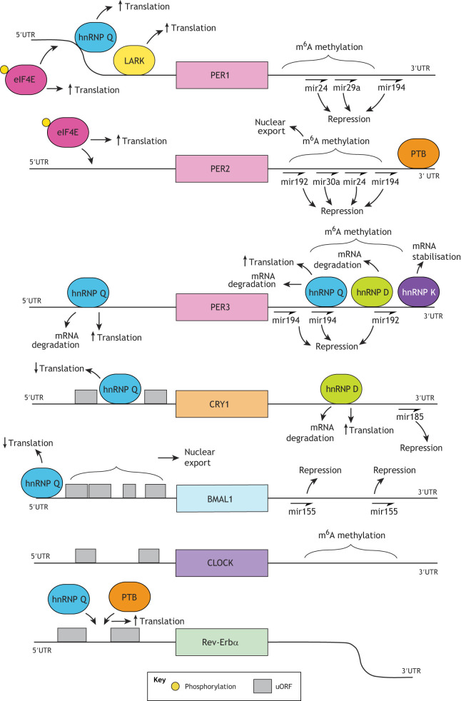
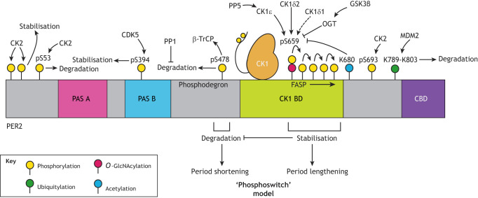
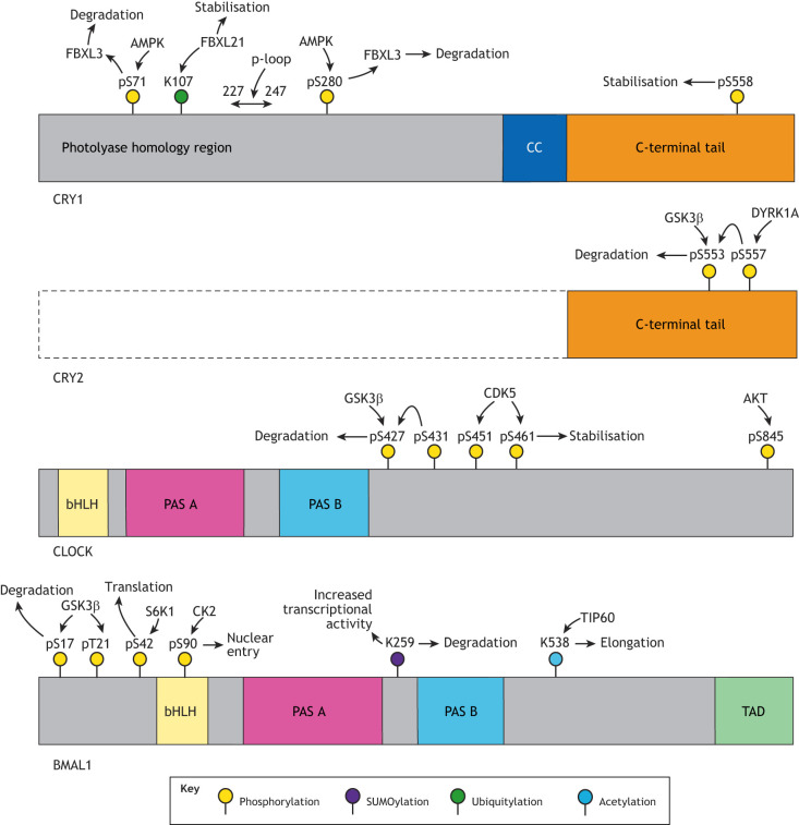
Mammalian circadian rhythms drive ∼24 h periodicity in a wide range of cellular processes, temporally coordinating physiology and behaviour within an organism, and synchronising this with the external day-night cycle. The canonical model for this timekeeping consists of a delayed negative-feedback loop, containing transcriptional activator complex CLOCK-BMAL1 (BMAL1 is also known as ARNTL) and repressors period 1, 2 and 3 (PER1, PER2 and PER3) and cryptochrome 1 and 2 (CRY1 and CRY2), along with a number of accessory factors. Although the broad strokes of this system are defined, the exact molecular mechanisms by which these proteins generate a self-sustained rhythm with such periodicity and fidelity remains a topic of much research. Recent studies have identified prominent roles for a number of crucial post-transcriptional, translational and, particularly, post-translational events within the mammalian circadian oscillator, providing an increasingly complex understanding of the activities and interactions of the core clock proteins. In this Review, we highlight such contemporary work on non-transcriptional events and set it within our current understanding of cellular circadian timekeeping.

Keywords: Cellular timekeeping; Circadian rhythm; Post-transcriptional modification; Post-translational modification; Translational regulation.


Conflict of interest statement

Competing interestsThe authors declare no competing or financial interests.

Figures

Fig. 1.
Fig. 1.
Schematic overview of factors regulating mRNA levels of core clock proteins. Myriad factors influence the stability of mRNA coding for core clock proteins. Coding sequences are represented as coloured blocks, flanked by 5′ and 3′ UTRs. Large coloured lozenge shapes represent proteins and half-arrows denote miRNA binding. Readers should be aware that the exact binding sites of some proteins are not currently strictly defined, and so their order relative to each other is not definitive.
Fig. 2.
Fig. 2.
Schematic illustration of post-translational regulation of PER2. PER2 abundance and stability is a crucial determinant of circadian period and phase, with PER stabilisation associated with increased cellular period, whereas destabilisation of PER2 shortens the period. Phosphorylation of PER2 by CK1 is integral to regulation of PER2 stability. CK1δ and CK1ε bind to PER2 at the CK1-binding domain (CK1 BD) and can either stabilise or promote degradation of PER2, depending on the exact modification site, with phosphorylation at S478 in the phosphodegron promoting recruitment of the E3 ubiquitin ligase β-TrCP and thus degradation, whereas phosphorylation at the FASP region delays degradation. This interplay constitutes the ‘phosphoswitch’ model, which proposes that the balance of phosphorylation between these stabilising and degrading regions determines overall PER2 half-life. Dephosphorylation of PER2 by protein phosphatase 1 (PP1) reduces its overall degradation. In addition, CK2-mediated phosphorylation at the extreme N-terminus is proposed to stabilise the protein, whereas CK2 phosphorylation at S53 is thought to promote degradation, although more recent work suggests that the most significant site of CK2 phosphorylation on PER2 is S693. O-GlcNAcylation by O-GlcNAc transferase (OGT) at the priming serine (S659 in mice/S662 in humans) of the FASP site competitively inhibits PER2 phosphorylation by CK1, with GSK3β being a positive regulator of OGT activity. Acetylation at K680 also inhibits phosphorylation at the priming serine residue. Furthermore, ubiquitylation by MDM2 at sites downstream of FASP promotes PER2 degradation in a phosphorylation-independent manner, whereas phosphorylation at S394 within the PAS-B domain by CDK5 promotes stability. Phosphorylation and dephosphorylation (e.g. by PP5) of the C-terminal tail of CK1 can also influence its activity on its PER2 substrate. Further modifications that are discussed in the text but which currently lack a defined site of action or function, including direct phosphorylation of PER2 by GSK3β, decacetylation by SIRT1 and some O-GlcNAcylated serine residues, are not shown.
Fig. 3.
Fig. 3.
Schematic illustration overview of post-translational modifications on other core clock proteins. Although post-translational modifications of PER2 are the most studied, other core clock proteins are also controlled by significant post-translational modification. Only the C-terminal tail of CRY2 is shown, but the high similarity between the CRY1 and CRY2 photolyase-homology region (PHR) likely means that post-translational events applicable to CRY1 also occur on CRY2. Owing to their large number, only selected CRY1 phosphosites are shown.

References

    1. Anafi R. C., Francey L. J., Hogenesch J. B. and Kim J. (2017). CYCLOPS reveals human transcriptional rhythms in health and disease. Proc. Natl. Acad. Sci. USA 114, 5312-5317. 10.1073/pnas.1619320114 - DOI - PMC - PubMed
    1. Aryal R. P., Kwak P. B., Tamayo A. G., Gebert M., Chiu P.-L., Walz T. and Weitz C. J. (2017). Macromolecular assemblies of the mammalian circadian clock. Mol. Cell 67, 770-782.e6. 10.1016/j.molcel.2017.07.017 - DOI - PMC - PubMed
    1. Asher G., Gatfield D., Stratmann M., Reinke H., Dibner C., Kreppel F., Mostoslavsky R., Alt F. W. and Schibler U. (2008). SIRT1 regulates circadian clock gene expression through PER2 deacetylation. Cell 134, 317-328. 10.1016/j.cell.2008.06.050 - DOI - PubMed
    1. Atger F., Gobet C., Marquis J., Martin E., Wang J., Weger B., Lefebvre G., Descombes P., Naef F. and Gachon F. (2015). Circadian and feeding rhythms differentially affect rhythmic mRNA transcription and translation in mouse liver. Proc. Natl. Acad. Sci. USA 112, E6579-E6588. 10.1073/pnas.1515308112 - DOI - PMC - PubMed
    1. Balsalobre A., Brown S. A., Marcacci L., Tronche F., Kellendonk C., Reichardt H. M., Schutz G. and Schibler U. (2000). Resetting of circadian time in peripheral tissues by glucocorticoid signaling. Science 289, 2344-2347. 10.1126/science.289.5488.2344 - DOI - PubMed

Publication types

MeSH terms