. 2020 Sep 15;11(5):e01882-20.
doi: 10.1128/mBio.01882-20.

MARCH8 Inhibits Ebola Virus Glycoprotein, Human Immunodeficiency Virus Type 1 Envelope Glycoprotein, and Avian Influenza Virus H5N1 Hemagglutinin Maturation

Affiliations

MARCH8 Inhibits Ebola Virus Glycoprotein, Human Immunodeficiency Virus Type 1 Envelope Glycoprotein, and Avian Influenza Virus H5N1 Hemagglutinin Maturation

Changqing Yu et al. mBio. .

Abstract

Membrane-associated RING-CH-type 8 (MARCH8) strongly blocks human immunodeficiency virus type 1 (HIV-1) envelope glycoprotein (Env) incorporation into virions by downregulating its cell surface expression, but the mechanism is still unclear. We now report that MARCH8 also blocks the Ebola virus (EBOV) glycoprotein (GP) incorporation via surface downregulation. To understand how these viral fusion proteins are downregulated, we investigated the effects of MARCH8 on EBOV GP maturation and externalization via the conventional secretion pathway. MARCH8 interacted with EBOV GP and furin when detected by immunoprecipitation and retained the GP/furin complex in the Golgi when their location was tracked by a bimolecular fluorescence complementation (BiFC) assay. MARCH8 did not reduce the GP expression or affect the GP modification by high-mannose N-glycans in the endoplasmic reticulum (ER), but it inhibited the formation of complex N-glycans on the GP in the Golgi. Additionally, the GP O-glycosylation and furin-mediated proteolytic cleavage were also inhibited. Moreover, we identified a novel furin cleavage site on EBOV GP and found that only those fully glycosylated GPs were processed by furin and incorporated into virions. Furthermore, the GP shedding and secretion were all blocked by MARCH8. MARCH8 also blocked the furin-mediated cleavage of HIV-1 Env (gp160) and the highly pathogenic avian influenza virus H5N1 hemagglutinin (HA). We conclude that MARCH8 has a very broad antiviral activity by prohibiting different viral fusion proteins from glycosylation and proteolytic cleavage in the Golgi, which inhibits their transport from the Golgi to the plasma membrane and incorporation into virions.IMPORTANCE Enveloped viruses express three classes of fusion proteins that are required for their entry into host cells via mediating virus and cell membrane fusion. Class I fusion proteins are produced from influenza viruses, retroviruses, Ebola viruses, and coronaviruses. They are first synthesized as a type I transmembrane polypeptide precursor that is subsequently glycosylated and oligomerized. Most of these precursors are cleaved en route to the plasma membrane by a cellular protease furin in the late secretory pathway, generating the trimeric N-terminal receptor-binding and C-terminal fusion subunits. Here, we show that a cellular protein, MARCH8, specifically inhibits the furin-mediated cleavage of EBOV GP, HIV-1 Env, and H5N1 HA. Further analyses uncovered that MARCH8 blocked the EBOV GP glycosylation in the Golgi and inhibited its transport from the Golgi to the plasma membrane. Thus, MARCH8 has a very broad antiviral activity by specifically inactivating different viral fusion proteins.

Keywords: Ebola; HIV; MARCH8; class I fusion protein; furin; glycosylation; influenza; viral envelope.


Figures

FIG 1
FIG 1
MARCH8 downregulates EBOV GP from the cell surface. (A) MARCH8 expressions in 293T, THP-1, Vero E6, HeLa, Huh7, Hep G2, and TZM-bI cells, or in 293T cells transfected with a human MARCH8 expression vector (293T+MARCH8), were analyzed by Western blotting (WB) using a specific anti-human MARCH8. (B) HIV-1 firefly luciferase-reporter pseudoviruses were produced from 293T cells in the presence of EBOV GPΔMLD and MARCH8 from different species or APOBEC3G. Viral infectivity was determined via infecting Vero E6 cells. Infectivity is shown as relative values, with the infectivity in the presence of a control (Ctrl) vector set as 100. Results were from three different experiments. h, human; b, cow (Bos taurus); m, mouse. (C) HIV-1 pseudoviruses were collected from cell culture supernatants in panel B and purified via ultracentrifugation. In addition, HIV-1 pseudoviruses containing the full-length EBOV GP were produced and purified similarly. The levels of GP1 expression in these VLPs were analyzed by WB using anti-FLAG, and the levels of VLPs were determined by anti-HIV-1 Gag (p24). Protein expressions in 293T cells are shown in Fig. 4D. (D) EBOV virus-like particles (VLPs) were produced from 293T cells after expressing EBOV VP40, human MARCH8 or its W114A mutant, and EBOV GP or GPΔMLD. After purification via ultracentrifugation, these VLPs were analyzed by WB. Protein expressions in viral producer cells are shown in Fig. 4C. (E) EBOV GPΔMLD that has a GFP tag was expressed in HeLa cells in the presence or absence of human MARCH8. After staining with DAPI, the GP localization was detected by confocal microscopy. (F) EBOV GPΔMLD was expressed with increasing amounts of human MARCH8 in 293T cells. The GP expression on cell surface was detected by flow cytometry using anti-FLAG followed by Alexa Fluor 488-conjugated goat anti-mouse IgG.
FIG 2
FIG 2
Tracking MARCH8, EBOV GP, and furin protein complexes by BiFC. (A) FLAG-tagged GPΔMLD was expressed with HA-tagged human MARCH8. In addition, FLAG-tagged furin was expressed with HA-tagged human MARCH8 and GPΔMLD in 293T cells. Proteins were immunoprecipitated with anti-FLAG and analyzed by WB. MARCH8, furin, and GP were detected by anti-HA, anti-FLAG, or anti-EBOV GP, respectively. (B) GFP, GP-VC, furin-VN, or human MARCH8-VN proteins were individually expressed in HeLa cells. After staining with DAPI, fluorescent signals were observed by confocal microscopy. (C) The MARCH8-VN-HA/GP-VC or furin-VN-HA/GP-VC BiFC pair was expressed in HeLa cells. Cells were stained with fluorescent anti-HA to detect MARCH8 or furin. In addition, furin-VN that does not express the HA tag was expressed with GP-VC and HA-tagged MARCH8 in HeLa cells. Cells were stained with fluorescent anti-HA to detect MARCH8. Fluorescent signals were observed by confocal microscopy. (D) Indicated BiFC fusion proteins were expressed individually or pairwise, and the levels of BiFC signals were determined by flow cytometry. SERINC5, serine incorporator 5. Experiments were repeated three times, and identical results were obtained.
FIG 3
FIG 3
MARCH8 retains EBOV GP in the Golgi. The furin-VN/GP-VC BiFC pair was expressed with CNX-mCherry or TGN-mCherry in the presence or absence of human MARCH8 in HeLa cells. After staining with DAPI, fluorescent signals were observed by confocal microscopy.
FIG 4
FIG 4
MARCH8 blocks the EBOV GP proteolytic cleavage. (A) GP was expressed with wild-type (WT) human MARCH8 or its W114A mutant in 293T cells. The GP and MARCH8 expression was detected by WB using anti-FLAG or anti-HA. Activities of MARCH8 proteins from different species were also compared. (B) GP was expressed with human MARCH8 in HeLa and Hep G2 cells, or in Huh7 cells in the presence of control (Ctrl) or MARCH8-specific siRNAs. The GP processing was determined by WB. (C) Similar experiments as in panel A were conducted to determine how MARCH8 and its W114A mutant affect GP cleavage in the presence of EBOV VP40. (D) Similar experiments as in panel A were conducted to determine how MARCH8 proteins from different species affect GP cleavage in the presence of HIV-1 proteins. (E) GPΔMLD was expressed with human MARCH8 in 293T cells and treated with NH4Cl, bafilomycin A1 (BafA1), MG132, or DBeQ. Protein expressions were determined by WB. (F) VSV-G was expressed with human MARCH8 or its W114A mutant in 293T cells and treated with NH4Cl or MG132. Protein expressions were determined by WB. Numbers beside gels in this and subsequent figures indicate molecular masses in kilodaltons.
FIG 5
FIG 5
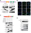
Mapping MARCH8 and furin active domains. (A) A schematic diagram of human MARCH8 protein with a RING domain and two TM domains is presented. Three MARCH8 C-terminal CT deletion mutants, 1–213, 1–247, and 1–272, were created and expressed with the full-length GP or GPΔMLD. MARCH8 and GP expression was detected by WB using anti-HA or anti-FLAG. (B) MARCH8 and its three deletion mutants that have a C-terminal GFP tag were expressed in HeLa cells. After staining with DAPI, their subcellular localizations were detected by confocal microscopy. (C) A schematic diagram of human furin functional domains is presented. FLAG-tagged CD, P domain, and CD subdomain-deletion mutants were created and were expressed with GPΔMLD. Proteins were immunoprecipitated by anti-FLAG and detected by WB. Furin was detected by anti-FLAG, and EBOV GP was detected by anti-EBOV GP antibodies. (D) The full-length GP was expressed with increasing amounts of furin in 293T cells. The GP and furin expression was determined by WB.
FIG 6
FIG 6
MARCH8 blocks the EBOV GP modification by Nc- and O-glycans. (A) GP, GPΔFR, GPΔMLD, or GPΔMLDΔFR was expressed with human MARCH8 in 293T cells. After lysis, cell lysate was treated with Endo H or PNGase F, or left untreated, and analyzed by WB. (B) GPΔFR and GPΔMLDΔFR were expressed with EBOV VP40 and human MARCH8 or furin in 293T cells. EBOV VLPs were purified from supernatants via ultracentrifugation. EBOV GP, MARCH8, or furin expressions in cells and/or VLPs were determined by WB.
FIG 7
FIG 7
MARCH8 blocks the EBOV GP shedding and secretion. (A) Full-length GP and GPΔMLD that have an N-terminal FLAG tag were expressed with MARCH8 or furin. GP proteins released into supernatants were immunoprecipitated using anti-FLAG. The GP and MARCH8 expression in cells or supernatants was detected by WB. (B) A schematic diagram for EBOV GP, secreted GP (sGP), and small secreted GP (ssGP) expression is presented. Their relative expression levels from nonedited (7A) and edited (8A, 9A) GP mRNAs and a novel furin cleavage site in GP1 are shown. (C) GP1, GP1ΔMLD, and sGP that have an N-terminal FLAG tag were expressed with indicated MARCH8 proteins in 293T cells. GP proteins released into supernatants were immunoprecipitated as in panel A. The GP and MARCH8 protein expression in cells and supernatants was detected by WB. Sup, supernatants.
FIG 8
FIG 8
MARCH8 blocks the HIV-1 Env and H5N1 HA maturation. (A) FLAG-tagged furin was expressed with MARCH8 and HIV-1 Env or H5N1 HA in 293T cells. Proteins were immunoprecipitated with anti-FLAG and analyzed by WB. (B) HIV-1 Env and H5N1 HA were expressed with MARCH8 in 293T cells, and their processing was determined by WB. (C) HIV-1 Env and H5N1 HA were expressed with MARCH8 in 293T cells. After lysis of cells, cell lysate was treated with Endo H or PNGase F, or left untreated, and analyzed by WB.

Similar articles

Cited by

References

    1. Ng F, Tang BL. 2016. Unconventional protein secretion in animal cells. Methods Mol Biol 1459:31–46. doi:10.1007/978-1-4939-3804-9_2. - DOI - PubMed
    1. Viotti C. 2016. ER to Golgi-dependent protein secretion: the conventional pathway. Methods Mol Biol 1459:3–29. doi:10.1007/978-1-4939-3804-9_1. - DOI - PubMed
    1. Reily C, Stewart TJ, Renfrow MB, Novak J. 2019. Glycosylation in health and disease. Nat Rev Nephrol 15:346–366. doi:10.1038/s41581-019-0129-4. - DOI - PMC - PubMed
    1. Frabutt DA, Zheng YH. 2016. Arms race between enveloped viruses and the host ERAD machinery. Viruses 8:255. doi:10.3390/v8090255. - DOI - PMC - PubMed
    1. Rey FA, Lok SM. 2018. Common features of enveloped viruses and implications for immunogen design for next-generation vaccines. Cell 172:1319–1334. doi:10.1016/j.cell.2018.02.054. - DOI - PMC - PubMed

Publication types

MeSH terms