. 2020 Sep 15;11(5):e02219-20.
doi: 10.1128/mBio.02219-20.

Evidence That VirS Is a Receptor for the Signaling Peptide of the Clostridium perfringens Agr-like Quorum Sensing System

Affiliations

Evidence That VirS Is a Receptor for the Signaling Peptide of the Clostridium perfringens Agr-like Quorum Sensing System

Jihong Li et al. mBio. .

Abstract

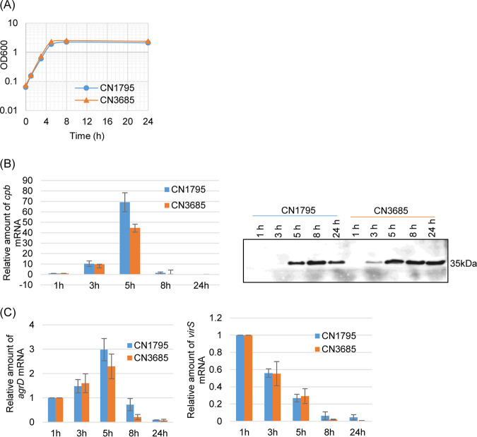
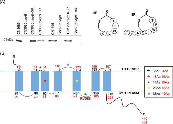
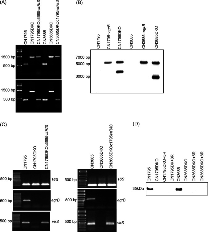
Since both the Agr (accessory gene regulator)-like quorum sensing (QS) system and VirS/VirR (VirS/R) two-component regulatory system of Clostridium perfringens positively regulate production of several toxins, including C. perfringens beta toxin (CPB), it has been hypothesized the VirS membrane sensor protein is an Agr-like QS signaling peptide (SP) receptor. To begin evaluating whether VirS is an SP receptor, this study sequenced the virS gene in C. perfringens strains CN3685 and CN1795 because it was reported that agrB mutants of both strains increase CPB production in response to the pentapeptide 5R, likely the natural SP, but only the CN3685 agrB mutant responds to 8R, which is 5R plus a 3-amino-acid tail. This sequencing identified differences between the predicted VirS extracellular loop 2 (ECL2) of CN3685 versus that of CN1795. To explore if those ECL2 differences explain strain-related variations in SP sensitivity and support VirS as an SP receptor, virS agrB double-null mutants of each strain were complemented to swap which VirS protein they produce. CPB Western blotting showed that this complementation changed the natural responsiveness of each strain to 8R. A pulldown experiment using biotin-5R demonstrated that VirS can bind SP. To further support VirS:SP binding and to identify a VirS binding site for SP, a 14-mer peptide corresponding to VirS ECL2 was synthesized. This ECL2 peptide inhibited 5R signaling to agrB mutant and wild-type strains. This inhibition was specific, since a single N to D substitution in the ECL2 peptide abrogated these effects. Collectively, these results support VirS as an important SP receptor and may assist development of therapeutics.IMPORTANCEC. perfringens beta toxin (CPB) is essential for the virulence of type C strains, a common cause of fatal necrotizing enteritis and enterotoxemia in humans and domestic animals. Production of CPB, as well as several other C. perfringens toxins, is positively regulated by both the Agr-like QS system and the VirS/R two-component regulatory system. This study presents evidence that the VirS membrane sensor protein is a receptor for the AgrD-derived SP and that the second extracellular loop of VirS is important for SP binding. Understanding interactions between SP and VirS improves knowledge of C. perfringens pathogenicity and may provide insights for designing novel strategies to reduce C. perfringens toxin production during infections.

Keywords: Agr-like quorum sensing; Clostridium perfringens; VirS/R two-component regulatory system; beta toxin; signal peptide receptor; toxin production regulation.


Figures

FIG 1
FIG 1
Growth curves and timing of Clostridium perfringens beta toxin (CPB) production versus virS, agrD, and cpb expression in wild-type CN1795 and CN3685 cultures. (A) Growth curves for wild-type CN1795 and CN3685 strains cultured at 37°C for up to 24 h in TY medium. Aliquots of each culture were measured for their optical density at 600 nm (OD600) at 1, 3, 5, 8, 10, and 24 h. This experiment was repeated three times, and a representative result is shown. (B) Reverse transcription-quantitative PCR (qRT-PCR) analyses of cpb expression levels (left) and CPB Western blot analysis (right) for CN1795 and CN3685. Transcript levels were determined using 10 ng of RNA isolated from TY cultures of these strains grown for 1, 3, 5, 8, 10, and 24 h at 37°C. Average threshold cycle (CT) values were normalized to that of the 16S rRNA housekeeping gene, and fold differences were calculated using the comparative CT method (2−ΔΔCT). Values of each bar indicate the fold change versus the 1-h culture. Western blot analysis for CPB production in supernatants of the two wild-type strains cultured in TY culture for 1, 3, 5, 8, 10, and 24 h. (C) qRT-PCR analyses of agrD and virS expression levels using the same cDNA preparation as that used for measuring cpb transcripts. Quantitative RT-PCR analyses shown in panels B and C were repeated three times, and mean values are shown. Error bars indicate standard deviations. A representative CPB Western blot based upon 3 repetitions is shown.
FIG 2
FIG 2
Modeling of the VirS proteins produced by CN1795 versus CN3685. (A) Comparison of signaling sensitivity of CN1795 and CN3658 to SP-based peptides 5R and 8R. CPB production (left) by wild-type or agrB null mutant (agrBKO) strains of CN3685 and CN1795 in the presence of 100 μM 5R and 8R (structures shown in the right panel). The samples for CPB Western blotting were prepared from the supernatants of overnight TY cultures (about 16 h) at 37°C. (B) The predicted topology of VirS protein produced by two wild-type strains. The predicted CN1795 VirS structure is shown in black numbers (total of 440 amino acids). The predicted CN3685 VirS structure is shown in red numbers (total of 446 amino acids). Asterisks (*) with different colors show the differences in this VirS region between these two strains. Amino acid (Aa) differences (indicated by asterisks) between the two strains are shown on the right side, with amino acid sequence numbers for CN1795 shown in black and those of CN3685 shown in red.
FIG 3
FIG 3
Preparation and characterization of CN1795 or CN3685 agrB virS double-null mutants and complementation of those double mutants to express a swapped VirS. (A) PCR confirmation of agrB virS double-null mutant construction by targeted intron-based mutagenesis and genetic complementation of those mutants with a swapped virR/S operon. Using DNA isolated from wild-type strains, a PCR assay amplified ∼500-bp products using internal agrB primers (upper) or internal virS primers (lower). After targeted insertion of a 900-bp intron, the same PCR assays amplified an ∼1.5-kb product using DNA isolated from the double-null mutant (DKO) strains. After complementation of the double mutants to carry the swapped virR/S operon (creating CN1795DKOc3685virR/S and CN3685DKOc1795virR/S), agrB PCR products remained the large size indicative of an intron insertion, but the virS PCR products were the smaller size present in wild-type strains, confirming genetic virS complementation. A 1-kb molecular ladder (Fisher Scientific) was used, with the size in bp shown at left. (B) Southern blot hybridization of an intron-specific probe with DNA from wild-type strains, the agrB single mutants, or agrB virS double-null mutants. DNA from each strain was digested with EcoRI and electrophoresed on a 1% agarose gel prior to blotting and hybridization with an intron-specific probe. Size of DNA fragments, in kb is shown at left. (C) RT-PCR analyses for expression of 16S RNA (top), the agrB gene (middle), or the virS gene (bottom) by wild-type CN1795 (left) or CN3685 (right), their agrB virS double-null mutants (DKO), or complementing strains of those double mutants with a swapped virR/S operon. Those samples did not amplify a product when subjected to PCR without reverse transcription, indicating that the RNA preparations from all strains were free from DNA contamination (data not shown). (D) Western blot analyses of CPB production by wild-type strains, agrB virS double-null mutants, or those mutants cultured in the presence of 100 μM 5R or 8R peptides. Size of proteins in kDa is shown at left. All experiments were repeated three times, and representative results of three repetitions are shown.
FIG 4
FIG 4
Western blot analyses of CPB production by wild-type, agrB virS double-null mutant, and swapped complementing strains, grown in the presence or absence of 100 μM 5R or 8R peptide. Cultures were grown for 5 h at 37°C in TY medium, and the OD600 values of each culture were adjusted to the same density. Supernatants were then collected, and equal volumes were subjected to CPB Western blotting. Size of proteins in kDa is shown at left. All experiments were repeated three times, and results representative of three repetitions are shown.
FIG 5
FIG 5
Preparation and characterization of CN1795 or CN3685 virS null mutants and complemented strains. (A) PCR confirmation of virS null mutants created by intron-based mutagenesis or complementing strains carrying a virR/S operon from CN1795 or CN3685 (indicated by c1795virR/S or c3685virR/S). Using DNA isolated from wild-type strains, an ∼500-bp product was PCR amplified using internal virS primers. However, after targeted insertion of a 900-bp intron into virS, the same PCR assay amplified an ∼1.5-kb product using DNA from the null mutant strains. Using DNA from the complemented strains, the same-size virS PCR products were amplified as when using DNA from wild-type strains. A 1-kb molecular ladder (Fisher Scientific) was also electrophoresed, and the size in bp is shown at left. (B) Southern blot hybridization of an intron-specific probe with DNA from wild type or the virS null mutants of CN1795 or CN3685. DNA from each strain was digested with EcoRI and then electrophoresed on a 1% agarose gel prior to blotting and hybridization with an intron-specific probe. Size of DNA fragments in kb is shown at right. (C) RT-PCR analyses of 16S RNA (top) and virS (bottom) transcription in wild-type CN1795 (left) or CN3685 (right), their virS null mutants, or complementing strains carrying the virR/S operon of either strain. To show that the RNA preparations from both strains were free from DNA contamination, the samples were also subjected to PCR without reverse transcription, but no products were amplified (data not shown). (D) Western blot analyses of CPB expression by wild type, virS null mutants, and complementing strains. Size of proteins in kDa is shown at left. All experiments were repeated three times, and results representative of three repetitions are shown.
FIG 6
FIG 6
Streptavidin beads containing bound B-5R pull down His6-tagged VirS. (A) CPB production by wild-type or agrB mutant CN1795 (upper), wild-type or agrB mutant CN3685 (lower), or those null mutants incubated in the presence of 100 μM B-5R. Size of proteins in kDa is shown at left. (B) His6 tag Western blot of B-PER buffer extracts of CN3685::virSc3685virR/Shis (lane 1) or CN3685::virSc3685virR/S (lane 3). After those extracts were incubated with streptavidin beads preincubated with B-5R or 5R, unbound supernatant material is shown in lanes 2 and 4. Pulldowns of extracts from complementing strains using beads preincubated with 5R or B-5R are shown in lanes 5 to 9. Note that in these lanes, using CN3685::virSc3685virR/Shis-containing extracts, but not CN3685::virSc3685virR/S-containing extracts, incubation of beads pretreated with B-5R, but not with 5R, resulted in pulldown of a protein that reacted with His6 tag antibody and was ∼50 kDa, the size of VirS. Size of protein markers in kDa is shown at left. All experiments were repeated three times, and results shown are representative of three repetitions.
FIG 7
FIG 7
The KIGK peptide corresponding to the major predicted VirS ECL2 inhibits the ability of the 5R SP to induce CPB production. (A) Western blot showing effects of KIGK preincubation with 5R on CPB production by agrB null mutant strains of CN1795 and CN3685. Cultures of these strains were grown for 5 h at 37°C in TY medium with DMSO, DMSO plus 5R (100 μM), or DMSO plus 5R (100 μM) that had been preincubated with KIGK (500 M). (B) Western blot showing effects of preincubation of 5R with the KIGK_D peptide, which corresponds to the mutated VirS 2nd ECL, on CPB production by agrB null mutant strains of CN1795 and CN3685. Cultures of those strains were grown for 5 h at 37°C in TY medium with DMSO, DMSO plus 5R (100 μM), or DMSO plus 5R (100 μM) that had been preincubated with the KIGK mutant peptide (KIGK_D; 500 μM). (C) Western blot showing effects of KIGK on CPB production by wild-type CN1795 and CN3685 cultures. Cultures of those strains were grown in TY medium with DMSO, DMSO plus KIGK (1 mM), or DMSO plus KIGK_D (1 mM) for 5 h at 37°C. For all panels, CPB Western blots of culture supernatants are shown. Size of proteins in kDa is shown at left. All experiments were repeated three times, and results representative of three repetitions are shown.
FIG 8
FIG 8
Quantitative RT-PCR analysis for virS and agrD gene expression in CN1795 (left) or CN3685 (right) wild-type, agrB, or virS null mutants, and complemented strains. Quantitative RT-PCR analyses of agrD (A) and virS (B) transcript levels were performed with 20 ng of the RNA isolated from 5-h (for agrD analyses) or 2-h (for virS analyses) TY medium cultures of the wild type, virS or agrB null mutants, and their complemented strains (comp). Average CT values were normalized to the value for the housekeeping 16S RNA gene, and the fold differences were calculated using the comparative CT (2−ΔΔCT) method. The value of each bar indicates the calculated fold change relative to the value for the wild-type strains. Shown are the mean values from three independent experiments. *, P < 0.05 compared to the wild-type culture by ordinary one-way analysis of variance (ANOVA).

Similar articles

Cited by

References

    1. McClane BA, Uzal FA, Mf M, D L, Td W. 2006. The enterotoxic clostridia, p 688–752. In Dworkin M, Falkow S, Rosenburg E, Schleifer H, Stackebrandt E (ed), The prokaryotes, 3rd ed Springer Press, New York, NY.
    1. Rood JI. 2006. Clostridium perfringens and histotoxic disease, p 753–770. In Dworkin M, Falkow S, Rosenburg E, Schleifer H, Stackebrandt E (ed), The prokaryotes, 3rd ed Springer Press, New York, NY.
    1. Uzal FA, Navarro MA, Li J, Freedman JC, Shrestha A, McClane BA. 2018. Comparative pathogenesis of enteric clostridial infections in humans and animals. Anaerobe 53:11–20. doi:10.1016/j.anaerobe.2018.06.002. - DOI - PMC - PubMed
    1. Navarro MA, McClane BA, Uzal FA. 2018. Mechanisms of action and cell death associated with Clostridium perfringens toxins. Toxins (Basel) 10:212. doi:10.3390/toxins10050212. - DOI - PMC - PubMed
    1. Uzal FA, Freedman JC, Shrestha A, Theoret JR, Garcia J, Awad MM, Adams V, Moore RJ, Rood JI, McClane BA. 2014. Towards an understanding of the role of Clostridium perfringens toxins in human and animal disease. Future Microbiol 9:361–377. doi:10.2217/fmb.13.168. - DOI - PMC - PubMed

Publication types

MeSH terms

LinkOut - more resources