

Emergence of anomalous dynamics in soft matter probed at the European XFEL
- PMID: 32934145
- PMCID: PMC7533660
- DOI: 10.1073/pnas.2003337117
Emergence of anomalous dynamics in soft matter probed at the European XFEL
Abstract
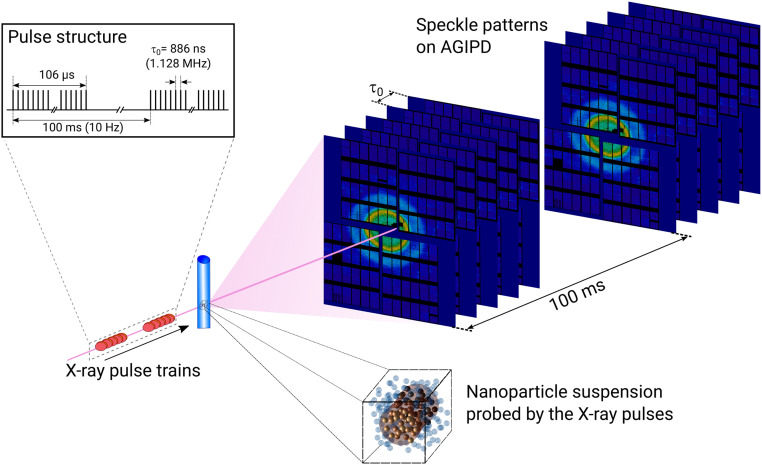
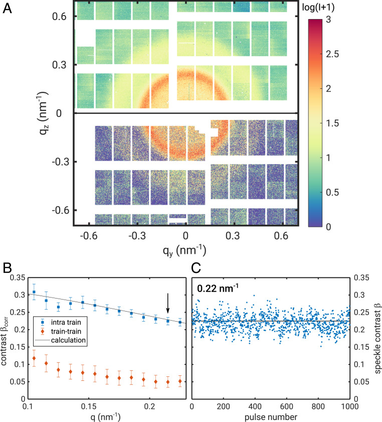
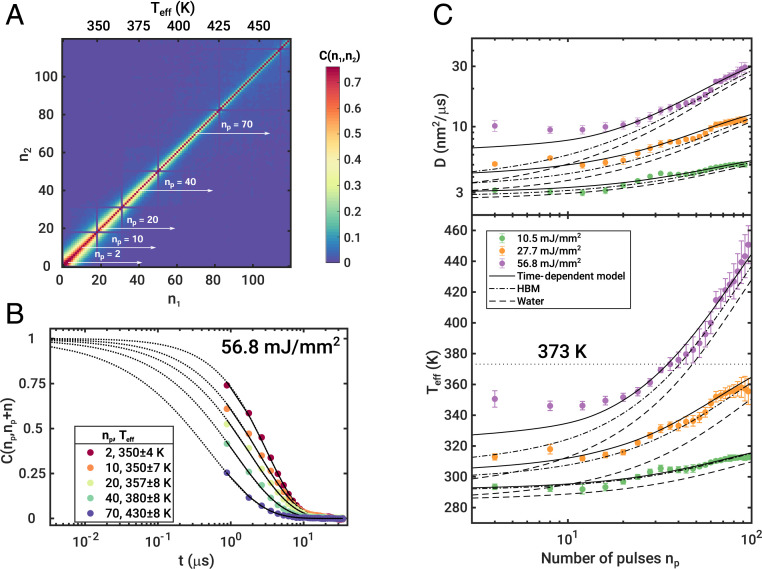
Dynamics and kinetics in soft matter physics, biology, and nanoscience frequently occur on fast (sub)microsecond but not ultrafast timescales which are difficult to probe experimentally. The European X-ray Free-Electron Laser (European XFEL), a megahertz hard X-ray Free-Electron Laser source, enables such experiments via taking series of diffraction patterns at repetition rates of up to 4.5 MHz. Here, we demonstrate X-ray photon correlation spectroscopy (XPCS) with submicrosecond time resolution of soft matter samples at the European XFEL. We show that the XFEL driven by a superconducting accelerator provides unprecedented beam stability within a pulse train. We performed microsecond sequential XPCS experiments probing equilibrium and nonequilibrium diffusion dynamics in water. We find nonlinear heating on microsecond timescales with dynamics beyond hot Brownian motion and superheated water states persisting up to 100 μs at high fluences. At short times up to 20 μs we observe that the dynamics do not obey the Stokes-Einstein predictions.
Keywords: Free-Electron Laser; X-ray photon correlation spectroscopy; diffusion; soft matter.
Conflict of interest statement
The authors declare no competing interest.
Figures

Similar articles
-
Microsecond hydrodynamic interactions in dense colloidal dispersions probed at the European XFEL.IUCrJ. 2021 Jul 28;8(Pt 5):775-783. doi: 10.1107/S2052252521006333. eCollection 2021 Sep 1. IUCrJ. 2021. PMID: 34584738 Free PMC article.
-
Resolving molecular diffusion and aggregation of antibody proteins with megahertz X-ray free-electron laser pulses.Nat Commun. 2022 Sep 21;13(1):5528. doi: 10.1038/s41467-022-33154-7. Nat Commun. 2022. PMID: 36130930 Free PMC article.
-
Megahertz-rate ultrafast X-ray scattering and holographic imaging at the European XFEL.J Synchrotron Radiat. 2022 Nov 1;29(Pt 6):1454-1464. doi: 10.1107/S1600577522008414. Epub 2022 Sep 29. J Synchrotron Radiat. 2022. PMID: 36345754 Free PMC article.
-
Megahertz data collection from protein microcrystals at an X-ray free-electron laser.Nat Commun. 2018 Aug 28;9(1):3487. doi: 10.1038/s41467-018-05953-4. Nat Commun. 2018. PMID: 30154468 Free PMC article.
-
MHz X-ray photon correlation spectroscopy using an acoustic levitator at the European XFEL.J Synchrotron Radiat. 2025 May 1;32(Pt 3):669-677. doi: 10.1107/S1600577525002875. Epub 2025 Apr 25. J Synchrotron Radiat. 2025. PMID: 40278848 Free PMC article.
Cited by
-
Exploring non-equilibrium processes and spatio-temporal scaling laws in heated egg yolk using coherent X-rays.Nat Commun. 2023 Sep 11;14(1):5580. doi: 10.1038/s41467-023-41202-z. Nat Commun. 2023. PMID: 37696830 Free PMC article.
-
Nanosecond X-ray photon correlation spectroscopy using pulse time structure of a storage-ring source.IUCrJ. 2021 Jan 1;8(Pt 1):124-130. doi: 10.1107/S2052252520015778. eCollection 2021 Jan 1. IUCrJ. 2021. PMID: 33520248 Free PMC article.
-
Theoretical Studies of Anisotropic Melting of Ice Induced by Ultrafast Nonthermal Heating.ACS Phys Chem Au. 2024 May 8;4(4):385-392. doi: 10.1021/acsphyschemau.3c00072. eCollection 2024 Jul 24. ACS Phys Chem Au. 2024. PMID: 39069981 Free PMC article.
-
Using coherent X-rays to follow dynamics in amorphous ices.Environ Sci Atmos. 2022 Sep 13;2(6):1314-1323. doi: 10.1039/d2ea00052k. eCollection 2022 Nov 10. Environ Sci Atmos. 2022. PMID: 36561555 Free PMC article.
-
Microsecond hydrodynamic interactions in dense colloidal dispersions probed at the European XFEL.IUCrJ. 2021 Jul 28;8(Pt 5):775-783. doi: 10.1107/S2052252521006333. eCollection 2021 Sep 1. IUCrJ. 2021. PMID: 34584738 Free PMC article.
References
-
- Emma P., et al. , First lasing and operation of an ångstrom-wavelength free-electron laser. Nat. Photonics 4, 641–647 (2010).
-
- Ishikawa T., et al. , A compact x-ray free-electron laser emitting in the sub-ångström region. Nat. Photon. 6, 540–544 (2012).
-
- Altarelli M., The European X-ray free-electron laser facility in Hamburg. Nucl. Instrum. Methods Phys. Res. Sect. B 269, 2845–2849 (2011).
-
- Yang H., Kim G., Kang H. S., First saturation of 14.5 keV free electron laser at PAL-XFEL. Nucl. Instrum. Methods Phys. Res. Sect. A 911, 51–54 (2018).
-
- Bostedt C., et al. , Linac coherent light source: The first five years. Rev. Mod. Phys. 88, 015007 (2016).
Publication types
LinkOut - more resources
Full Text Sources
