. 2020 Sep 15;12(1):24.
doi: 10.1038/s41368-020-00092-3.

Prognostic value of tertiary lymphoid structure and tumour infiltrating lymphocytes in oral squamous cell carcinoma

Affiliations

Prognostic value of tertiary lymphoid structure and tumour infiltrating lymphocytes in oral squamous cell carcinoma

Qunxing Li et al. Int J Oral Sci. .

Abstract

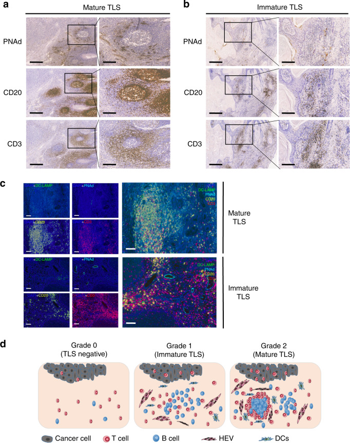
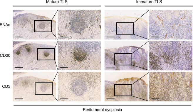
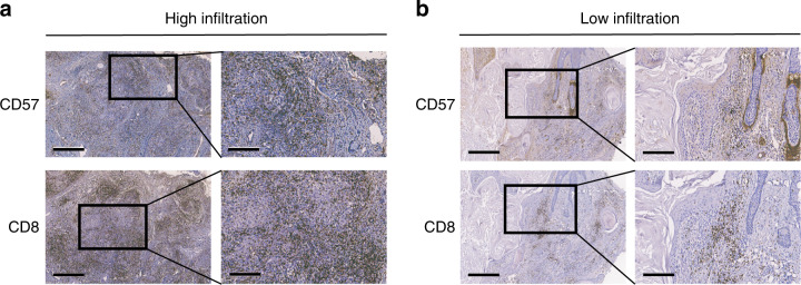
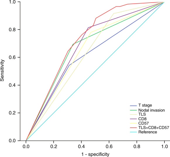
Tertiary lymphoid structures (TLS) are ectopic lymphoid structures in cancers that are largely associated with favourable prognosis. However, the prognostic value of TLSs in oral squamous cell carcinoma (OSCC) is largely unknown, and the association between tumour infiltrating lymphocytes (TILs) and TLSs has been rarely explored in OSCC. In this study, associated markers of TLS, including peripheral node address (PNAd) in high endothelial venules, CD20 in B cells and CD3 in T cells, were examined in 168 OSCC patients, and survival analysis was performed between TLS-positive and TLS-negative cohorts. We detected the presence of TILs by staining CD8+ cytotoxic T cells and CD57+ NK cells as well. TLSs appeared as highly organized structures in 45 (26.8%) cases. TLS-positive patients had a better 5-year overall survival (OS) rate (88.9% vs. 56.1%, P < 0.001) and relapse-free survival (RFS) rate (88.9% vs. 63.4%, P = 0.002). Moreover, the presence of TLS was an independent prognostic factor for both the 5-year OS rate (hazard ratio [HR] = 3.784; 95% confidence interval [CI], 1.498-9.562) and RFS rate (HR = 3.296; 95% CI, 1.279-8.490) in multivariate analysis. Furthermore, a higher density of CD8+ T cells and CD57+ NK cells was found in TLS-positive sections than in TLS-negative counterparts (P < 0.001), and their combination provided a higher predictive accuracy (AUC = 0.730; 95% CI, 0.654-0.805). In conclusion, our results suggest that TLS is an independent positive prognostic factor for OSCC patients. These findings provide a theoretical basis for the future diagnostic and therapeutic value of TLSs in OSCC treatment.


Conflict of interest statement

The authors declare no competing interests.

Figures

Fig. 1
Fig. 1
TLSs are highly organized structures in OSCC tissues and appear as clusters of B-cell follicles surrounded by T-cell zones and HEVs. a Mature TLS, similar to lymph node; CD20+ B cells form a follicular structure. The CD3+ T-cell zone is located near the follicle, and HEV are diffusely distributed around the follicle. b Immature TLS; CD20+ B cells do not form any follicular structure within the PNAd+ HEV area. CD3+ T cells were more diffusely distributed than mature TLSs. c Representative multiplex IHC images showing immune cells of immature TLS and mature TLS samples by simultaneous staining of B cells (CD20, yellow), T cells (CD3, red), mature dendritic cells (DC-LAMP, green), high endothelial venules (PNAd, cyan) and the nuclear stain DAPI (blue) (×200, original magnification). d TLS grading in OSCC tissues. Grade 0, no TLS present; grade 1, immature TLS; grade 2, mature TLS. Scale bar, 500 μm in “4X” pictures and 200 μm in “10X” pictures
Fig. 2
Fig. 2
Mature and immature TLSs are also located in OSCC peritumoural dysplasia tissues. The structure is similar to those located in the cancer nests. Scale bar, 500 μm in “4X” pictures and 200 μm in “10X” pictures
Fig. 3
Fig. 3
The presence of TLS could be a good prognostic factor in OSCC tissues. a, b Graphs showing the 5-year survival rates of TLS in 168 OSCC patients using the Kaplan–Meier method and log-rank test. a TLS-positive patients had a higher 5-year OS rate than TLS-negative patients (88.9% vs. 56.1%, P < 0.001). b TLS-positive patients had a higher 5-year RFS rate than TLS-negative patients (88.9% vs. 63.4%, P = 0.002). c, d The outcome of different subtypes of TLS in 45 TLS-positive OSCC patients using the Kaplan–Meier method and log-rank test. There were no significant differences in the 5-year OS and RFS rates between mature TLS and immature TLS in TLS-positive OSCC patients (94.1% vs. 85.7%, P = 0.411 and 88.2% vs. 89.3%, P = 0.896, respectively). e, f TCGA database showed that high TLS signature predicted better survival than low TLS signature in overall survival rate but no significance in relapse-free survival rate in head and neck cancer (P = 0.0081 and 0.63, respectively)
Fig. 4
Fig. 4
The distribution of CD8+ and CD57+ immune cells in OSCC tissues. a High infiltration of CD8+ and CD57+ immune cells in OSCC tissues. b Low infiltration of CD8+ and CD57+ immune cells in OSCC tissues. Scale bar, 500 μm in “4X” pictures and 200 μm in “10X” pictures
Fig. 5
Fig. 5
TLS combined with TIL had high predictive accuracy for 5-year OS. ROC curves indicating the predictive accuracy, sensitivity and specificity of each potential parameter. The AUCs of TLS, CD8 and CD57 were 0.641 (95% CI 0.558–0.725), 0.682 (95% CI 0.599–0.765) and 0.656 (95% CI 0.571–0.741, respectively), but TLS combined with CD8+ T cells and CD57+ NK cells provided the highest predictive accuracy (AUC = 0.730; 95% CI, 0.654–0.805)

References

    1. Bray F, et al. Global cancer statistics 2018: GLOBOCAN estimates of incidence and mortality worldwide for 36 cancers in 185 countries. CA Cancer J. Clin. 2018;68:394–424. - PubMed
    1. Chi AC, Day TA, Neville BW. Oral cavity and oropharyngeal squamous cell carcinoma–an update. CA Cancer J. Clin. 2015;65:401–421. - PubMed
    1. Siegel RL, Miller KD, Jemal A. Cancer statistics, 2020. CA Cancer J. Clin. 2020;70:7–30. - PubMed
    1. Ribas A, Wolchok JD. Cancer immunotherapy using checkpoint blockade. Science. 2018;359:1350–1355. - PMC - PubMed
    1. Ferris RL, et al. Nivolumab for recurrent squamous-cell carcinoma of the head and neck. N. Engl. J. Med. 2016;375:1856–1867. - PMC - PubMed

Publication types