

Voxel-based morphometry and task functional magnetic resonance imaging in essential tremor: evidence for a disrupted brain network
- PMID: 32934259
- PMCID: PMC7493988
- DOI: 10.1038/s41598-020-69514-w
Voxel-based morphometry and task functional magnetic resonance imaging in essential tremor: evidence for a disrupted brain network
Abstract
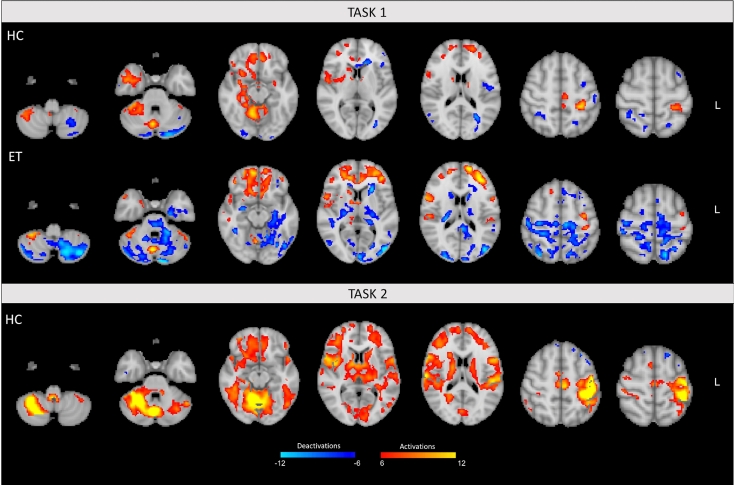
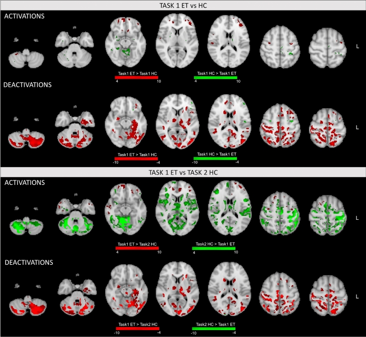
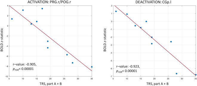
The pathophysiology of essential tremor (ET) is controversial and might be further elucidated by advanced neuroimaging. Focusing on homogenous ET patients diagnosed according to the 2018 consensus criteria, this study aimed to: (1) investigate whether task functional MRI (fMRI) can identify networks of activated and deactivated brain areas, (2) characterize morphometric and functional modulations, relative to healthy controls (HC). Ten ET patients and ten HC underwent fMRI while performing two motor tasks with their upper limb: (1) maintaining a posture (both groups); (2) simulating tremor (HC only). Activations/deactivations were obtained from General Linear Model and compared across groups/tasks. Voxel-based morphometry and linear regressions between clinical and fMRI data were also performed. Few cerebellar clusters of gray matter loss were found in ET. Conversely, widespread fMRI alterations were shown. Tremor in ET (task 1) was associated with extensive deactivations mainly involving the cerebellum, sensory-motor cortex, and basal ganglia compared to both tasks in HC, and was negatively correlated with clinical tremor scales. Homogeneous ET patients demonstrated deactivation patterns during tasks triggering tremor, encompassing a network of cortical and subcortical regions. Our results point towards a marked cerebellar involvement in ET pathophysiology and the presence of an impaired cerebello-thalamo-cortical tremor network.
Conflict of interest statement
The authors declare no competing interests.
Figures
Similar articles
-
Deep brain stimulation in the caudal zona incerta modulates the sensorimotor cerebello-cerebral circuit in essential tremor.Neuroimage. 2020 Apr 1;209:116511. doi: 10.1016/j.neuroimage.2019.116511. Epub 2019 Dec 31. Neuroimage. 2020. PMID: 31901420
-
Functional disconnection of the dentate nucleus in essential tremor.J Neurol. 2020 May;267(5):1358-1367. doi: 10.1007/s00415-020-09711-9. Epub 2020 Jan 23. J Neurol. 2020. PMID: 31974808
-
Role of altered cerebello-thalamo-cortical network in the neurobiology of essential tremor.Neuroradiology. 2017 Feb;59(2):157-168. doi: 10.1007/s00234-016-1771-1. Epub 2017 Jan 6. Neuroradiology. 2017. PMID: 28062908
-
Mapping Essential Tremor to a Common Brain Network Using Functional Connectivity Analysis.Neurology. 2023 Oct 10;101(15):e1483-e1494. doi: 10.1212/WNL.0000000000207701. Epub 2023 Aug 18. Neurology. 2023. PMID: 37596042 Free PMC article. Review.
-
Understanding the pathophysiology of essential tremor through advanced neuroimaging: a review.J Neurol Sci. 2013 Dec 15;335(1-2):9-13. doi: 10.1016/j.jns.2013.09.003. Epub 2013 Sep 10. J Neurol Sci. 2013. PMID: 24060292 Review.
Cited by
-
The Role of the Cerebellum in Tremor - Evidence from Neuroimaging.Tremor Other Hyperkinet Mov (N Y). 2021 Nov 15;11:49. doi: 10.5334/tohm.660. eCollection 2021. Tremor Other Hyperkinet Mov (N Y). 2021. PMID: 34820148 Free PMC article. Review.
-
Imaging the Pathophysiology of Essential Tremor-A Systematic Review.Front Neurol. 2021 Jun 16;12:680254. doi: 10.3389/fneur.2021.680254. eCollection 2021. Front Neurol. 2021. PMID: 34220687 Free PMC article.
-
Radiomics based on diffusion tensor imaging and 3D T1-weighted MRI for essential tremor diagnosis.Front Neurol. 2024 Aug 27;15:1460041. doi: 10.3389/fneur.2024.1460041. eCollection 2024. Front Neurol. 2024. PMID: 39263276 Free PMC article.
-
Oscillatory Coupling Between Thalamus, Cerebellum, and Motor Cortex in Essential Tremor.Mov Disord. 2025 May;40(5):896-905. doi: 10.1002/mds.30165. Epub 2025 Mar 3. Mov Disord. 2025. PMID: 40028845 Free PMC article.
-
Essential tremor disrupts rhythmic brain networks during naturalistic movement.Neurobiol Dis. 2025 Apr;207:106858. doi: 10.1016/j.nbd.2025.106858. Epub 2025 Feb 25. Neurobiol Dis. 2025. PMID: 40015653 Free PMC article.
References
Publication types
MeSH terms
LinkOut - more resources
Full Text Sources
Medical
